天津医药 ›› 2023, Vol. 51 ›› Issue (2): 124-130.doi: 10.11958/20220960
柴小兵( ), 张利, 褚菲菲, 吴慧丽△()
收稿日期:2022-06-21
修回日期:2022-07-13
出版日期:2023-02-15
发布日期:2023-02-24
通讯作者:
△E-mail:作者简介:柴小兵(1979),男,副主任医师,主要从事胃肠道肿瘤方面研究。E-mail:基金资助:
CHAI Xiaobing(), ZHANG Li, CHU Feifei, WU Huili△()
Received:2022-06-21
Revised:2022-07-13
Published:2023-02-15
Online:2023-02-24
Contact:
△E-mail:柴小兵, 张利, 褚菲菲, 吴慧丽. m6A识别蛋白HuR调控lncRNA TRG-AS1抑制结直肠癌生长的机制研究[J]. 天津医药, 2023, 51(2): 124-130.
CHAI Xiaobing, ZHANG Li, CHU Feifei, WU Huili. The mechanism of m6A recognition protein HuR inhibiting the growth of colorectal cancer by regulating lncRNA TRG-AS1[J]. Tianjin Medical Journal, 2023, 51(2): 124-130.
摘要:
目的 探讨N6-甲基腺苷(m6A)识别蛋白人类抗原R(HuR)调控长链非编码RNA T细胞受体γ位点反义RNA 1(lncRNA TRG-AS1)对结直肠癌(CRC)生长的影响。方法 比色法检测CRC患者的癌组织、癌旁组织及正常结肠上皮细胞NCM460及CRC细胞HCT116、SW480、LOVO中m6A含量;实时荧光定量PCR(qPCR)检测TRG-AS1表达;Western blot检测HuR蛋白表达。将HCT116细胞分为Ct组、OE-NC组、OE-HuR组、si-NC组、si-HuR组、si-HuR+pcDNA组、si-HuR+pcDNA-TRG-AS1组,CCK-8法检测细胞增殖;平板克隆实验检测细胞克隆形成能力;流式细胞术检测细胞凋亡率;划痕愈合实验检测细胞迁移;Transwell检测细胞侵袭;裸鼠体内移植瘤实验观察肿瘤生长情况;采用甲基化RNA免疫共沉淀(MeRIP)检测TRG-AS1上是否存在m6A位点;RNA pull-down实验和RNA免疫共沉淀(RIP)检测TRG-AS1与HuR蛋白的相互作用。结果 在CRC组织和细胞中,HuR蛋白、TRG-AS1高表达,m6A含量降低,且在HCT116细胞中HuR蛋白、TRG-AS1表达最高,m6A含量最低(P<0.05),选择HCT116细胞为研究对象。与si-NC组比较,si-HuR组HuR蛋白、TRG-AS1表达降低,m6A含量升高(P<0.05);与OE-NC组比较,OE-HuR组HuR蛋白、TRG-AS1表达升高,m6A含量降低(P<0.05);与si-HuR组、si-HuR+pcDNA组比较,si-HuR+pcDNA-TRG-AS1组HuR蛋白、m6A含量变化差异无统计学意义,TRG-AS1表达升高(P<0.05);下调HuR可抑制HCT116细胞增殖、迁移、侵袭及体内移植瘤的生长,促进细胞凋亡,而上调HuR则呈相反趋势;过表达TRG-AS1减弱了沉默HuR对HCT116细胞增殖、划痕愈合率、侵袭、体内移植瘤生长的抑制作用以及对细胞凋亡的促进作用;TRG-AS1上存在m6A位点,且TRG-AS1能与HuR蛋白相互作用。结论 沉默m6A识别蛋白HuR可通过抑制TRG-AS1表达进而抑制HCT116细胞增殖、迁移与侵袭,促进细胞凋亡。
中图分类号:
| 基因名称 | 引物序列(5→3?) | 产物大小(bp) |
|---|---|---|
| TRG-AS1 | 上游:GGAGTCTGCTCTAAGAGCTG 下游:CAGAGCAAAGATGCTCTGC | 145 |
| GAPDH | 上游:TGACTTCAACAGCGACACCCA 下游:CACCCTGTTGCTGTAGCCAAA | 126 |
表1 qPCR引物序列
Tab.1 qPCR primer sequences
| 基因名称 | 引物序列(5→3?) | 产物大小(bp) |
|---|---|---|
| TRG-AS1 | 上游:GGAGTCTGCTCTAAGAGCTG 下游:CAGAGCAAAGATGCTCTGC | 145 |
| GAPDH | 上游:TGACTTCAACAGCGACACCCA 下游:CACCCTGTTGCTGTAGCCAAA | 126 |
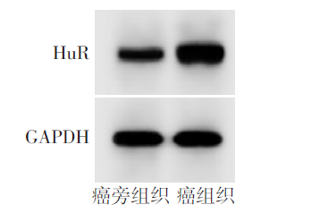
图1 Western blot检测组织中HuR蛋白表达
Fig.1 Western blot detection of HuR protein expression in tissue
| 组别 | HuR蛋白 | m6A含量(%) | TRG-AS1 |
|---|---|---|---|
| 癌旁组织 | 0.42±0.03 | 0.92±0.08 | 1.00±0.00 |
| 癌组织 | 1.35±0.21 | 0.31±0.02 | 2.22±0.21 |
| t | 19.110** | 32.244** | 25.323** |
表2 Comparison of HuR protein, m6A content and TRG-AS1 expression in cancer and adjacent tissues
Tab.2 癌组织和癌旁组织HuR蛋白、m6A含量和TRG-AS1表达比较 (n=19,$\bar{x}±s$)
| 组别 | HuR蛋白 | m6A含量(%) | TRG-AS1 |
|---|---|---|---|
| 癌旁组织 | 0.42±0.03 | 0.92±0.08 | 1.00±0.00 |
| 癌组织 | 1.35±0.21 | 0.31±0.02 | 2.22±0.21 |
| t | 19.110** | 32.244** | 25.323** |
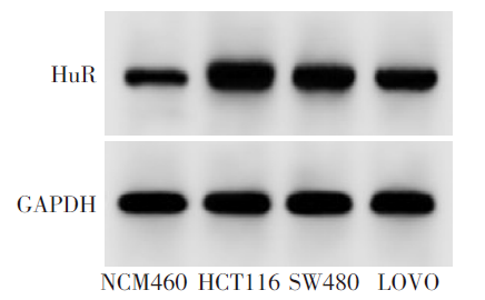
图2 Western blot检测细胞中HuR蛋白表达
Fig.2 Western blot detection of HuR protein expression in cells
| 组别 | HuR | m6A含量(%) | TRG-AS1 |
|---|---|---|---|
| NCM460 | 0.36±0.03 | 1.23±0.11 | 1.00±0.00 |
| HCT116 | 1.48±0.23a | 0.27±0.02a | 2.58±0.24a |
| SW480 | 1.21±0.20ab | 0.38±0.03ab | 2.27±0.22ab |
| LOVO | 1.01±0.09ab | 0.46±0.04ab | 2.04±0.23ab |
| F | 53.645** | 305.547** | 70.881** |
表3 Comparison of m6A content, HuR protein and TRG-AS1 expression between normal colonic epithelial cells and CRC cells
Tab.3 正常结肠上皮细胞及CRC细胞m6A含量、HuR蛋白、TRG-AS1表达比较 (n=6,$\bar{x}±s$)
| 组别 | HuR | m6A含量(%) | TRG-AS1 |
|---|---|---|---|
| NCM460 | 0.36±0.03 | 1.23±0.11 | 1.00±0.00 |
| HCT116 | 1.48±0.23a | 0.27±0.02a | 2.58±0.24a |
| SW480 | 1.21±0.20ab | 0.38±0.03ab | 2.27±0.22ab |
| LOVO | 1.01±0.09ab | 0.46±0.04ab | 2.04±0.23ab |
| F | 53.645** | 305.547** | 70.881** |
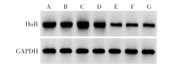
图3 Western blot检测HCT116细胞中HuR蛋白表达 A:Ct组;B:OE-NC组;C:OE-HuR组;D:si-NC组;E:si-HuR组;F:si-HuR+pcDNA组;G:si-HuR+pcDNA-TRG-AS1组。
Fig.3 Western blot detection of HuR protein expression in HCT116 cells
| 组别 | HuR | m6A含量 (%) | TRG-AS1 |
|---|---|---|---|
| Ct组 | 1.46±0.21 | 0.28±0.03 | 1.00±0.00 |
| OE-NC组 | 1.47±0.22 | 0.26±0.02 | 1.02±0.08 |
| OE-HuR组 | 1.98±0.25ab | 0.10±0.01ab | 2.34±0.22ab |
| si-NC组 | 1.45±0.22 | 0.27±0.01 | 1.01±0.09 |
| si-HuR组 | 0.35±0.03ac | 1.12±0.08ac | 0.71±0.12ac |
| si-HuR+pcDNA组 | 0.35±0.04 | 1.14±0.09 | 0.69±0.13 |
| si-HuR+pcDNA-TRG-AS1组 | 0.36±0.03 | 1.13±0.10 | 0.94±0.11de |
| F | 95.617** | 381.869** | 125.637** |
表4 Comparison of HuR protein, m6A content and TRG-AS1 expression in HCT116 cells between the seven groups
Tab.4 各组HCT116细胞中的HuR蛋白、m6A含量、TRG-AS1表达比较 (n=6,$\bar{x}±s$)
| 组别 | HuR | m6A含量 (%) | TRG-AS1 |
|---|---|---|---|
| Ct组 | 1.46±0.21 | 0.28±0.03 | 1.00±0.00 |
| OE-NC组 | 1.47±0.22 | 0.26±0.02 | 1.02±0.08 |
| OE-HuR组 | 1.98±0.25ab | 0.10±0.01ab | 2.34±0.22ab |
| si-NC组 | 1.45±0.22 | 0.27±0.01 | 1.01±0.09 |
| si-HuR组 | 0.35±0.03ac | 1.12±0.08ac | 0.71±0.12ac |
| si-HuR+pcDNA组 | 0.35±0.04 | 1.14±0.09 | 0.69±0.13 |
| si-HuR+pcDNA-TRG-AS1组 | 0.36±0.03 | 1.13±0.10 | 0.94±0.11de |
| F | 95.617** | 381.869** | 125.637** |
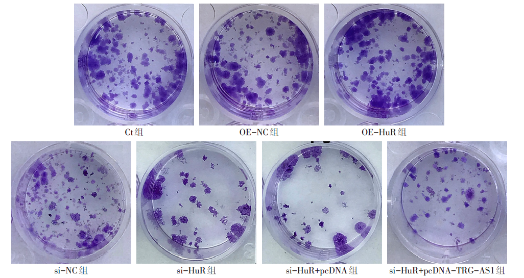
图4 沉默或过表达HuR对HCT116细胞克隆形成的影响
Fig.4 Effects of silencing or overexpression of HuR on HCT116 cell clone formation
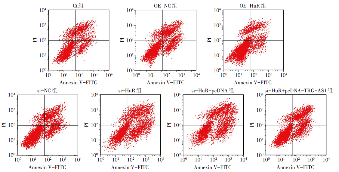
图5 Annexin V-FITC/PI双重染色检测沉默或过表达HuR对HCT116细胞凋亡的影响
Fig.5 Effects of silencing or overexpression of HuR on apoptosis of HCT116 cells detected by Annexin V-FITC/PI double staining
| 组别 | OD450值 | 克隆形成 率(%) | 细胞凋亡 率(%) |
|---|---|---|---|
| Ct组 | 0.82±0.08 | 62.23±5.14 | 41.27±3.68 |
| OE-NC组 | 0.83±0.09 | 63.15±5.09 | 42.26±3.71 |
| OE-HuR组 | 1.25±0.12ab | 83.36±6.05ab | 24.48±2.39ab |
| si-NC组 | 0.84±0.08 | 62.78±5.16 | 42.33±5.08 |
| si-HuR组 | 0.32±0.02ac | 26.69±2.44ac | 65.54±5.62ac |
| si-HuR+pcDNA组 | 0.33±0.03 | 26.75±2.38 | 64.57±5.41 |
| si-HuR+pcDNA-TRG-AS1组 | 0.69±0.05de | 48.87±4.12de | 46.67±3.08de |
| F | 112.818** | 126.394** | 67.001** |
表5 Comparison of OD450 value, clone formation rate and apoptosis rate between different groups of HCT116 cells
Tab.5 各组HCT116细胞OD450值、克隆形成率、细胞凋亡率比较 (n=6,$\bar{x}±s$)
| 组别 | OD450值 | 克隆形成 率(%) | 细胞凋亡 率(%) |
|---|---|---|---|
| Ct组 | 0.82±0.08 | 62.23±5.14 | 41.27±3.68 |
| OE-NC组 | 0.83±0.09 | 63.15±5.09 | 42.26±3.71 |
| OE-HuR组 | 1.25±0.12ab | 83.36±6.05ab | 24.48±2.39ab |
| si-NC组 | 0.84±0.08 | 62.78±5.16 | 42.33±5.08 |
| si-HuR组 | 0.32±0.02ac | 26.69±2.44ac | 65.54±5.62ac |
| si-HuR+pcDNA组 | 0.33±0.03 | 26.75±2.38 | 64.57±5.41 |
| si-HuR+pcDNA-TRG-AS1组 | 0.69±0.05de | 48.87±4.12de | 46.67±3.08de |
| F | 112.818** | 126.394** | 67.001** |
| 组别 | 划痕愈合率(%) | 侵袭细胞数目(个) |
|---|---|---|
| Ct组 | 51.17±4.66 | 74.43±6.25 |
| OE-NC组 | 51.28±5.02 | 74.58±6.12 |
| OE-HuR组 | 69.44±6.24ab | 113.35±10.04ab |
| si-NC组 | 51.34±5.13 | 73.96±6.11 |
| si-HuR组 | 21.25±2.06ac | 27.75±2.16ac |
| si-HuR+pcDNA组 | 21.36±2.09 | 28.14±2.02 |
| si-HuR+pcDNA-TRG-AS1组 | 36.67±3.24de | 51.14±4.33de |
| F | 100.512** | 159.505** |
表6 Comparison of wound healing rate and the number of invasive cells between different groups of HCT116 cells
Tab.6 各组HCT116细胞划痕愈合率、侵袭细胞数目比较 (n=6,$\bar{x}±s$)
| 组别 | 划痕愈合率(%) | 侵袭细胞数目(个) |
|---|---|---|
| Ct组 | 51.17±4.66 | 74.43±6.25 |
| OE-NC组 | 51.28±5.02 | 74.58±6.12 |
| OE-HuR组 | 69.44±6.24ab | 113.35±10.04ab |
| si-NC组 | 51.34±5.13 | 73.96±6.11 |
| si-HuR组 | 21.25±2.06ac | 27.75±2.16ac |
| si-HuR+pcDNA组 | 21.36±2.09 | 28.14±2.02 |
| si-HuR+pcDNA-TRG-AS1组 | 36.67±3.24de | 51.14±4.33de |
| F | 100.512** | 159.505** |
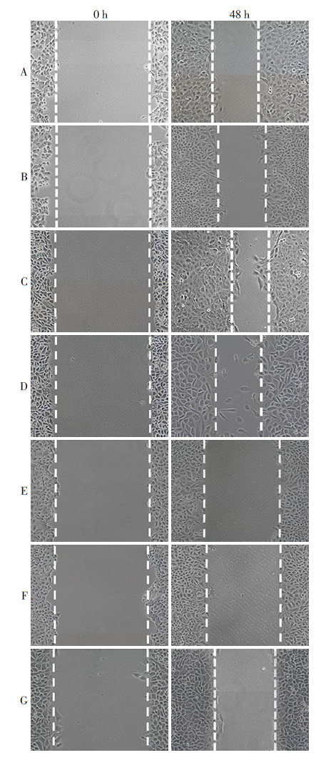
图6 沉默或过表达HuR对HCT116细胞迁移的影响 A:Ct组;B:OE-NC组;C:OE-HuR组;D:si-NC组;E:si-HuR组;F:si-HuR+pcDNA组;G:si-HuR+pcDNA-TRG-AS1组。
Fig.6 The effect of silencing or overexpressing HuR on the migration of HCT116 cells
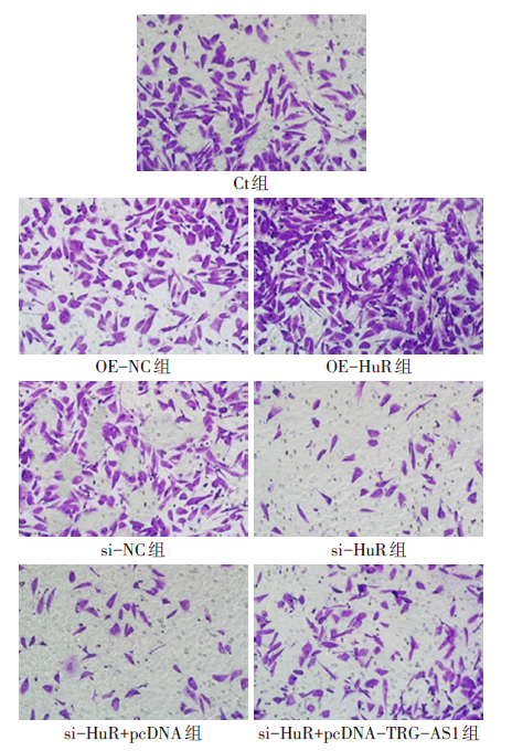
图7 沉默或过表达HuR对HCT116细胞侵袭的影响(结晶紫染色,×200)
Fig.7 The effect of silencing or overexpressing HuR on the invasion of HCT116 cells (crystal violet dyeing, ×200)
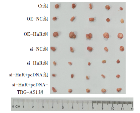
图8 各组移植瘤质量比较
Fig.8 Comparison of tumor quality in each group
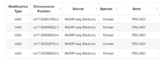
图9 RMVar数据库显示TRG-AS1上存在潜在的m6A位点
Fig.9 RMVar database showing potential m6A sites on TRG-AS1
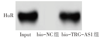
图10 RNA下拉实验表明HuR与TRG-AS1结合
Fig.10 RNA pull-down experiments showing HuR binds to TRG-AS1
| [1] | BRAY F, FERLAY J, SOERJOMATARAM I, et al. Global cancer statistics 2018:GLOBOCAN estimates of incidence and mortality worldwide for 36 cancers in 185 countries[J]. CA Cancer J Clin, 2018, 68(6):394-424. doi:10.3322/caac.21492. |
| [2] | RAWLA P, SUNKARA T, BARSOUK A. Epidemiology of colorectal cancer:incidence,mortality,survival,and risk factors[J]. Prz Gastroenterol, 2019, 14(2):89-103. doi:10.1055/s-0029-1242458. |
| [3] | MATTIUZZI C, SANCHIS-GOMAR F, LIPPI G. Concise update on colorectal cancer epidemiology[J]. Ann Transl Med, 2019, 7(21):609. doi:10.21037/atm.2019.07.91. |
| [4] | 王月帆, 葛春梅, 尹昊瓒, 等. m6A甲基化修饰识别蛋白YTHDF2在肝癌组织中的表达及临床意义[J]. 现代生物医学进展, 2021, 21(9):1601-1606. |
| WANG Y F, GE C M, YIN H Z, et al. Expression and clinical significance of m6A binding-protein YTHDF2 in hepatocellular carcinoma[J]. Progress in Modern Biomedicine, 2021, 21(9):1601-1606. doi:10.13241/j.cnki.pmb.2021.09.001. | |
| [5] | CHEN J, WU Y, LUO X, et al. Circular RNA circRHOBTB3 represses metastasis by regulating the HuR-mediated mRNA stability of PTBP1 in colorectal cancer[J]. Theranostics, 2021, 11(15):7507-7526. doi:10.7150/thno.59546. |
| [6] | SUN X, QIAN Y, WANG X, et al. LncRNA TRG-AS1 stimulates hepatocellular carcinoma progression by sponging miR-4500 to modulate BACH1[J]. Cancer Cell Int, 2020, 20:367. doi:10.1186/s12935-020-01440-3. |
| [7] | MA S, CHEN C, JI X, et al. The interplay between m6A RNA methylation and noncoding RNA in cancer[J]. J Hematol Oncol, 2019, 12(1):121. doi:10.1186/s13045-019-0805-7. |
| [8] | MA J Z, YANG F, ZHOU C C, et al. METTL14 suppresses the metastatic potential of hepatocellular carcinoma by modulating N(6)-methyladenosine-dependent primary microRNA processing[J]. Hepatology, 2017, 65(2):529-543. doi:10.1002/hep.28885. |
| [9] | 陈莉, 吉慧娟, 任利彬, 等. 烷基化修复蛋白B同源物5调控果蝇Zeste基因增强子人类同源物2对K562/阿霉素细胞耐药的影响[J]. 实用临床医药杂志, 2022, 26(7):87-92,97. |
| CHEN L, JI H J, REN L B, et al. Effect of alkylation repair protein B homolog 5 on drug resistance of K562/adriamycin cells by regulating enhancer of Zeste homolog 2[J]. Journal of Clinical Medicine in Practice, 2022, 26(7):87-92,97. doi:10.7619/jcmp.20213953. | |
| [10] | 许琼冠, 欧阳一彬, 谢镇明, 等. 胶质瘤中HuR通过上调PTBP1表达激活Akt通路对胶质瘤细胞的作用研究[J]. 临床神经外科杂志, 2021, 18(6):663-669. |
| XU Q G, OUYANG Y B, XIE Z M, et al. Effect of HuR in glioma cells on up-regulating expression of PTBP1 to activate Akt pathway[J]. Journal of Clinical Neurosurgery, 2021, 18(6):663-669. doi:10.3969/j.issn.1672-7770.2021.06.013. | |
| [11] | 湛钊, 周莉莉, 高义沙, 等. shRNA沉默HuR抑制肝细胞癌细胞系SMMC-7721的增殖、迁移及侵袭能力[J]. 中国生物化学与分子生物学报, 2020, 36(8):952-960. |
| ZHAN Z, ZHOU L L, GAO Y S, et al. shRNA silencing HuR inhibits proliferation, migration and invasion of SMMC-7721 hepatocellular carcinoma cells[J]. Chinese Journal of Biochemistry and Molecular Biology, 2020, 36(8):952-960. doi:10.13865/j.cnki.cjbmb.2020.07.1544. | |
| [12] | 曹小梅, 曹楠婧, 王福花, 等. 癌基因HuR调节胃癌功能的研究[J]. 中国病理生理杂志, 2019, 35(7):1328-1332. |
| CAO X M, CAO N J, WANG F H, et al. Effects of HuR on cell viability, migration and invasion of gastric cancer cell line MGC-803[J]. Chinese Journal of Pathophysiology, 2019, 35(7):1328-1332. doi:10.3969/j.issn.1000-4718.2019.07.028. | |
| [13] | WANG H, WEI W, ZHANG Z Y, et al. TCF4 and HuR mediated-METTL14 suppresses dissemination of colorectal cancer via N6-methyladenosine-dependent silencing of ARRDC4[J]. Cell Death Dis, 2021, 13(1):3. doi:10.1038/s41419-021-04459-0. |
| [14] | 费奕雯. m6A识别蛋白YTHDF2促慢性髓细胞白血病细胞生长的研究[D]. 苏州: 苏州大学, 2020. |
| FEI Y W. The m6A recognition protein YTHDF2 promotes the growth of chronic myeloid leukemia cells[D]. Suzhou: Soochow University, 2020. | |
| [15] | HE S, WANG X, ZHANG J, et al. TRG-AS1 is a potent driver of oncogenicity of tongue squamous cell carcinoma through microRNA-543/Yes-associated protein 1 axis regulation[J]. Cell Cycle, 2020, 19(15):1969-1982. doi:10.1080/15384101.2020.1786622. |
| [16] | XIE H, SHI S, CHEN Q, et al. LncRNA TRG-AS1 promotes glioblastoma cell proliferation by competitively binding with miR-877-5p to regulate SUZ12 expression[J]. Pathol Res Pract, 2019, 215(8):152476. doi:10.1016/j.prp.2019.152476. |
| [17] | ZHANG M, ZHU W, HAERYFAR M, et al. Long non-coding RNA TRG-AS1 promoted proliferation and invasion of lung cancer cells through the miR-224-5p/SMAD4 axis[J]. Onco Targets Ther, 2021, 14:4415-4426. doi:10.2147/OTT.S297336. |
| [1] | 杨晓芳, 贾新燕, 丰文君. miR-181a-5p通过HMGB1/NF-κB信号通路调控狼疮性肾炎小鼠肾小球系膜细胞增殖和凋亡[J]. 天津医药, 2026, 54(3): 232-237. |
| [2] | 王喆, 邱林, 马贲. 番茄来源胞外囊泡样颗粒对口腔鳞状细胞癌的作用效果研究[J]. 天津医药, 2026, 54(2): 145-150. |
| [3] | 李志伟, 张会超, 杨凤鸣, 曾垂义. 基于miR-144-3p/MAPK1通路探讨红参总皂苷对扩张型心肌病小鼠心肌细胞凋亡的影响[J]. 天津医药, 2026, 54(1): 23-29. |
| [4] | 赵兰君, 李良惠, 马馨, 巩娇娇, 郑臣辉, 石琳. 穿心莲内酯调控STAT3/GPX4通路对骨髓瘤细胞增殖和凋亡的影响[J]. 天津医药, 2026, 54(1): 8-13. |
| [5] | 孔翠文, 路延双, 孙丽萍, 于芬芬. LncRNA SNHG14靶向miR-30a-5p对高糖诱导的足细胞损伤的影响[J]. 天津医药, 2025, 53(9): 903-909. |
| [6] | 贾薇, 田志. Furin启动子甲基化水平与社区非糖尿病人群血压的相关性[J]. 天津医药, 2025, 53(9): 987-992. |
| [7] | 刘虹, 张玥玥, 王一琳, 王彩丽, 王晓敏, 毛敏, 李燕. MicroRNA-34a通过调控Wnt途径影响慢性淋巴细胞白血病进展的机制探讨[J]. 天津医药, 2025, 53(8): 785-790. |
| [8] | 曹振振, 叶睿, 刘佳瑶, 孟彤, 孙荣, 徐菱遥. 血清Hsp90α联合β2-MG检测在结直肠癌早期诊断和预后评估中的应用价值[J]. 天津医药, 2025, 53(7): 756-760. |
| [9] | 韩建存, 周谊. 川陈皮素调节FAK/AKT信号通路对喉鳞状细胞癌细胞增殖和凋亡的影响[J]. 天津医药, 2025, 53(6): 561-565. |
| [10] | 吴宾, 杨自更, 金玲, 张婧, 韦红梅, 蔡冰冰, 魏玉英. miRNA-381-3p/MuRF1轴对低氧性肺动脉高压小鼠心肺损伤的影响[J]. 天津医药, 2025, 53(6): 571-577. |
| [11] | 余朝霞, 马贲, 邱林, 高倩, 尼娜. 基于网络药理学和实验验证探究鲍式层孔菌多酚的抗头颈鳞癌机制[J]. 天津医药, 2025, 53(5): 456-461. |
| [12] | 李晨, 李占恩, 苏宏伟, 侯彩云, 董少文. KRT17调节Wnt/β-catenin信号通路对膀胱癌细胞增殖、凋亡及上皮间质转化的影响[J]. 天津医药, 2025, 53(5): 462-467. |
| [13] | 周美娟, 缪小祥. 帕金森病患者血清miR-214-3p、miR-124-3p表达水平与病情严重程度的关系及其早期诊断价值[J]. 天津医药, 2025, 53(5): 498-502. |
| [14] | 苏红见, 张春艳, 张卫东, 韩利, 乔亚红. 鸢尾素调控EBF3/ALOX15通路影响肺腺癌细胞增殖和迁移[J]. 天津医药, 2025, 53(4): 337-342. |
| [15] | 祁卫华, 黄广磊, 张媛媛, 班宏英, 毛诏旭. 连翘脂素调节cAMP/EPAC1/RAP1信号通路对肺癌细胞恶性进展的影响[J]. 天津医药, 2025, 53(4): 343-348. |
| 阅读次数 | ||||||
|
全文 |
|
|||||
|
摘要 |
|
|||||