
天津医药 ›› 2026, Vol. 54 ›› Issue (8): 874-880.doi: 10.11958/20260542
杨立恒( ), 李毅, 刘爽, 孔楠, 张冬睿, 贾玮△()
收稿日期:2026-03-05
修回日期:2026-04-15
出版日期:2026-08-15
发布日期:2026-08-07
通讯作者:
△E-mail:作者简介:杨立恒(1987),女,主治医师,主要从事肺部感染性疾病、慢性气道疾病、肺部肿瘤方面研究。E-mail:基金资助:
YANG Liheng(), LI Yi, LIU Shuang, KONG Nan, ZHANG Dongrui, JIA Wei△()
Received:2026-03-05
Revised:2026-04-15
Published:2026-08-15
Online:2026-08-07
Contact:
△E-mail: 杨立恒, 李毅, 刘爽, 孔楠, 张冬睿, 贾玮. 1例肺癌患者免疫检查点抑制剂治疗后肺结核潜伏感染激活的多学科诊疗分析[J]. 天津医药, 2026, 54(8): 874-880.
YANG Liheng, LI Yi, LIU Shuang, KONG Nan, ZHANG Dongrui, JIA Wei. Multidisciplinary diagnosis and treatment analysis of pulmonary tuberculosis latent infection activation in a lung cancer patient after immune checkpoint inhibitor therapy[J]. Tianjin Medical Journal, 2026, 54(8): 874-880.
摘要:
免疫检查点抑制剂(ICIs)已在肺癌中广泛应用,ICIs在提高患者总生存期的同时,会导致一系列与免疫治疗相关的不良反应。文章报告1例ⅢA期肺鳞癌患者在启用程序性细胞死亡蛋白1抑制剂替雷利珠单抗联合TP方案化疗后短期内发生活动性肺结核的诊疗经过,并分析该病例在诊断、治疗决策与全程管理中各专科的协作与贡献,阐明多学科诊疗在识别ICIs相关结核风险,以及精准鉴别、制定序贯治疗策略和改善患者预后中的价值,以期为临床实践提供参考。
中图分类号:
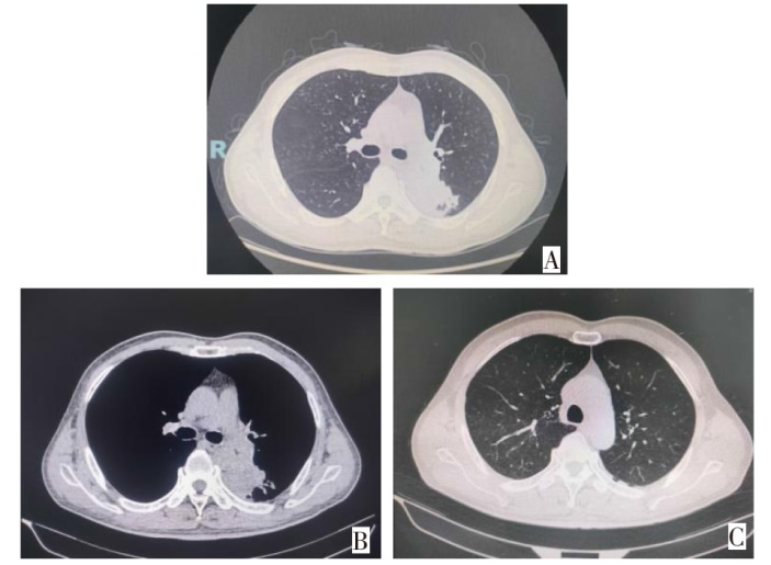
图1 患者入院前门诊胸部平扫CT表现 A、B:左肺下叶背段不规则软组织密度团块影,左肺下叶背段阻塞性炎症,左肺门及主肺动脉窗纵隔淋巴结增大;C:双肺上叶胸膜下散在囊状透亮影。
Fig.1 Chest plain CT findings of a patient on outpatient examination prior to admission
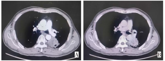
图2 患者胸部增强CT表现 A:左肺下叶背段不规则软组织密度团块于增强后呈不均匀、延迟强化,动脉期ΔCT值约30 HU;B:左肺下叶背段不规则软组织密度团块于增强后呈不均匀、延迟强化,延迟期ΔCT值约36 HU。
Fig.2 Chest enhanced CT findings of the patient
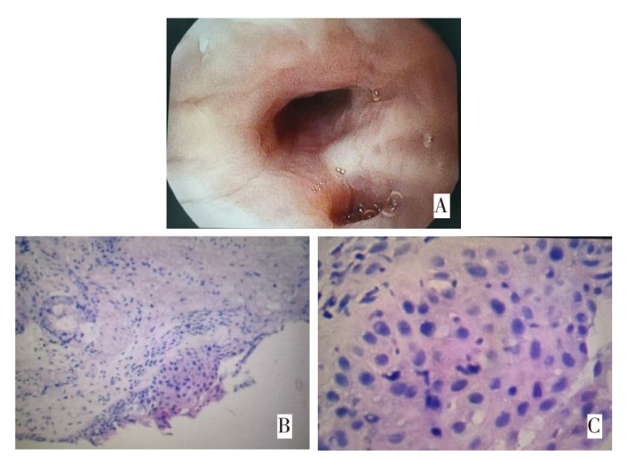
图3 患者支气管镜检查表现及病理活检结果 A:支气管镜检查示左下叶背段支气管黏膜水肿、充血,管腔略狭窄,于左下叶背段支气管亚段间嵴及远端黏膜活检;B、C:病理检测(HE染色,×100/×400),呈慢性炎症,部分上皮鳞化、不典型增生。
Fig.3 Bronchoscopic findings and pathological biopsy results of the patient
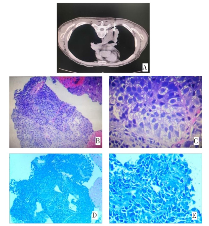
图4 患者行CT引导下经皮肺穿刺活检及其病理结果 A:CT引导下肺穿刺活检影像;B、C:左肺下叶穿刺活检结果(HE染色,×100/×400),为鳞状细胞癌(低分化);D、E:肺穿刺活检抗酸染色(Ziehl-Neelsen染色,×100/×400),未见抗酸杆菌。
Fig.4 CT findings and pathological results of percutaneous lung biopsy of the patient
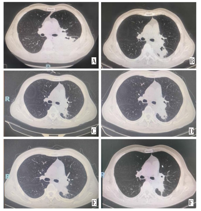
图5 患者治疗过程中影像学评估 A:免疫联合化疗1周期后见左下叶空洞形成,新发胸腔积液(2024年7月4日);B:抗结核治疗3周后左肺下叶空洞减小,胸腔积液明显减少(2024年7月22日);C:继续化疗4周期同时抗结核治疗,肿块减小,空洞进一步减小(2024年10月16日);D:左肺下叶肿块较前无著变,痰结核菌培养阴性,重启免疫治疗(2025年3月26日);E:免疫维持治疗中左肺下叶肿块影较前无著变(2025年10月23日);F:胸部放疗后左肺下叶肿块较前减小(2026年1月5日)。
Fig.5 Imaging evaluation of the patient during treatment process
| 序号 | 作者 | 发表 年份 | 患者性别/年龄 | 肺癌 类型 | 免疫治疗 药物 | 感染出现时间 | 影像学/ 临床表现 | 确诊 方式 | 抗结核 治疗时间 | 免疫治疗是否中断 | 结核病情 转归 | 肿瘤病情 转归 |
|---|---|---|---|---|---|---|---|---|---|---|---|---|
| 1 | Suliman等[ | 2021 | 女/58岁 | 肺腺癌 | 帕博利珠单抗 | 6个周期后 | 左肺实变伴空洞 | 支气管灌洗液涂片、PCR | 计划9个月 | 暂停,未重新启动 | 痰菌阴转 | 免疫治疗进展后,二线化疗获得接近完全缓解 |
| 2 | Murakami等[ | 2021 | 男/73岁 | 肺腺癌 | 帕博利珠单抗 | 约18个月后 | 右肺实变、结节 | 痰培养、PCR | 6个月 | 中断,完成抗结核治疗后重启 | 痰菌转阴(PCR和培养均阴性),肺部阴影消失 | 重启帕博利珠单抗后获得部分缓解 |
| 3 | Nakahama等[ | 2022 | 男/68岁 | 肺腺癌 | 帕博利珠单抗+化疗 | 4个月出现,9个月确诊 | 右肺S3 小结节 | 痰培养、PCR | 12个月(报道时仍在治疗中) | 未中断 | 右上肺结节缩小,痰培养转阴 | 肿瘤稳定 |
| 4 | Hirano等[ | 2024 | 男/67岁 | 肺腺癌 | 纳武利尤+伊匹木单抗 | 1个月后 | 空洞、左肺弥漫浸润 | 痰培养、PCR、VNTR证实复发 | 9个月 | 中断2个月后重启 | 完成抗结核治疗后2个月复发;再次治疗后痰菌转阴 | 免疫治疗10个月后进展,患者最终因呼吸衰竭死亡 |
| 5 | Takata等[ | 2019 | 男/75岁 | 肺腺癌 | 纳武利尤单抗 (PD-1) | 免疫治疗15个周期后 | 新发肺部浸润影 | 痰涂片、PCR、痰培养 | 9个月 | 中断,抗结核治疗4个月后重启 | 痰培养转为 阴性 | 重启纳武利尤后获得部分缓解 |
| 6 | Nikola等[ | 2023 | 女/52岁 | 肺腺癌 | 帕博利珠单抗 (PD-1) | 免疫治疗33个周期后 | 双肺粟粒样结节、树芽征 | 支气管镜肺泡灌洗液培养 | 未明确(治疗1个月后因肝毒性暂停) | 中断 | 症状好转,CT显示病灶吸收;结核病原学转阴 | 患者最终因肝衰竭死亡 |
| 7 | van Eeden等[ | 2019 | 女/56岁 | 肺腺癌 | 纳武利尤单抗 (PD-1) | 数月后 | 双肺新发结节、淋巴管炎 | 痰涂片阳性(大量) | 未明确 | 中断 | 抗结核治疗后临床改善,但病原学具体转归未明确报告 | 肿瘤进展,患者最终死亡 |
| 8 | Kato等[ | 2020 | 女/75岁 | 肺鳞癌 | 度伐利尤单抗 (PD-L1) | 巩固治疗5周期后 | 双肺浸润影、树芽征 | 痰涂片、T-SPOT、PCR | 未明确(报道时仍在治疗中) | 中断 | 痰PCR和涂片在治疗后转为阴性,病情得到控制 | 肿瘤控制 良好 |
| 9 | Chu等[ | 2017 | 男/59岁 | 肺腺癌 | 纳武利尤单抗 (PD-1) | 3个 周期后 | 心包积液、心包增厚 | 心包积液培养、活检 | 6个月 | 中断1个周期后重启 | 心包及胸腔积液消退,心包积液培养转阴 | 纳武利尤治疗6个月后进展 |
| 10 | Sirgiovanni等[ | 2021 | 男/44岁 | 小细胞肺癌 | 纳武利尤+伊匹木单抗 | 3个 周期后 | 双肺上叶空洞、纵隔淋巴结大 | 支气管镜肺泡灌洗液涂片、PCR | 未明确 | 中断 | 抗感染治疗后,痰菌转阴(具体时间未明确),肺部病灶显著缩小 | 获得长期缓解,停药24个月仍稳定 |
| 11 | 李亚坤等[ | 2025 | 男/65岁 | 肺鳞癌 | 帕博利珠单抗+化疗 | 治疗irAE过程中 | 双肺粟粒样结节;合并肺曲霉菌病 | 血NGS、痰X-pert、血培养 | 未明确(出院时仍在治疗中) | 中断 | 临床症状好转,胸部CT示结节吸收,病原学未明确 | 免疫联合化疗后获得部分 缓解 |
表1 肺癌免疫检查点抑制剂治疗后并发结核分枝杆菌感染患者特征总结
Tab.1 Summary of case characteristics of mycobacterium tuberculosis infection after immunotherapy with checkpoint inhibitors in lung cancer patients
| 序号 | 作者 | 发表 年份 | 患者性别/年龄 | 肺癌 类型 | 免疫治疗 药物 | 感染出现时间 | 影像学/ 临床表现 | 确诊 方式 | 抗结核 治疗时间 | 免疫治疗是否中断 | 结核病情 转归 | 肿瘤病情 转归 |
|---|---|---|---|---|---|---|---|---|---|---|---|---|
| 1 | Suliman等[ | 2021 | 女/58岁 | 肺腺癌 | 帕博利珠单抗 | 6个周期后 | 左肺实变伴空洞 | 支气管灌洗液涂片、PCR | 计划9个月 | 暂停,未重新启动 | 痰菌阴转 | 免疫治疗进展后,二线化疗获得接近完全缓解 |
| 2 | Murakami等[ | 2021 | 男/73岁 | 肺腺癌 | 帕博利珠单抗 | 约18个月后 | 右肺实变、结节 | 痰培养、PCR | 6个月 | 中断,完成抗结核治疗后重启 | 痰菌转阴(PCR和培养均阴性),肺部阴影消失 | 重启帕博利珠单抗后获得部分缓解 |
| 3 | Nakahama等[ | 2022 | 男/68岁 | 肺腺癌 | 帕博利珠单抗+化疗 | 4个月出现,9个月确诊 | 右肺S3 小结节 | 痰培养、PCR | 12个月(报道时仍在治疗中) | 未中断 | 右上肺结节缩小,痰培养转阴 | 肿瘤稳定 |
| 4 | Hirano等[ | 2024 | 男/67岁 | 肺腺癌 | 纳武利尤+伊匹木单抗 | 1个月后 | 空洞、左肺弥漫浸润 | 痰培养、PCR、VNTR证实复发 | 9个月 | 中断2个月后重启 | 完成抗结核治疗后2个月复发;再次治疗后痰菌转阴 | 免疫治疗10个月后进展,患者最终因呼吸衰竭死亡 |
| 5 | Takata等[ | 2019 | 男/75岁 | 肺腺癌 | 纳武利尤单抗 (PD-1) | 免疫治疗15个周期后 | 新发肺部浸润影 | 痰涂片、PCR、痰培养 | 9个月 | 中断,抗结核治疗4个月后重启 | 痰培养转为 阴性 | 重启纳武利尤后获得部分缓解 |
| 6 | Nikola等[ | 2023 | 女/52岁 | 肺腺癌 | 帕博利珠单抗 (PD-1) | 免疫治疗33个周期后 | 双肺粟粒样结节、树芽征 | 支气管镜肺泡灌洗液培养 | 未明确(治疗1个月后因肝毒性暂停) | 中断 | 症状好转,CT显示病灶吸收;结核病原学转阴 | 患者最终因肝衰竭死亡 |
| 7 | van Eeden等[ | 2019 | 女/56岁 | 肺腺癌 | 纳武利尤单抗 (PD-1) | 数月后 | 双肺新发结节、淋巴管炎 | 痰涂片阳性(大量) | 未明确 | 中断 | 抗结核治疗后临床改善,但病原学具体转归未明确报告 | 肿瘤进展,患者最终死亡 |
| 8 | Kato等[ | 2020 | 女/75岁 | 肺鳞癌 | 度伐利尤单抗 (PD-L1) | 巩固治疗5周期后 | 双肺浸润影、树芽征 | 痰涂片、T-SPOT、PCR | 未明确(报道时仍在治疗中) | 中断 | 痰PCR和涂片在治疗后转为阴性,病情得到控制 | 肿瘤控制 良好 |
| 9 | Chu等[ | 2017 | 男/59岁 | 肺腺癌 | 纳武利尤单抗 (PD-1) | 3个 周期后 | 心包积液、心包增厚 | 心包积液培养、活检 | 6个月 | 中断1个周期后重启 | 心包及胸腔积液消退,心包积液培养转阴 | 纳武利尤治疗6个月后进展 |
| 10 | Sirgiovanni等[ | 2021 | 男/44岁 | 小细胞肺癌 | 纳武利尤+伊匹木单抗 | 3个 周期后 | 双肺上叶空洞、纵隔淋巴结大 | 支气管镜肺泡灌洗液涂片、PCR | 未明确 | 中断 | 抗感染治疗后,痰菌转阴(具体时间未明确),肺部病灶显著缩小 | 获得长期缓解,停药24个月仍稳定 |
| 11 | 李亚坤等[ | 2025 | 男/65岁 | 肺鳞癌 | 帕博利珠单抗+化疗 | 治疗irAE过程中 | 双肺粟粒样结节;合并肺曲霉菌病 | 血NGS、痰X-pert、血培养 | 未明确(出院时仍在治疗中) | 中断 | 临床症状好转,胸部CT示结节吸收,病原学未明确 | 免疫联合化疗后获得部分 缓解 |
| [1] | Haanen J, Obeid M, Spain L, et al. Management of toxicities from Immunotherapy: ESMO clinical practice guideline for diagnosis,treatment and follow-up[J]. Ann Oncol 2022, 33:1217-1238. doi:10.1016/j.annonc.2022.10.001. |
| [2] | Postow M A, Sidlow R, Hellmann M D. Immune-related adverse events associated with immune Checkpoint blockade[J]. N Engl J Med, 2018, 378(2):158-168. doi:10.1056/NEJMra1703481. |
| [3] | Wang S L, Chan T A. Navigating established and emerging biomarkers for immune checkpoint inhibitor therapy[J]. Cancer Cell, 2025, 43(4):641-664. doi:10.1016/j.ccell.2025.03.006. |
| [4] | 崔语恒, 赵少荣, 刘晶晶, 等. PD-1/PD-L1抑制剂联合其他免疫检查点抑制剂治疗三阴性乳腺癌的研究进展[J]. 天津医药, 2020, 48(12):1230-1235. |
| Cui Y H, Zhao S R, Liu J J, et al. Research progress of PD-1/PD-L1 inhibitors combined with other immune checkpoint inhibitors in the treatment of triple negative breast cancer[J]. Tianjin Med J, 2020, 48(12):1230-1235. doi:10.11958/20200705. | |
| [5] | Sharma P, Goswami S, Raychaudhuri D, et al. Immune checkpoint therapy-current perspectives and future directions[J]. Cell, 2023, 186(8):1652-1669. doi:10.1016/j.cell.2023.03.006. |
| [6] | World Health Organization. Global tuberculosis report 2025[R]. Geneva:WHO, 2025. |
| [7] | Liu K W, Wang D P, Yao C, et al. Increased tuberculosis incidence due to immunotherapy based on PD-1 and PD-L1 blockade:a systematic review and meta-analysis[J]. Front Immunol, 2022, 13:27220. doi:10.3389/fimmu.2022.727220. |
| [8] | Suliman A M, Bek S A, Elkhatim M S, et al. Tuberculosis following programmed cell death receptor-1(PD-1)inhibitor in a patient with non-small cell lung cancer. Case report and literature review[J]. 2021, 70(4):935-944. doi:10.1007/s00262-020-02726-1. |
| [9] | Zaemes J, Kim C. Immune checkpoint inhibitor use and tuberculosis:a systematic review of the literature[J]. Eur J Cancer, 2020, 132:168-175. doi:10.1016/j.ejca.2020.03.015. |
| [10] | Murakami S, Usui R, Nakahara Y, et al. Readministration of pembrolizumab after treatment of tuberculosis activated by initial pembrolizumab therapy[J]. Intern Med, 2021, 60(11):1743-1746. doi:10.2169/internalmedicine.6002-20. |
| [11] | Nakahama K, Kaneda H, Ogawa K, et al. Safe concurrent use of anti-tuberculosis drugs and pembrolizumab in a patient with non-small-cell lung cancer who was infected with mycobacterium tuberculosis[J]. Intern Med, 2022, 61(7):1039-1042. doi:10.2169/internalmedicine.6811-20. |
| [12] | Hirano S, Takahashi H, Nakamura S, et al. A case of pulmonary tuberculosis that developed during nivolumab and ipilimumab treatment for pulmonary adenocarcinoma that recurred two months after completion of anti-tuberculous treatment[J]. Chin Clin Oncol, 2024, 13(3):43. doi:10.21037/cco-23-153. |
| [13] | Takata S, Koh G, Han Y, et al. Paradoxical response in a patient with non-small cell lung cancer who received nivolumab followed by anti-Mycobacterium tuberculosis agents[J]. J Infect Chemother, 2019, 25(1):54-58. doi:10.1016/j.jiac.2018.06.016. |
| [14] | Nikola N, Natasa D, Milica K. A rare case of miliary tuberculosis in a patient with NSCLC receiving PD1 inhibitor therapy,mimicking autoimmune pneumonitis[J]. J Cancer Res Ther, 2023, 19(Suppl 2):S909-S911. doi:10.4103/jcrt.jcrt_410_22. |
| [15] | Van Eeden R, Rapoport B L, Smit T, et al. Tuberculosis infection in a patient treated with nivolumab for non-small cell lung cancer:case report and literature review[J]. Front Oncol, 2019, 9:659. doi:10.3389/fonc.2019.00659. |
| [16] | Kato Y, Watanabe Y, Yamane Y, et al. Reactivation of TB during administration of durvalumab after chemoradiotherapy for non-small-cell lung cancer:a case report[J]. Immunotherapy, 2020, 12(6):373-378. doi:10.2217/imt-2020-0061. |
| [17] | Chu Y C, Fang K C, Chen H C, et al. Pericardial tamponade caused by a hypersensitivity response to tuberculosis reactivation after anti-PD-1 treatment in a patient with advanced pulmonary adenocarcinoma[J]. J Thorac Oncol, 2017, 12(8):e111-e114. doi:10.1016/j.jtho.2017.03.012. |
| [18] | Sirgiovanni M, Hinterleitner C, Horger M, et al. Long-term remission of small cell lung cancer after reactivation of tuberculosis following immune-checkpoint blockade:a case report[J]. Thorac Cancer, 2021, 12(5):699-702. doi:10.1111/1759-7714.13821. |
| [19] | 李亚坤, 谭蓓, 秦岭, 等. 《免疫检查点抑制剂相关结肠炎治疗过程中发生致命性机会性感染1例[J]. 中华结核和呼吸杂志, 2025, 48(3):272-276. |
| Li Y K, Tan B, Qin L, et al. Fatal opportunistic infection occurred during the treatment of immune checkpoint inhibitor-associated colitis:case report[J]. Chin J Tuberc Respir Dis, 2025, 48(3):272-276. doi:10.3760/cma.j.cn112147-20240920-00553. | |
| [20] | Zhan M, Zhou L Y, Liu R, et al. Tuberculosis infection following administration of immune checkpoint inhibitors:a real world observational study[J]. Front Pharmacol, 2025, 16:1703831. doi:10.3389/fphar.2025.1703831. |
| [21] | Dhar C. Testing for latent tuberculosis before starting patients on immune checkpoint inhibitors[J]. Indian Cancer, 2021, 58(3):469-470. doi:10.4103/ijc.IJC_283_20. |
| [22] | Luo T Y, Chen W S, Huang D H, et al. Preclinical models of immune checkpoint inhibitors-related interstitial pneumonia for anti-PD1 tumor immunotherapy[J]. Immunobiology, 2025, 230(2):152884. doi:10.1016/j.imbio.2025.152884. |
| [23] | 张文宏, 李太生. 免疫抑制剂与结核潜伏感染激活的临床专家共识(2025版)[J]. 中华传染病杂志, 2024, 42(12):715-735. |
| Zhang W H, Li T S. Clinical expert consensus on immunosuppressants and the activation of latent tuberculosis infection(2025 edition)[J]. Chinese Journal of Infectious Diseases, 2024, 42(12):715-735. doi:10.3760/cma.j.cn311365-20250228-00064. | |
| [24] | Zhou G, Luo Q, Luo S, et al. Interferon-γ release assays or tuberculin skin test for detection and management of latent tuberculosis infection:a systematic review and meta-analysis[J]. Lancet Infect Dis, 2020, 20(12):1457-1469. doi:10.1016/S1473-3099(20)30276-0. |
| [25] | Langan E A, Graetz V, Allerheiligen J, et al. Immune checkpoint inhibitors and tuberculosis: an old disease in a new context[J]. Lancet Oncol, 2020, 21(1):e55-65. doi:10.1016/S1470-2045(19)30674-6. |
| [26] | Fujita K, Elkington P, Redelman-Sidi G, et al. Serial interferon-gamma release assay in lung cancer patients receiving immune checkpoint inhibitors:a prospective cohort study[J]. Cancer Immunol Immunother, 2022, 71(11):2757-2764. doi:10.1007/s00262-022-03198-1. |
| [27] | Shi J, Li J, Wang Q, et al. The safety and efffcacy of immunotherapy with anti-programmed cell death 1 monoclonal antibody for lung cancer complicated with Mycobacterium tuberculosis infection[J]. Transl Lung Cancer Res, 2021, 10(10):3929-3942. doi:10.21037/tlcr-21-524. |
| [28] | Zhu J, He Z C, Liang D, et al. Pulmonary tube:rculosis associated with immune checkpoint inhibitors: a pharmacovigilance study[J]. Thorax, 2022, 77(7):721-723. doi:10.1136/thoraxjnl-2021-217575. |
| [29] | Yin J Q, Wu Y J, Yang X, et al. Checkpoint inhibitor pneumonitis induced by anti-PD-1/PD-L1 therapy in non-small-cell lung cancer:occurrence and mechanism[J]. Front Immunol, 2022, 13:830631. doi:10.3389/fimmu.2022.830631. |
| [1] | 梁东阁, 曹欢, 张婷, 何倩倩, 黄晗, 仝瑞防. 地榆皂苷Ⅰ调节RhoA/ROCK信号通路对支原体肺炎幼鼠肺组织炎性损伤的影响[J]. 天津医药, 2026, 54(8): 808-814. |
| [2] | 张晶, 桂晓玲, 李金鹏, 陈新. 右束支传导阻滞联合血清HMGB1在COPD患者并发肺心病监测中的临床价值[J]. 天津医药, 2026, 54(8): 842-847. |
| [3] | 张渤, 徐伟, 张静, 马肖. GFPT1通过调节PD-L1通路对非小细胞肺癌细胞的影响[J]. 天津医药, 2026, 54(7): 687-692. |
| [4] | 张轶轶, 刘帝亮, 陈飞, 龚媛媛, 王贵生. MTA1、ARL2和VEGF-C在非小细胞肺癌组织中的表达[J]. 天津医药, 2026, 54(7): 705-710. |
| [5] | 钱进, 魏夕阳, 徐冲. 血磷、血镁水平与老年COPD急性加重患者病情及预后的关系[J]. 天津医药, 2026, 54(7): 765-770. |
| [6] | 崔志刚, 英策, 张岑, 李棣华, 卓玉珍, 杨磊. 土贝母来源外泌体对脓毒症诱导急性肺损伤的保护作用[J]. 天津医药, 2026, 54(6): 591-597. |
| [7] | 杨丹, 刘翠翠, 赵东芳, 刘政, 朱亚芳. CTI联合CALLY指数辅助诊断老年慢性阻塞性肺疾病急性加重合并心力衰竭的临床价值[J]. 天津医药, 2026, 54(6): 603-607. |
| [8] | 尚葛础, 王冠. 衰弱导向的老年神经介入术后肺炎风险管理的研究进展[J]. 天津医药, 2026, 54(6): 663-667. |
| [9] | 李士欣, 王佳佳, 何其瑾, 刘思琦, 姜潇思, 范习凯, 宋淑美, 翟建华. 1例脓毒性心肌病继发心室电风暴患者多学科诊疗分析[J]. 天津医药, 2026, 54(5): 544-549. |
| [10] | 姚国华, 刘杰, 张雯, 马翠安, 魏博涛, 高娜. 基于BP神经网络构建儿童肺炎支原体混合腺病毒感染的重症肺炎预测模型[J]. 天津医药, 2026, 54(4): 369-373. |
| [11] | 张润春, 李树华, 王玉珍, 王巧文. 肺炎支原体肺炎患儿血清LXA4和KLF5表达的临床意义[J]. 天津医药, 2026, 54(3): 269-274. |
| [12] | 朱海燕, 王烨, 尹艳. 老年NSCLC患者术后胃肠功能紊乱的危险因素研究[J]. 天津医药, 2026, 54(3): 289-294. |
| [13] | 熊炳贤, 王景坤, 刘衬云, 陈惠群. 不同浓度罗哌卡因竖脊肌平面阻滞对胸腔镜肺叶切除术患者的影响[J]. 天津医药, 2026, 54(3): 314-318. |
| [14] | 黄熷远, 付靖, 赵亚, 王龙灏, 仓顺东. 非小细胞肺癌EGFR-TKI耐药与p53基因突变的研究进展[J]. 天津医药, 2026, 54(3): 333-336. |
| [15] | 张庆玲, 刘仕琦, 付殷, 李冀. 泻白散对LPS诱导幼龄大鼠急性肺损伤的调控作用机制[J]. 天津医药, 2026, 54(2): 151-157. |
| 阅读次数 | ||||||
|
全文 |
|
|||||
|
摘要 |
|
|||||
