天津医药 ›› 2022, Vol. 50 ›› Issue (10): 1014-1019.doi: 10.11958/20220649
郭玉静( ), 胡英, 龙启福, 许玉珍, 李积东, 永胜(),△
收稿日期:2022-04-27
修回日期:2022-06-07
出版日期:2022-10-15
发布日期:2022-10-20
通讯作者:
△永胜
E-mail:ly47757@163.com;yongsheng@qhu.edu.cn
作者简介:郭玉静(1998),女,硕士在读,主要从事低氧对免疫功能改变及适应机制方面研究。E-mail: 基金资助:
GUO Yujing(), HU Ying, LONG Qifu, XU Yuzhen, LI Jidong, YONG Sheng(),△
Received:2022-04-27
Revised:2022-06-07
Published:2022-10-15
Online:2022-10-20
Contact:
△YONG Sheng
E-mail:ly47757@163.com;yongsheng@qhu.edu.cn
郭玉静, 胡英, 龙启福, 许玉珍, 李积东, 永胜. 低氧暴露对小鼠脾淋巴细胞增殖和凋亡的影响[J]. 天津医药, 2022, 50(10): 1014-1019.
GUO Yujing, HU Ying, LONG Qifu, XU Yuzhen, LI Jidong, YONG Sheng. Effects of hypoxia exposure on proliferation and apoptosis of mouse spleen lymphocytes[J]. Tianjin Medical Journal, 2022, 50(10): 1014-1019.
摘要:
目的 研究低氧暴露诱导淋巴细胞数量减少与细胞增殖和凋亡的关系。方法 从C57BL/6小鼠脾中分离淋巴细胞,将淋巴细胞分别在低氧(1%O2)和常氧(21%O2)环境中培养12、24、48 h。用流式细胞术检测T、B淋巴细胞数量;羧基荧光素乙酰乙酸和AnnexinⅤ-FITC/PI方法分别检测淋巴细胞的增殖和凋亡;扫描电镜观察淋巴细胞形态;实时荧光定量逆转录PCR和Western blot检测凋亡相关因子鼠源B细胞淋巴瘤/白血病-2(bcl-2)、bcl-2同源拮抗剂-杀伤蛋白(Bak)和胱天蛋白酶-3(caspase-3)的mRNA和蛋白表达水平。结果 低氧暴露12、24、48h,可降低T、B淋巴细胞数量,抑制淋巴细胞增殖,促进细胞凋亡;扫描电镜观察显示,同一时间下,低氧组淋巴细胞更早出现凋亡特征形态学变化;低氧暴露12、24、48 h,淋巴细胞Bak和caspase-3的mRNA和蛋白表达水平均上调,而bcl-2的mRNA和蛋白表达水平均下调。结论 低氧暴露通过抑制淋巴细胞增殖、促进凋亡,从而介导淋巴细胞数量下降。
中图分类号:
| 基因 名称 | 引物序列(5′→3′) | 产物大小 (bp) |
|---|---|---|
| β-actin | 上游:CATCCGTAAAGACCTCTATGCCAAC | 171 |
| 下游:ATGGAGCCACCGATCCACA | ||
| Bak | 上游:GGTCTTTCGAAGCTACGTTTTT | 243 |
| 下游:ATCTTGGTGAAGAGTTCGTAGG | ||
| bcl-2 | 上游:GATGACTTCTCTCGTCGCTAC | 156 |
| 下游:GAACTCAAAGAAGGCCACAATC | ||
| caspase-3 | 上游:GGCCTGAAATACCAAGTCAGGAA | 128 |
| 下游:CCATGGCTTAGAATCACACACACA |
表1 qPCR引物序列
Tab. 1 Sequence of the primers in qPCR
| 基因 名称 | 引物序列(5′→3′) | 产物大小 (bp) |
|---|---|---|
| β-actin | 上游:CATCCGTAAAGACCTCTATGCCAAC | 171 |
| 下游:ATGGAGCCACCGATCCACA | ||
| Bak | 上游:GGTCTTTCGAAGCTACGTTTTT | 243 |
| 下游:ATCTTGGTGAAGAGTTCGTAGG | ||
| bcl-2 | 上游:GATGACTTCTCTCGTCGCTAC | 156 |
| 下游:GAACTCAAAGAAGGCCACAATC | ||
| caspase-3 | 上游:GGCCTGAAATACCAAGTCAGGAA | 128 |
| 下游:CCATGGCTTAGAATCACACACACA |
| 组别 | 12 h | 24 h | 48 h | |||
|---|---|---|---|---|---|---|
| T淋巴细胞 | B淋巴细胞 | T淋巴细胞 | B淋巴细胞 | T淋巴细胞 | B淋巴细胞 | |
| 常氧组 | 40.17±1.90 | 33.47±2.38 | 32.65±4.60 | 27.96±1.50 | 26.30±1.11 | 24.62±2.88 |
| 低氧组 | 32.26±3.30 | 27.42±2.73 | 23.77±2.61 | 23.39±2.31 | 23.16±1.33 | 18.71±1.31 |
| t | 3.603* | 2.895* | 2.904* | 2.873* | 3.140* | 3.229* |
表2 2组不同时间T、B淋巴细胞百分比比较 (n=3,%,$\bar{x} \pm s$)
Tab. 2 Comparison of the percentages of T and B lymphocytes at different time points between the two groups
| 组别 | 12 h | 24 h | 48 h | |||
|---|---|---|---|---|---|---|
| T淋巴细胞 | B淋巴细胞 | T淋巴细胞 | B淋巴细胞 | T淋巴细胞 | B淋巴细胞 | |
| 常氧组 | 40.17±1.90 | 33.47±2.38 | 32.65±4.60 | 27.96±1.50 | 26.30±1.11 | 24.62±2.88 |
| 低氧组 | 32.26±3.30 | 27.42±2.73 | 23.77±2.61 | 23.39±2.31 | 23.16±1.33 | 18.71±1.31 |
| t | 3.603* | 2.895* | 2.904* | 2.873* | 3.140* | 3.229* |
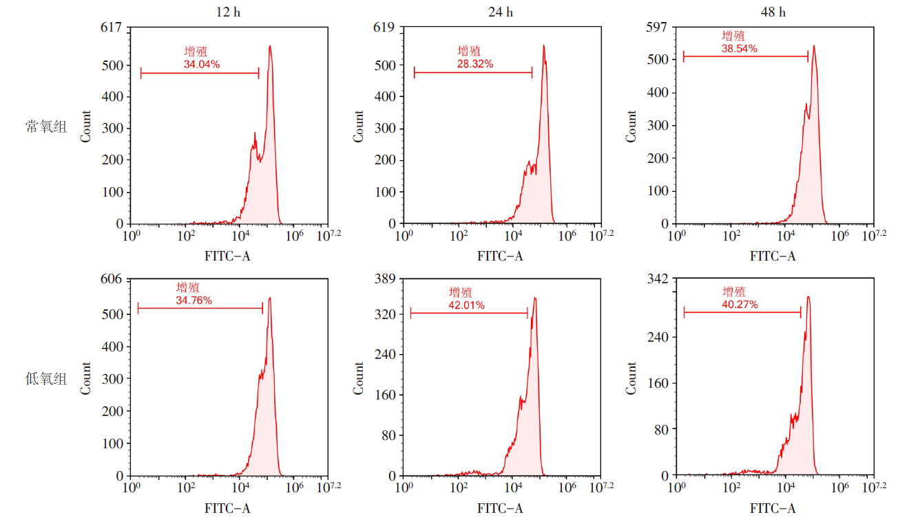
图1 常氧和低氧暴露不同时间对淋巴细胞增殖的影响
Fig.1 Effects of normoxia and hypoxia exposure on lymphocyte proliferation at different time points
| 组别 | 12 h | 24 h | 48 h |
|---|---|---|---|
| 常氧组 | 31.56±1.63 | 38.50±0.31 | 43.32±1.35 |
| 低氧组 | 27.31±2.05 | 34.74±0.82 | 40.66±0.49 |
| t | 4.749** | 11.324** | 5.566** |
表3 2组不同时间淋巴细胞增殖率比较 (n=3,%,$\bar{x} \pm s$)
Tab. 3 Comparison of lymphocyte proliferation rates at different time points between two groups
| 组别 | 12 h | 24 h | 48 h |
|---|---|---|---|
| 常氧组 | 31.56±1.63 | 38.50±0.31 | 43.32±1.35 |
| 低氧组 | 27.31±2.05 | 34.74±0.82 | 40.66±0.49 |
| t | 4.749** | 11.324** | 5.566** |
| 组别 | 12 h | 24 h | 48 h |
|---|---|---|---|
| 常氧组 | 17.05±1.33 | 30.66±3.11 | 82.01±1.66 |
| 低氧组 | 22.28±1.13 | 36.34±1.64 | 86.99±1.42 |
| t | 5.446** | 3.172* | 6.033** |
表4 2组不同时间淋巴细胞凋亡率比较 (n=3,%,$\bar{x} \pm s$)
Tab. 4 Comparison of apoptosis rate of lymphocyte at different time points between the two groups
| 组别 | 12 h | 24 h | 48 h |
|---|---|---|---|
| 常氧组 | 17.05±1.33 | 30.66±3.11 | 82.01±1.66 |
| 低氧组 | 22.28±1.13 | 36.34±1.64 | 86.99±1.42 |
| t | 5.446** | 3.172* | 6.033** |
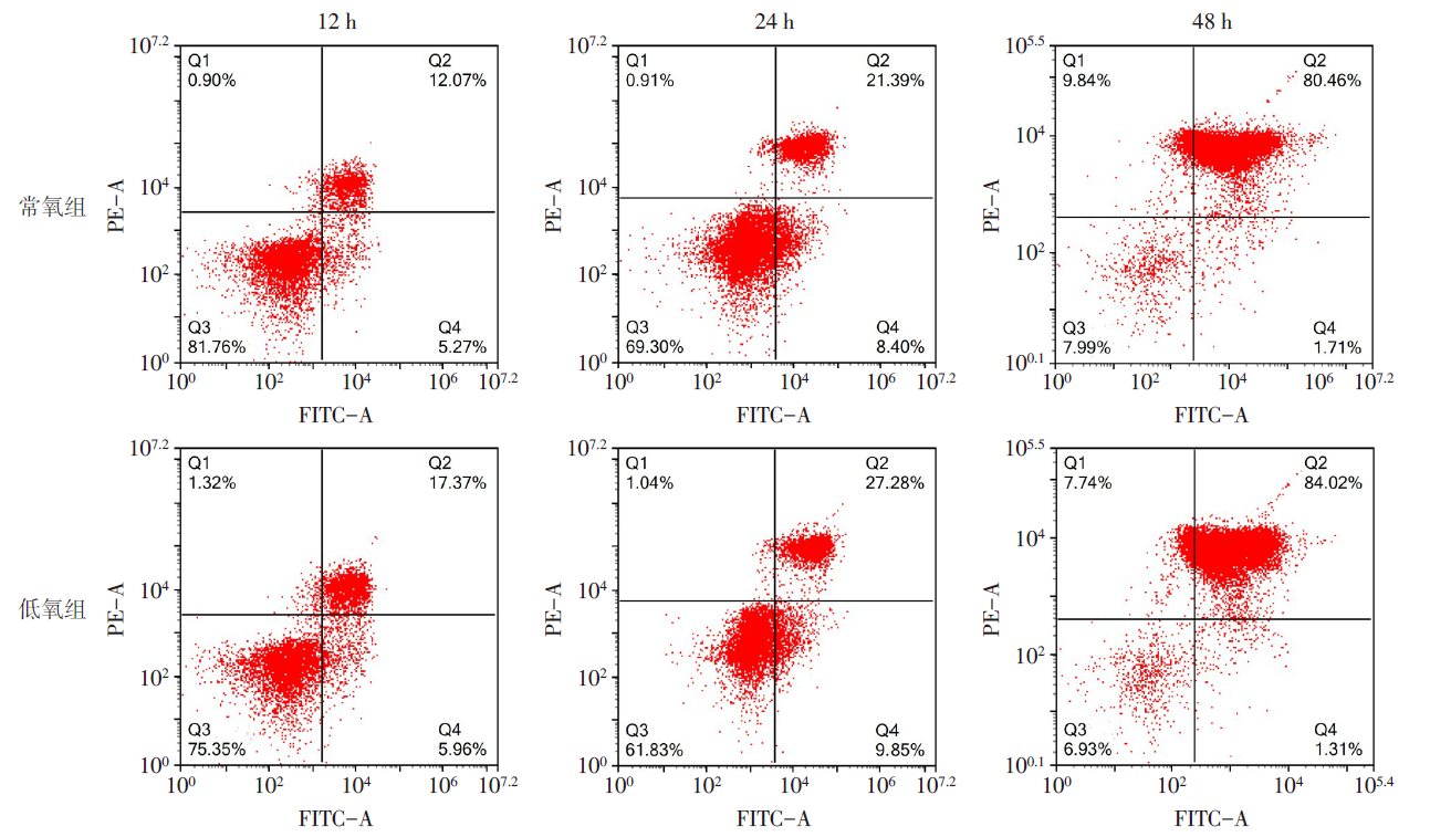
图2 常氧和低氧暴露不同时间对淋巴细胞凋亡的影响
Fig.2 Effects of normoxia and hypoxia exposure on lymphocyte apoptosis at different time points
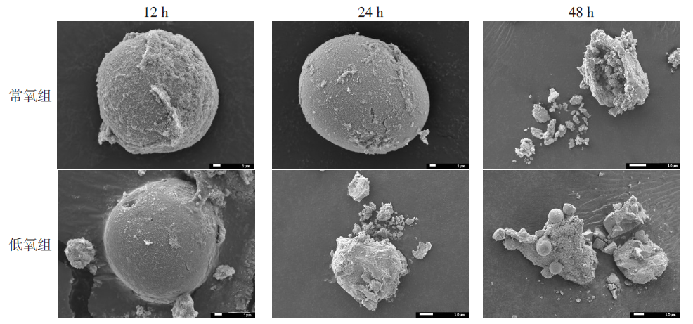
图3 SEM观察低氧暴露不同时间淋巴细胞形态变化
Fig.3 Lymphocyte morphological changes of hypoxia exposure at different time points observed by SEM
| 组别 | 12 h | 24 h | 48 h | ||||||
|---|---|---|---|---|---|---|---|---|---|
| Bak | bcl-2 | caspase-3 | Bak | bcl-2 | caspase-3 | Bak | bcl-2 | caspase-3 | |
| 常氧组 | 0.98±0.12 | 1.00±0.10 | 0.96±0.08 | 1.01±0.36 | 1.10±0.30 | 0.94±0.08 | 0.95±0.49 | 1.16±0.47 | 0.98±0.16 |
| 低氧组 | 1.11±0.18 | 0.66±0.21 | 1.25±0.35 | 1.43±0.28 | 0.52±0.22 | 1.15±0.19 | 1.44±0.42 | 0.74±0.39 | 2.21±0.56 |
| t | 2.248* | 4.527** | 2.171* | 2.724* | 4.440** | 2.532* | 2.721* | 2.516* | 5.973** |
表5 2组低氧暴露不同时间淋巴细胞Bak、bcl-2、caspase-3 mRNA表达水平比较 (n=3,$\bar{x} \pm s$)
Tab. 5 Comparison of mRNA expression levels of Bak, bcl-2 and caspase-3 in lymphocytes at different time points between the two groups
| 组别 | 12 h | 24 h | 48 h | ||||||
|---|---|---|---|---|---|---|---|---|---|
| Bak | bcl-2 | caspase-3 | Bak | bcl-2 | caspase-3 | Bak | bcl-2 | caspase-3 | |
| 常氧组 | 0.98±0.12 | 1.00±0.10 | 0.96±0.08 | 1.01±0.36 | 1.10±0.30 | 0.94±0.08 | 0.95±0.49 | 1.16±0.47 | 0.98±0.16 |
| 低氧组 | 1.11±0.18 | 0.66±0.21 | 1.25±0.35 | 1.43±0.28 | 0.52±0.22 | 1.15±0.19 | 1.44±0.42 | 0.74±0.39 | 2.21±0.56 |
| t | 2.248* | 4.527** | 2.171* | 2.724* | 4.440** | 2.532* | 2.721* | 2.516* | 5.973** |
| 组别 | 12 h | 24 h | 48 h | ||||||
|---|---|---|---|---|---|---|---|---|---|
| Bak | bcl-2 | caspase-3 | Bak | bcl-2 | caspase-3 | Bak | bcl-2 | caspase-3 | |
| 常氧组 | 0.31±0.11 | 0.62±0.06 | 0.45±0.08 | 0.23±0.15 | 0.90±0.14 | 0.71±0.06 | 0.37±0.13 | 0.36±0.04 | 0.36±0.03 |
| 低氧组 | 0.54±0.09 | 0.44±0.09 | 0.72±0.15 | 0.48±0.03 | 0.50±0.17 | 0.94±0.13 | 0.73±0.17 | 0.23±0.02 | 0.66±0.10 |
| t | 2.828* | 2.819* | 2.794* | 2.799* | 3.117* | 2.787* | 2.867* | 4.658** | 4.950** |
表6 2组低氧暴露不同时间淋巴细胞Bak、bcl-2、caspase-3蛋白表达水平比较 (n=3,$\bar{x} \pm s$)
Tab. 6 Comparison of Bak, bcl-2 and caspase-3 protein expression levels in lymphocytes at different time points between the two groups of mice
| 组别 | 12 h | 24 h | 48 h | ||||||
|---|---|---|---|---|---|---|---|---|---|
| Bak | bcl-2 | caspase-3 | Bak | bcl-2 | caspase-3 | Bak | bcl-2 | caspase-3 | |
| 常氧组 | 0.31±0.11 | 0.62±0.06 | 0.45±0.08 | 0.23±0.15 | 0.90±0.14 | 0.71±0.06 | 0.37±0.13 | 0.36±0.04 | 0.36±0.03 |
| 低氧组 | 0.54±0.09 | 0.44±0.09 | 0.72±0.15 | 0.48±0.03 | 0.50±0.17 | 0.94±0.13 | 0.73±0.17 | 0.23±0.02 | 0.66±0.10 |
| t | 2.828* | 2.819* | 2.794* | 2.799* | 3.117* | 2.787* | 2.867* | 4.658** | 4.950** |
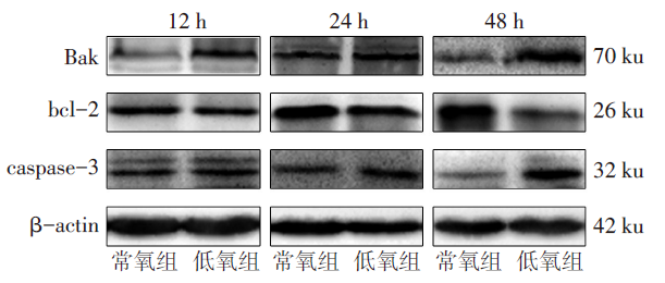
图4 低氧暴露不同时间淋巴细胞Bak、bcl-2、caspase-3的蛋白表达水平
Fig.4 Protein expression levels of Bak, bcl-2 and caspase-3 in lymphocytes at different time points between the two groups of mice
| [1] | ZHANG T, SUO C, ZHENG C, et al. Hypoxia and metabolism in metastasis[J]. Adv Exp Med Biol, 2019, 1136:87-95. doi: 10.1007/978-3-030-12734-3_6. |
| [2] | LUNDEBERG J, FEINER J R, SCHOBER A, et al. Increased cytokines at high altitude:Lack of effect of ibuprofen on acute mountain sickness,physiological variables,or cytokine levels[J]. High Alt Med Biol, 2018, 19(3):249-258. doi: 10.1089/ham.2017.0144. |
| [3] | 王新, 孙蓓, 刘芳, 等. 间歇低氧下小鼠血管内皮功能障碍机制的研究[J]. 天津医药, 2017, 45(2):160-163. |
| WANG X, SUN B, LIU F, et al. Studies of the mechanism of endothelial dysfunction in rats under intermittent hypoxia[J]. Tianjin Med J, 2017, 45(2):160-163. doi: 10.11958/20161305. | |
| [4] | WANG J, JIANG R, TAN Y, et al. Human pulmonary artery smooth muscle cell dysfunction is regulated by miR-509-5p in hypoxic environment[J]. Cell Cycle, 2022, 21(11):1212-1221. doi: 10.1080/15384101.2022.2044147. |
| [5] | NOMAN M Z, DESANTIS G, JANJI B, et al. PD-L1 is a novel direct target of HIF-1α,and its blockade under hypoxia enhanced MDSC-mediated T cell activation[J]. J Exp Med, 2014, 211(5):781-790. doi: 10.1084/jem.20131916. |
| [6] | BURROWS N, BASHFORD-ROGERS R J M, BHUTE V J, et al. Dynamic regulation of hypoxia-inducible factor-1α activity is essential for normal B cell development[J]. Nat Immunol, 2020, 21(11):1408-1420. doi: 10.1038/s41590-020-0772-8. |
| [7] | JIANG X, TIAN W, KIM D, et al. Hypoxia and hypoxia-inducible factors in lymphedema[J]. Front Pharmacol, 2022, 13:851057. doi: 10.3389/fphar.2022.851057. |
| [8] | 张晓娜, 李积东, 胡方杰, 等. 高原低氧暴露小鼠脾脏T淋巴细胞数量减少且免疫活性下降[J]. 细胞与分子免疫学杂志, 2017, 33(2):164-167. |
| ZHANG X N, LI J D, HU F J, et al. Decreased number and immune activity of splenic T lymphocytes in mice exposed to hypoxia at high altitude[J]. Chinese Journal of Cellular and Molecular Immunology, 2017, 33(2):164-167. doi: 10.13423/j.cnki.cjcmi.008007. | |
| [9] | XIE L, LIN Y, DENG Y, et al. The effect of SARS-CoV-2 on the spleen and T lymphocytes[J]. Viral Immunol, 2021, 34(6):416-420. doi: 10.1089/vim.2020.0320. |
| [10] | HU F, LIU H, XU L, et al. Hypoxia-inducible factor-1α perpetuates synovial fibroblast interactions with T cells and B cells in rheumatoid arthritis[J]. Eur J Immunol, 2016, 46(3):742-751. doi: 10.1002/eji.201545784. |
| [11] | LIU Y N, YANG J F, HUANG D J, et al. Hypoxia induces mitochondrial defect that promotes T cell exhaustion in tumor microenvironment through MYC-regulated pathways[J]. Front Immunol, 2020, 11:1906. doi: 10.3389/fimmu.2020.01906. |
| [12] | BYRNES J R, WEEKS A M, SHIFRUT E, et al. Hypoxia is a dominant remodeler of the effector T cell surface proteome relative to activation and regulatory T cell suppression[J]. Mol Cell Proteomics, 2022, 21(4):100217. doi: 10.1016/j.mcpro.2022.100217. |
| [13] | READ K A, POWELL M D, SREEKUMAR B K, et al. In vitro differentiation of effector CD4+ T helper cell subsets[J]. Methods Mol Biol, 2019, 1960:75-84. doi: 10.1007/978-1-4939-9167-9_6. |
| [14] | ZHU J. T helper cell differentiation,heterogeneity,and plasticity[J]. Cold Spring Harb Perspect Biol, 2018, 10(10):a030338. doi: 10.1101/cshperspect.a030338. |
| [15] | RUTERBUSCH M, PRUNER K B, SHEHATA L, et al. In vivo CD4+ T cell differentiation and function:Revisiting the Th1/Th2 paradigm[J]. Annu Rev Immunol, 2020, 38:705-725. doi: 10.1146/ annurev-immunol-103019-085803. |
| [16] | DZHALILOVA D S, KOSYREVA A M, DIATROPTOV M E, et al. Morphological characteristics of the thymus and spleen and the subpopulation composition of lymphocytes in peripheral blood during systemic inflammatory response in male rats with different resistance to hypoxia[J]. Int J Inflam, 2019, 2019:7584685. doi: 10.1155/2019/7584685. |
| [17] | CHEN Y, GABER T. Hypoxia/HIF modulates immune responses[J]. Biomedicines, 2021, 9(3):260. doi: 10.3390/biomedicines9030260. |
| [18] | LEE H S, JEONG G S. Salinosporamide A,a marine-derived proteasome inhibitor,inhibits T cell activation through regulating proliferation and the cell cycle[J]. Molecules, 2020, 25(21):5031. doi: 10.3390/molecules25215031. |
| [19] | HUBBI M E, SEMENZA G L. Regulation of cell proliferation by hypoxia-inducible factors[J]. Am J Physiol Cell Physiol, 2015, 309(12):C775-C782. doi: 10.1152/ajpcell.00279.2015. |
| [20] | CHO S H, RAYBUCK A L, STENGEL K, et al. Germinal centre hypoxia and regulation of antibody qualities by a hypoxia response system[J]. Nature, 2016, 537(7619):234-238. doi: 10.1038/nature19334. |
| [21] | SHAN T, CHEN S, CHEN X, et al. M2-TAM subsets altered by lactic acid promote T cell apoptosis through the PDL1/PD1 pathway[J]. Oncol Rep, 2020, 44(5):1885-1894. doi: 10.3892/or.2020.7767. |
| [22] | DHURIYA Y K, SHARMA D, NAIK A A. Cellular demolition:Proteins as molecular players of programmed cell death[J]. Int J Biol Macromol, 2019, 138:492-503. doi: 10.1016/j.ijbiomac.2019.07.113. |
| [23] | JENG P S, INOUE-YAMAUCHI A, HSIEH J J, et al. BH3-dependent and independent activation of BAX and BAK in mitochondrial apoptosis[J]. Curr Opin Physiol, 2018, 3:71-81. doi: 10.1016/j.cophys.2018.03.005. |
| [24] | YAMAZAKI T, GALLUZZI L. Bax and bak dynamics control mitochondrial DNA release during apoptosis[J]. Cell Death Differ, 2022, 29(6):1296-1298. doi: 10.1038/s41418-022-00985-2. |
| [25] | MUSAOGULLARI A, MANDATO A, CHAI Y C. Role of glutathione depletion and reactive oxygen species generation on caspase-3 activation:A study with the kinase inhibitor staurosporine[J]. Front Physiol, 2020, 11:998. doi: 10.3389/fphys.2020.00998. |
| [26] | WANG Y, LIU Y, FEI A, et al. Lncrna xist facilitates hypoxia-induced myocardial cell injury through targeting miR-191-5p/TRAF3 axis[J]. Mol Cell Biochem, 2022, 477(6):1697-1707. doi: 10.1007/s11010-022-04385-5. |
| [27] | ZHAO X, LIU L, LI R, et al. Hypoxia-inducible factor 1-α(HIF-1α)induces apoptosis of human uterosacral ligament fibroblasts through the death receptor and mitochondrial pathways[J]. Med Sci Monit, 2018, 24:8722-8733. doi: 10.12659/MSM.913384. |
| [1] | 杨晓芳, 贾新燕, 丰文君. miR-181a-5p通过HMGB1/NF-κB信号通路调控狼疮性肾炎小鼠肾小球系膜细胞增殖和凋亡[J]. 天津医药, 2026, 54(3): 232-237. |
| [2] | 张婧, 魏玉英, 宁海虹, 韦红梅, 王嘉玮, 曹薇, 吴宾. DUSP9在2型糖尿病心肌病小鼠心肌损伤中的保护作用及其机制[J]. 天津医药, 2026, 54(3): 238-244. |
| [3] | 王喆, 邱林, 马贲. 番茄来源胞外囊泡样颗粒对口腔鳞状细胞癌的作用效果研究[J]. 天津医药, 2026, 54(2): 145-150. |
| [4] | 李志伟, 张会超, 杨凤鸣, 曾垂义. 基于miR-144-3p/MAPK1通路探讨红参总皂苷对扩张型心肌病小鼠心肌细胞凋亡的影响[J]. 天津医药, 2026, 54(1): 23-29. |
| [5] | 赵兰君, 李良惠, 马馨, 巩娇娇, 郑臣辉, 石琳. 穿心莲内酯调控STAT3/GPX4通路对骨髓瘤细胞增殖和凋亡的影响[J]. 天津医药, 2026, 54(1): 8-13. |
| [6] | 孔翠文, 路延双, 孙丽萍, 于芬芬. LncRNA SNHG14靶向miR-30a-5p对高糖诱导的足细胞损伤的影响[J]. 天津医药, 2025, 53(9): 903-909. |
| [7] | 刘虹, 张玥玥, 王一琳, 王彩丽, 王晓敏, 毛敏, 李燕. MicroRNA-34a通过调控Wnt途径影响慢性淋巴细胞白血病进展的机制探讨[J]. 天津医药, 2025, 53(8): 785-790. |
| [8] | 万艳波, 刘明, 王勇. 秦皮甲素调节HMGB1/RAGE信号通路对缺氧/复氧诱导的心肌细胞损伤的影响[J]. 天津医药, 2025, 53(8): 796-801. |
| [9] | 刘海威, 杨洁, 王力, 蒙诗波, 唐旭松, 刘成仁, 王永旺. 木犀草素通过NFE2L2/x-CT/GPX4信号轴调控ROS水平抑制胶质母细胞瘤[J]. 天津医药, 2025, 53(7): 673-678. |
| [10] | 韩建存, 周谊. 川陈皮素调节FAK/AKT信号通路对喉鳞状细胞癌细胞增殖和凋亡的影响[J]. 天津医药, 2025, 53(6): 561-565. |
| [11] | 余朝霞, 马贲, 邱林, 高倩, 尼娜. 基于网络药理学和实验验证探究鲍式层孔菌多酚的抗头颈鳞癌机制[J]. 天津医药, 2025, 53(5): 456-461. |
| [12] | 李晨, 李占恩, 苏宏伟, 侯彩云, 董少文. KRT17调节Wnt/β-catenin信号通路对膀胱癌细胞增殖、凋亡及上皮间质转化的影响[J]. 天津医药, 2025, 53(5): 462-467. |
| [13] | 苏红见, 张春艳, 张卫东, 韩利, 乔亚红. 鸢尾素调控EBF3/ALOX15通路影响肺腺癌细胞增殖和迁移[J]. 天津医药, 2025, 53(4): 337-342. |
| [14] | 祁卫华, 黄广磊, 张媛媛, 班宏英, 毛诏旭. 连翘脂素调节cAMP/EPAC1/RAP1信号通路对肺癌细胞恶性进展的影响[J]. 天津医药, 2025, 53(4): 343-348. |
| [15] | 闫玲新, 李森, 郭改莉, 孟婉秋, 徐超. 异牡荆素通过miR-339-5p/HSPA8轴调节胰腺癌细胞的生物学行为[J]. 天津医药, 2025, 53(3): 230-235. |
| 阅读次数 | ||||||
|
全文 |
|
|||||
|
摘要 |
|
|||||