天津医药 ›› 2023, Vol. 51 ›› Issue (12): 1293-1299.doi: 10.11958/20221929
吴德建1( ), 杨秋2, 谢桂丹1, 彭鑫1,△()
收稿日期:2022-11-22
修回日期:2023-03-20
出版日期:2023-12-15
发布日期:2023-12-22
通讯作者:
△ E-mail:作者简介:吴德建(1993),男,主治医师,主要从事肝炎、肝硬化诊疗方面研究。E-mail:
WU Dejian1(), YANG Qiu2, XIE Guidan1, PENG Xin1,△()
Received:2022-11-22
Revised:2023-03-20
Published:2023-12-15
Online:2023-12-22
Contact:
△ E-mail:吴德建, 杨秋, 谢桂丹, 彭鑫. 紫草素调节Notch信号通路对肝癌细胞恶性生物学活性的影响[J]. 天津医药, 2023, 51(12): 1293-1299.
WU Dejian, YANG Qiu, XIE Guidan, PENG Xin. Impact of shikonin on the malignant biological activity of liver cancer cells by regulating Notch signaling pathway[J]. Tianjin Medical Journal, 2023, 51(12): 1293-1299.
摘要:
目的 探讨紫草素(SHI)调节Notch信号通路对肝癌细胞恶性生物学活性的影响。方法 采用Western blot分别检测肝癌组织、癌旁组织、肝癌细胞(HepG2、Hep3B、HCCLM3、Huh-7、SMMC-7721细胞)及正常肝细胞(HL-7702细胞)中Notch、发状分裂相关增强子-1(HES1)、hairy相关转录因子1(HEY1)蛋白表达;将Huh-7细胞分为对照组、L-SHI组(1 μmol/L SHI)、M-SHI组(2 μmol/L SHI)、H-SHI组(4 μmol/L SHI)、DAPT组(50 μmol/L Notch信号抑制剂DAPT)、H-SHI+丙戊酸(VPA)组(4 μmol/L SHI和8 mmol/L Notch通路激活剂VPA)。CCK-8法和平板克隆实验检测Huh-7细胞增殖;流式细胞术检测Huh-7细胞凋亡以及细胞周期;细胞划痕实验以及Transwell侵袭实验分别检测Huh-7细胞迁移和侵袭情况;Western blot检测上皮间质转化(EMT)及凋亡相关蛋白表达。结果 Notch、HES1、HEY1在肝癌组织和细胞中表达水平明显升高,且Huh-7细胞差异最明显,以Huh-7细胞为研究对象。与对照组相比,L-SHI组、M-SHI组、H-SHI组、DAPT组Notch、HES1、HEY1、Bcl-2蛋白表达水平下调,S期和G2/M期细胞比例、OD450值及克隆数、迁移率、侵袭细胞数目、N-cadherin、Vimentin水平降低(P<0.05),G1/G0期细胞比例、凋亡率、Bax、cleaved-Caspase-3、E-cadherin水平升高(P<0.05),且SHI的作用效果呈现剂量依赖性;与H-SHI组相比,H-SHI+VPA组以上指标趋势相反,VPA减弱了SHI降低肝癌细胞恶性生物学活性的作用。结论 SHI可能通过抑制Notch信号通路抑制Huh-7细胞增殖、迁移和侵袭,促进Huh-7细胞凋亡。
中图分类号:
| 组别 | Notch | HES1 | HEY1 |
|---|---|---|---|
| 癌旁组织 | 0.78±0.09 | 0.63±0.07 | 0.39±0.05 |
| 癌组织 | 1.41±0.13 | 1.24±0.11 | 1.37±0.14 |
| t | 17.819** | 10.633** | 29.481** |
表1 Notch、HES1、HEY1蛋白在癌组织及癌旁组织中的表达
Tab.1 Expression of Notch, HES1 and HEY1 proteins in liver cancer tissue and adjacent tissue (n=20,$\bar{x}±s$)
| 组别 | Notch | HES1 | HEY1 |
|---|---|---|---|
| 癌旁组织 | 0.78±0.09 | 0.63±0.07 | 0.39±0.05 |
| 癌组织 | 1.41±0.13 | 1.24±0.11 | 1.37±0.14 |
| t | 17.819** | 10.633** | 29.481** |
| 组别 | Notch | HES1 | HEY1 |
|---|---|---|---|
| HL-7702细胞 | 0.60±0.06 | 0.51±0.05 | 0.35±0.04 |
| HepG2细胞 | 1.41±0.13a | 1.05±0.09a | 1.16±0.12a |
| Hep3B细胞 | 1.33±0.12a | 1.13±0.12a | 1.22±0.13a |
| HCCLM3细胞 | 1.35±0.14a | 1.07±0.11a | 1.26±0.14a |
| SMMC-7721细胞 | 1.37±0.15a | 1.09±0.13a | 1.24±0.16a |
| Huh-7细胞 | 1.60±0.17a | 1.23±0.14a | 1.42±0.17a |
| F | 67.663** | 52.880** | 81.609** |
表2 Notch、HES1、HEY1蛋白在正常肝细胞及肝癌细胞中的表达
Tab.2 Expression of Notch, HES1 and HEY1 proteins in normal liver cells and liver cancer cells (n=6,$\bar{x}±s$)
| 组别 | Notch | HES1 | HEY1 |
|---|---|---|---|
| HL-7702细胞 | 0.60±0.06 | 0.51±0.05 | 0.35±0.04 |
| HepG2细胞 | 1.41±0.13a | 1.05±0.09a | 1.16±0.12a |
| Hep3B细胞 | 1.33±0.12a | 1.13±0.12a | 1.22±0.13a |
| HCCLM3细胞 | 1.35±0.14a | 1.07±0.11a | 1.26±0.14a |
| SMMC-7721细胞 | 1.37±0.15a | 1.09±0.13a | 1.24±0.16a |
| Huh-7细胞 | 1.60±0.17a | 1.23±0.14a | 1.42±0.17a |
| F | 67.663** | 52.880** | 81.609** |
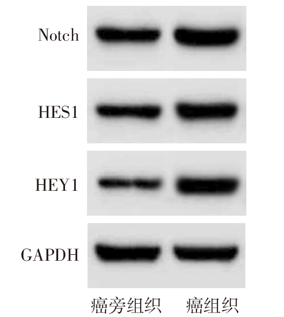
图1 Western blot检测肝癌组织及癌旁组织中Notch、HES1、HEY1蛋白表达
Fig.1 Western blot detection of Notch, HES1 and HEY1 protein expression in liver cancer tissue and adjacent tissue
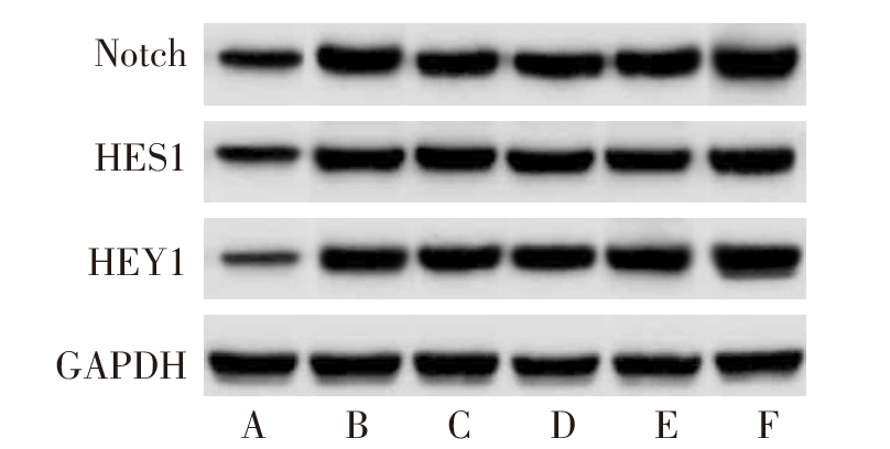
图2 Western blot检测正常肝细胞及肝癌细胞中Notch、HES1、HEY1蛋白水平 A:HL-7702细胞;B:HepG2细胞;C:Hep3B细胞;D:HCCLM3细胞;E:SMMC-7721细胞;F:Huh-7细胞。
Fig.2 Western blot detection of Notch, HES1 and HEY1 protein levels in normal hepatocytes and hepatoma cells
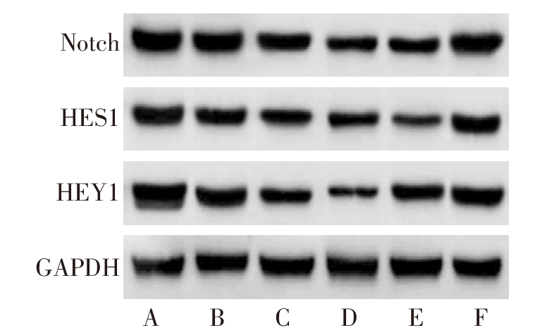
图3 Western blot检测SHI对Huh-7细胞中Notch、HES1、HEY1蛋白的影响 A:对照组;B:L-SHI组;C:M-SHI组;D:H-SHI组;E:DAPT组;F:H-SHI+VPA组。
Fig.3 Western blot detection of effects of SHI on Notch, HES1 and HEY1 proteins in Huh-7 cells
| 组别 | Notch | HES1 | HEY1 |
|---|---|---|---|
| 对照组 | 1.63±0.19 | 1.26±0.14 | 1.48±0.17 |
| L-SHI组 | 1.31±0.15a | 1.03±0.12a | 1.15±0.13a |
| M-SHI组 | 0.98±0.10ab | 0.80±0.09ab | 0.79±0.09ab |
| H-SHI组 | 0.64±0.06abc | 0.63±0.08abc | 0.38±0.04abc |
| DAPT组 | 0.72±0.07a | 0.39±0.03a | 0.89±0.09a |
| H-SHI+VPA组 | 1.39±0.14d | 1.11±0.14d | 1.27±0.15d |
| F | 96.630** | 91.860** | 106.778** |
表3 SHI对Huh-7细胞中Notch、HES1、HEY1蛋白表达的影响
Tab.3 Effects of SHI on Notch, HES1 and HEY1 proteins in Huh-7 cells (n=6,$\bar{x}±s$)
| 组别 | Notch | HES1 | HEY1 |
|---|---|---|---|
| 对照组 | 1.63±0.19 | 1.26±0.14 | 1.48±0.17 |
| L-SHI组 | 1.31±0.15a | 1.03±0.12a | 1.15±0.13a |
| M-SHI组 | 0.98±0.10ab | 0.80±0.09ab | 0.79±0.09ab |
| H-SHI组 | 0.64±0.06abc | 0.63±0.08abc | 0.38±0.04abc |
| DAPT组 | 0.72±0.07a | 0.39±0.03a | 0.89±0.09a |
| H-SHI+VPA组 | 1.39±0.14d | 1.11±0.14d | 1.27±0.15d |
| F | 96.630** | 91.860** | 106.778** |
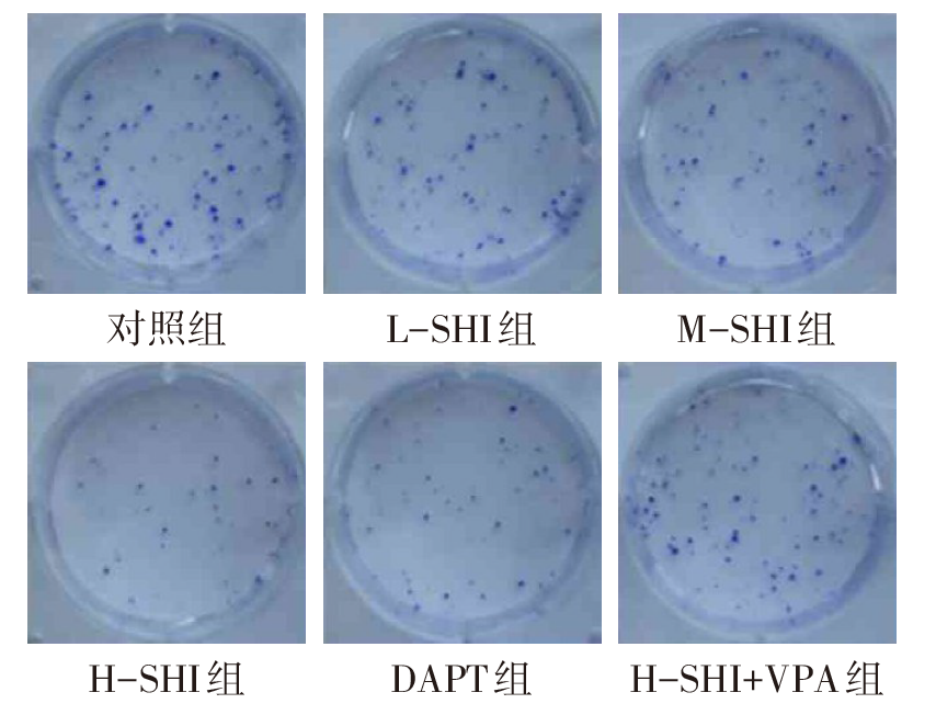
图4 SHI对Huh-7细胞克隆形成的影响
Fig.4 Effects of SHI on the formation of Huh-7 cell clone
| 组别 | OD450值 | 克隆数/个 |
|---|---|---|
| 对照组 | 1.33±0.14 | 56.76±5.94 |
| L-SHI组 | 1.12±0.11a | 44.83±5.54a |
| M-SHI组 | 0.89±0.09ab | 35.37±4.65ab |
| H-SHI组 | 0.67±0.07abc | 24.16±2.21abc |
| DAPT组 | 0.70±0.06a | 26.73±2.02a |
| H-SHI+VPA组 | 1.18±0.12d | 45.17±5.47d |
| F | 69.795** | 32.725** |
表4 SHI对Huh-7细胞OD450值及克隆数的影响
Tab.4 Effects of SHI on OD450 value and clone number of Huh-7 cells
| 组别 | OD450值 | 克隆数/个 |
|---|---|---|
| 对照组 | 1.33±0.14 | 56.76±5.94 |
| L-SHI组 | 1.12±0.11a | 44.83±5.54a |
| M-SHI组 | 0.89±0.09ab | 35.37±4.65ab |
| H-SHI组 | 0.67±0.07abc | 24.16±2.21abc |
| DAPT组 | 0.70±0.06a | 26.73±2.02a |
| H-SHI+VPA组 | 1.18±0.12d | 45.17±5.47d |
| F | 69.795** | 32.725** |
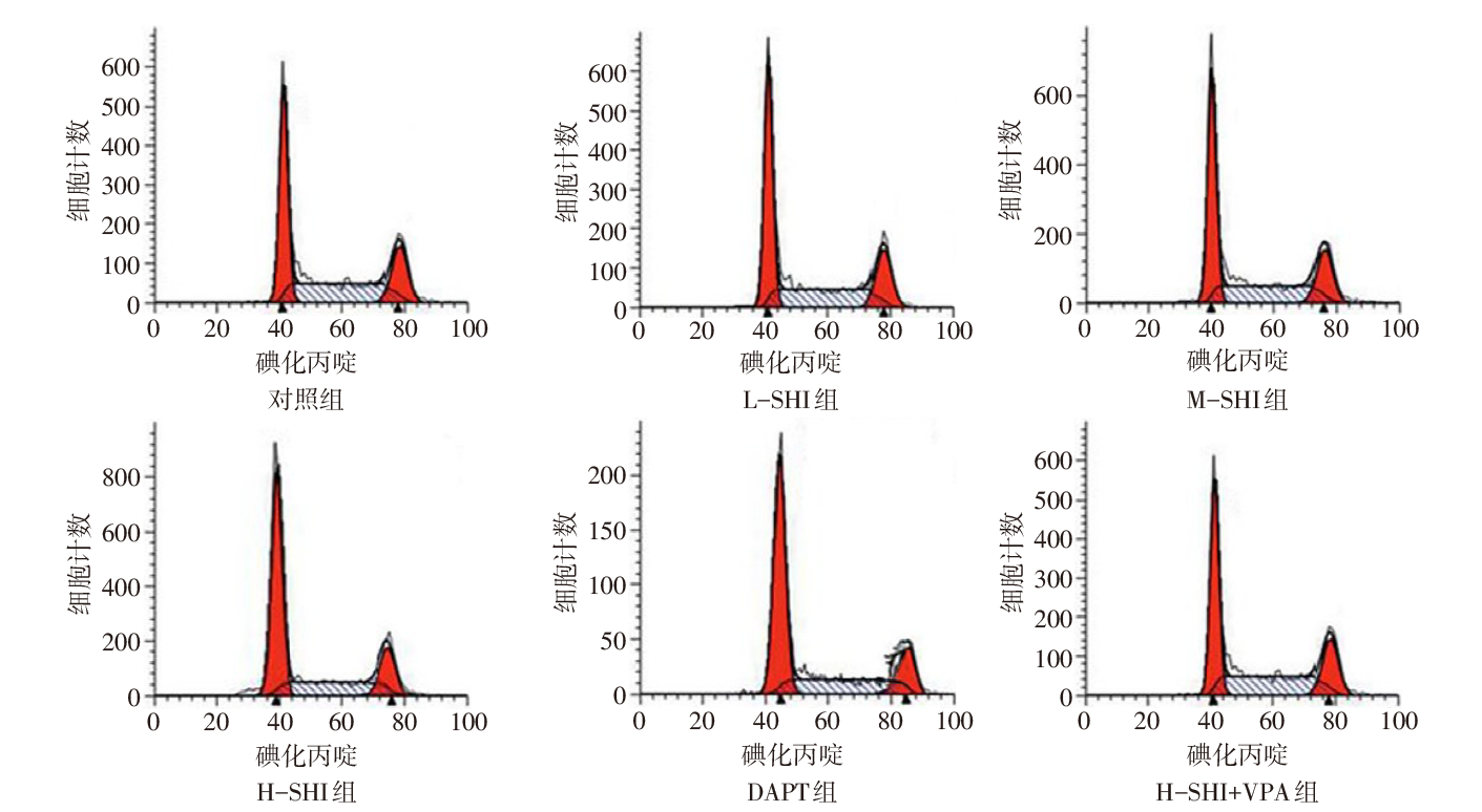
图5 流式细胞术检测SHI对Huh-7细胞细胞周期的影响
Fig.5 Effects of SHI on cell cycle of Huh-7 cells by flow cytometry
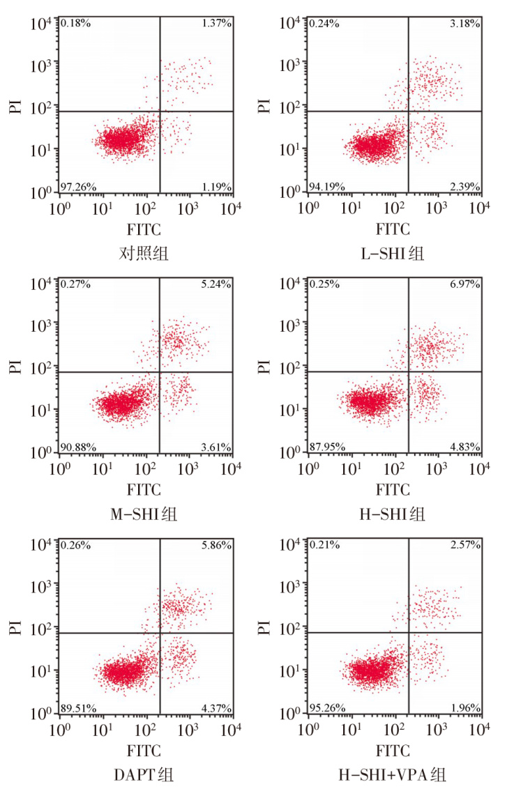
图6 流式细胞术检测Huh-7细胞凋亡
Fig.6 Detection of Huh-7 cell apoptosis by flow cytometry
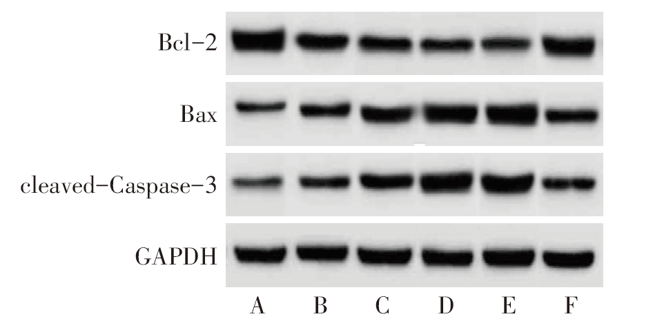
图7 SHI对Huh-7细胞中Bcl-2、Bax、cleaved-Caspase-3蛋白水平的影响 A:对照组;B:L-SHI组;C:M-SHI组;D:H-SHI组;E:DAPT组;F:H-SHI+VPA组。
Fig.7 Effects of SHI on Bcl-2, Bax, cleaved-Caspase-3 protein levels in Huh-7 cells
| 组别 | 细胞周期分布 | ||
|---|---|---|---|
| G1/G0 | S | G2/M | |
| 对照组 | 44.13±4.27 | 38.45±3.56 | 17.42±1.69 |
| L-SHI组 | 51.01±5.53a | 33.65±3.16a | 15.34±1.79a |
| M-SHI组 | 52.89±5.76a | 31.92±3.03a | 15.19±1.57a |
| H-SHI组 | 58.34±5.65a | 26.53±2.45a | 15.13±1.35a |
| DAPT组 | 57.23±5.34a | 27.96±2.67a | 14.81±1.23a |
| H-SHI+VPA组 | 50.78±5.56d | 33.12±3.32d | 16.10±1.37d |
| F | 9.124** | 20.200** | 7.609** |
表5 SHI对Huh-7细胞细胞周期的影响
Tab.5 Effects of SHI on cell cycle of Huh-7 cells
| 组别 | 细胞周期分布 | ||
|---|---|---|---|
| G1/G0 | S | G2/M | |
| 对照组 | 44.13±4.27 | 38.45±3.56 | 17.42±1.69 |
| L-SHI组 | 51.01±5.53a | 33.65±3.16a | 15.34±1.79a |
| M-SHI组 | 52.89±5.76a | 31.92±3.03a | 15.19±1.57a |
| H-SHI组 | 58.34±5.65a | 26.53±2.45a | 15.13±1.35a |
| DAPT组 | 57.23±5.34a | 27.96±2.67a | 14.81±1.23a |
| H-SHI+VPA组 | 50.78±5.56d | 33.12±3.32d | 16.10±1.37d |
| F | 9.124** | 20.200** | 7.609** |
| 组别 | 凋亡率/% | Bcl-2 | Bax | cleaved- Caspase-3 |
|---|---|---|---|---|
| 对照组 | 2.43±0.27 | 1.45±0.17 | 0.32±0.02 | 0.33±0.03 |
| L-SHI组 | 5.35±0.52a | 1.11±0.12a | 0.73±0.06a | 0.69±0.07a |
| M-SHI组 | 8.65±0.98ab | 0.82±0.09ab | 1.04±0.11ab | 0.99±0.09ab |
| H-SHI组 | 11.65±1.23abc | 0.53±0.05abc | 1.36±0.14abc | 1.37±0.14abc |
| DAPT组 | 9.97±0.96a | 0.46±0.04a | 1.30±0.13a | 1.28±0.12a |
| H-SHI+ VPA组 | 4.78±0.46d | 1.17±0.12d | 0.67±0.07d | 0.57±0.06d |
| F | 187.108** | 129.293** | 168.904** | 197.238** |
表6 SHI对Huh-7细胞凋亡率和凋亡蛋白的影响
Tab.6 Effects of SHI on apoptosis rate and apoptotic protein of Huh-7 cells
| 组别 | 凋亡率/% | Bcl-2 | Bax | cleaved- Caspase-3 |
|---|---|---|---|---|
| 对照组 | 2.43±0.27 | 1.45±0.17 | 0.32±0.02 | 0.33±0.03 |
| L-SHI组 | 5.35±0.52a | 1.11±0.12a | 0.73±0.06a | 0.69±0.07a |
| M-SHI组 | 8.65±0.98ab | 0.82±0.09ab | 1.04±0.11ab | 0.99±0.09ab |
| H-SHI组 | 11.65±1.23abc | 0.53±0.05abc | 1.36±0.14abc | 1.37±0.14abc |
| DAPT组 | 9.97±0.96a | 0.46±0.04a | 1.30±0.13a | 1.28±0.12a |
| H-SHI+ VPA组 | 4.78±0.46d | 1.17±0.12d | 0.67±0.07d | 0.57±0.06d |
| F | 187.108** | 129.293** | 168.904** | 197.238** |

图8 SHI对Huh-7细胞侵袭细胞数量的影响(×200)
Fig.8 Effects of SHI on the number of invasive cells of Huh-7 cells(×200)
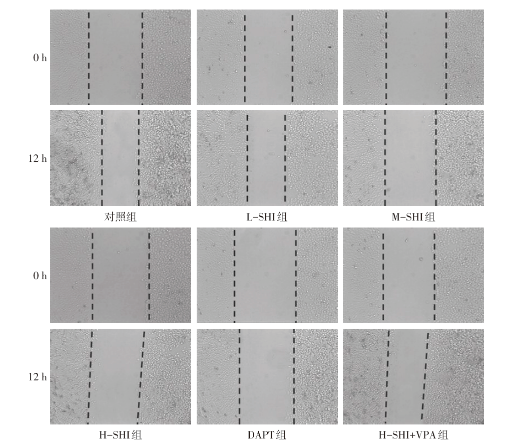
图9 SHI对Huh-7细胞迁移率的影响
Fig.9 Effects of SHI on the migration rate of Huh-7 cells
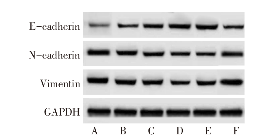
图10 Western blot检测Huh-7细胞中E-cadherin、N-cadherin、Vimentin蛋白水平 A:对照组;B:L-SHI组;C:M-SHI组;D:H-SHI组;E:DAPT组;F:H-SHI+VPA组。
Fig.10 Western blot detection of E-cadherin, N-cadherin and Vimentin protein levels in Huh-7 cells
| 组别 | 迁移率/% | 侵袭细胞数目/(个/视野) |
|---|---|---|
| 对照组 | 30.22±3.32 | 205.43±20.25 |
| L-SHI组 | 23.94±2.37a | 183.35±19.48a |
| M-SHI组 | 16.74±1.70ab | 166.33±16.46ab |
| H-SHI组 | 10.86±1.23abc | 141.57±15.13abc |
| DAPT组 | 12.13±1.26a | 150.73±15.97a |
| H-SHI+VPA组 | 25.48±2.76d | 199.87±20.42d |
| F | 121.414** | 20.769** |
表7 SHI对Huh-7细胞迁移率和侵袭细胞数目的影响
Tab.7 Effects of SHI on migration rate and number of invasive cells of Huh-7 cells
| 组别 | 迁移率/% | 侵袭细胞数目/(个/视野) |
|---|---|---|
| 对照组 | 30.22±3.32 | 205.43±20.25 |
| L-SHI组 | 23.94±2.37a | 183.35±19.48a |
| M-SHI组 | 16.74±1.70ab | 166.33±16.46ab |
| H-SHI组 | 10.86±1.23abc | 141.57±15.13abc |
| DAPT组 | 12.13±1.26a | 150.73±15.97a |
| H-SHI+VPA组 | 25.48±2.76d | 199.87±20.42d |
| F | 121.414** | 20.769** |
| 组别 | E-cadherin | N-cadherin | Vimentin |
|---|---|---|---|
| 对照组 | 0.22±0.02 | 0.90±0.09 | 1.18±0.21 |
| L-SHI组 | 0.39±0.04a | 0.71±0.07a | 0.98±0.09a |
| M-SHI组 | 0.57±0.06ab | 0.52±0.05ab | 0.74±0.08ab |
| H-SHI组 | 0.78±0.09abc | 0.30±0.02abc | 0.52±0.05abc |
| DAPT组 | 0.72±0.07a | 0.35±0.03a | 0.58±0.06a |
| H-SHI+VPA组 | 0.36±0.03d | 0.79±0.09d | 1.04±0.13d |
| F | 148.635** | 143.108** | 52.235** |
表8 SHI对Huh-7细胞E-cadherin、N-cadherin、Vimentin表达水平的影响
Tab.8 Effects of SHI on levels of E-cadherin, N-cadherin and Vimentin in Huh-7 cells
| 组别 | E-cadherin | N-cadherin | Vimentin |
|---|---|---|---|
| 对照组 | 0.22±0.02 | 0.90±0.09 | 1.18±0.21 |
| L-SHI组 | 0.39±0.04a | 0.71±0.07a | 0.98±0.09a |
| M-SHI组 | 0.57±0.06ab | 0.52±0.05ab | 0.74±0.08ab |
| H-SHI组 | 0.78±0.09abc | 0.30±0.02abc | 0.52±0.05abc |
| DAPT组 | 0.72±0.07a | 0.35±0.03a | 0.58±0.06a |
| H-SHI+VPA组 | 0.36±0.03d | 0.79±0.09d | 1.04±0.13d |
| F | 148.635** | 143.108** | 52.235** |
| [1] | CAO W, CHEN H D, YU Y W, et al. Changing profiles of cancer burden worldwide and in China:a secondary analysis of the global cancer statistics 2020[J]. Chin Med J (Engl), 2021, 134(7):783-791. doi:10.1097/CM9.0000000000001474. |
| [2] | CHIDAMBARANATHAN-REGHUPATY S, FISHER P B, SARKAR D. Hepatocellular carcinoma(HCC):epidemiology,etiology and molecular classification[J]. Adv Cancer Res, 2021, 149:1-61. doi:10.1016/bs.acr.2020.10.001. |
| [3] | WALLACE M C, PREEN D B, SHORT M W, et al. Hepatocellular carcinoma in Australia 1982-2014:increasing incidence and improving survival[J]. Liver Int, 2019, 39(3):522-530. doi:10.1111/liv.13966. |
| [4] | ZHANG S, GAO Q, LI W, et al. Shikonin inhibits cancer cell cycling by targeting Cdc25s[J]. BMC Cancer, 2019, 19(1):20. doi:10.1186/s12885-018-5220-x. |
| [5] | BAO C, LIU T, QIAN L, et al. Shikonin inhibits migration and invasion of triple-negative breast cancer cells by suppressing epithelial-mesenchymal transition via miR-17-5p/PTEN/Akt pathway[J]. J Cancer, 2021, 12(1):76-88. doi:10.7150/jca.47553. |
| [6] | XU Z, HUANG L, ZHANG T, et al. Shikonin inhibits the proliferation of cervical cancer cells via FAK/AKT/GSK3β signalling[J]. Oncol Lett, 2022, 24(3):304. doi:10.3892/ol.2022.13424. |
| [7] | LIU T, LI S, WU L, et al. Experimental study of hepatocellular carcinoma treatment by Shikonin through regulating PKM2[J]. J Hepatocell Carcinoma, 2020, 7:19-31. doi:10.2147/JHC.S237614. |
| [8] | LUIKEN S, FRAAS A, BIEG M, et al. NOTCH target gene HES5 mediates oncogenic and tumor suppressive functions in hepatocarcinogenesis[J]. Oncogene, 2020, 39(15):3128-3144. doi:10.1038/s41388-020-1198-3. |
| [9] | SHI J, HAN G, WANG J, et al. Matrine promotes hepatic oval cells differentiation into hepatocytes and alleviates liver injury by suppression of Notch signalling pathway[J]. Life Sci, 2020, 261:118354. doi:10.1016/j.lfs.2020.118354. |
| [10] | LEE J H, HAN S H, KIM Y M, et al. Shikonin inhibits proliferation of melanoma cells by MAPK pathway-mediated induction of apoptosis[J]. Biosci Rep, 2021, 41(1):BSR20203834. doi:10.1042/BSR20203834. |
| [11] | XIANG Z, MIAO Q, ZHANG J, et al. AB4 inhibits Notch signaling and promotes cancer cell apoptosis in liver cancer[J]. Oncol Rep, 2021, 45(6):112. doi:10.3892/or.2021.8063. |
| [12] | YANG X, LIU J, LIANG Q, et al. Valproic acid reverses sorafenib resistance through inhibiting activated Notch/Akt signaling pathway in hepatocellular carcinoma[J]. Fundam Clin Pharmacol, 2021, 35(4):690-699. doi:10.1111/fcp.12608. |
| [13] | ARMENGOL C, SARRIAS M R, SALA M. Hepatocellular carcinoma:present and future[J]. Med Clin (Barc), 2018, 150(10):390-397. doi:10.1016/j.medcli.2017.08.010. |
| [14] | MARKOWITSCH S D, JUETTER K M, SCHUPP P, et al. Shikonin reduces growth of docetaxel-resistant prostate cancer cells mainly through necroptosis[J]. Cancers (Basel), 2021, 13(4):882. doi:10.3390/cancers13040882. |
| [15] | TSAI M F, CHEN S M, ONG A Z, et al. Shikonin induced program cell death through generation of reactive oxygen species in renal cancer cells[J]. Antioxidants (Basel), 2021, 10(11):1831-1844. doi:10.3390/antiox10111831. |
| [16] | LI M Y, MI C, WANG K S, et al. Shikonin suppresses proliferation and induces cell cycle arrest through the inhibition of hypoxia-inducible factor-1α signaling[J]. Chem Biol Interact, 2017, 274:58-67. doi:10.1016/j.cbi.2017.06.029. |
| [17] | LI Y, ZHANG T, QIN S, et al. Effects of UPF1 expression on EMT process by targeting E-cadherin,N-cadherin,Vimentin and Twist in a hepatocellular carcinoma cell line[J]. Mol Med Rep, 2019, 19(3):2137-2143. doi:10.3892/mmr.2019.9838. |
| [18] | LI X, LIU W, GENG C, et al. Ginsenoside Rg3 suppresses epithelial-mesenchymal transition via downregulating Notch-Hes1 signaling in colon cancer cells[J]. Am J Chin Med, 2021, 49(1):217-235. doi:10.1142/S0192415X21500129. |
| [19] | ZHANG H S, ZHANG Z G, DU G Y, et al. Nrf2 promotes breast cancer cell migration via up-regulation of G6PD/HIF-1α/Notch1 axis[J]. J Cell Mol Med, 2019, 23(5):3451-3463. doi:10.1111/jcmm.14241. |
| [1] | 姜天佑, 李敏, 孙碧文, 李越洋, 邢丽静, 田晨. Let-7b诱导白血病相关巨噬细胞复极抑制AML的发展[J]. 天津医药, 2026, 54(3): 225-231. |
| [2] | 杨晓芳, 贾新燕, 丰文君. miR-181a-5p通过HMGB1/NF-κB信号通路调控狼疮性肾炎小鼠肾小球系膜细胞增殖和凋亡[J]. 天津医药, 2026, 54(3): 232-237. |
| [3] | 张婧, 魏玉英, 宁海虹, 韦红梅, 王嘉玮, 曹薇, 吴宾. DUSP9在2型糖尿病心肌病小鼠心肌损伤中的保护作用及其机制[J]. 天津医药, 2026, 54(3): 238-244. |
| [4] | 朱海燕, 王烨, 尹艳. 老年NSCLC患者术后胃肠功能紊乱的危险因素研究[J]. 天津医药, 2026, 54(3): 289-294. |
| [5] | 黄熷远, 付靖, 赵亚, 王龙灏, 仓顺东. 非小细胞肺癌EGFR-TKI耐药与p53基因突变的研究进展[J]. 天津医药, 2026, 54(3): 333-336. |
| [6] | 王喆, 邱林, 马贲. 番茄来源胞外囊泡样颗粒对口腔鳞状细胞癌的作用效果研究[J]. 天津医药, 2026, 54(2): 145-150. |
| [7] | 李志伟, 张会超, 杨凤鸣, 曾垂义. 基于miR-144-3p/MAPK1通路探讨红参总皂苷对扩张型心肌病小鼠心肌细胞凋亡的影响[J]. 天津医药, 2026, 54(1): 23-29. |
| [8] | 赵兰君, 李良惠, 马馨, 巩娇娇, 郑臣辉, 石琳. 穿心莲内酯调控STAT3/GPX4通路对骨髓瘤细胞增殖和凋亡的影响[J]. 天津医药, 2026, 54(1): 8-13. |
| [9] | 黄慧琦, 伍秋苑, 张昆, 李佩贤, 熊亚明, 叶国麟, 周丹. 川楝素联合奥拉帕尼在三阴性乳腺癌中的抗肿瘤机制研究[J]. 天津医药, 2025, 53(9): 897-902. |
| [10] | 孔翠文, 路延双, 孙丽萍, 于芬芬. LncRNA SNHG14靶向miR-30a-5p对高糖诱导的足细胞损伤的影响[J]. 天津医药, 2025, 53(9): 903-909. |
| [11] | 周鹏鹏, 丁烁, 姚卫康, 罗祎. 原发性肝癌患者术前免疫因素及与病理特征的关系[J]. 天津医药, 2025, 53(9): 952-956. |
| [12] | 杨桃, 全艳, 张加孟, 谢清耘, 黄麟洲. 甲状腺结节细针穿刺细胞学联合BRAF基因检测在甲状腺良恶性肿瘤鉴别诊断中的应用价值[J]. 天津医药, 2025, 53(9): 972-975. |
| [13] | 刘虹, 张玥玥, 王一琳, 王彩丽, 王晓敏, 毛敏, 李燕. MicroRNA-34a通过调控Wnt途径影响慢性淋巴细胞白血病进展的机制探讨[J]. 天津医药, 2025, 53(8): 785-790. |
| [14] | 万艳波, 刘明, 王勇. 秦皮甲素调节HMGB1/RAGE信号通路对缺氧/复氧诱导的心肌细胞损伤的影响[J]. 天津医药, 2025, 53(8): 796-801. |
| [15] | 刘海威, 杨洁, 王力, 蒙诗波, 唐旭松, 刘成仁, 王永旺. 木犀草素通过NFE2L2/x-CT/GPX4信号轴调控ROS水平抑制胶质母细胞瘤[J]. 天津医药, 2025, 53(7): 673-678. |
| 阅读次数 | ||||||
|
全文 |
|
|||||
|
摘要 |
|
|||||