| [1] |
MASOLA V, BONOMINI M, BORRELLI S, et al. Fibrosis of peritoneal membrane as target of new therapies in peritoneal dialysis[J]. Int J Mol Sci, 2022, 23(9):4831. doi:10.3390/ijms23094831.
|
| [2] |
WANG R, GUO T, LI J. Mechanisms of peritoneal mesothelial cells in peritoneal adhesion[J]. Biomolecules, 2022, 12(10):1498. doi:10.3390/biom12101498.
|
| [3] |
ROUMELIOTIS S, DOUNOUSI E, SALMAS M, et al. Unfavorable effects of peritoneal dialysis solutions on the peritoneal membrane: the role of oxidative stress[J]. Biomolecules, 2020, 10(5):768. doi:10.3390/biom10050768.
|
| [4] |
LOTFY A, ABOQUELLA N M, WANG H. Mesenchymal stromal/stem cell(MSC)-derived exosomes in clinical trials[J]. Stem Cell Res Ther, 2023, 14(1):66. doi:10.1186/s13287-023-03287-7.
|
| [5] |
HOANG D M, PHAM P T, BACH T Q, et al. Stem cell-based therapy for human diseases[J]. Signal Transduct Target Ther, 2022, 7(1):272. doi:10.1038/s41392-022-01134-4.
|
| [6] |
SUN J, ZHAO F, ZHANG W, et al. BMSCs and miR-124a ameliorated diabetic nephropathy via inhibiting notch signalling pathway[J]. J Cell Mol Med, 2018, 22(10):4840-4855. doi:10.1111/jcmm.13747.
|
| [7] |
LIN M, LIU X, ZHENG H, et al. IGF-1 enhances BMSC viability,migration,and anti-apoptosis in myocardial infarction via secreted frizzled-related protein 2 pathway[J]. Stem Cell Res Ther, 2020, 11(1):22. doi:10.1186/s13287-019-1544-y.
|
| [8] |
赵君谊, 单云, 朱晓琳, 等. 黄芪多糖对高糖腹透液诱导HMrSV5细胞凋亡的影响[J]. 中华中医药学刊, 2020, 38(10):113-117,280-281.
|
|
ZHAO J Y, SHAN Y, ZHU X L, et al. Effect of astragalus polysaccharide on apoptosis of hmrsv5 induced by peritoneal dialysis solution[J]. Chin Arch Tradit Chin Med, 2020, 38(10):113-117,280-281. doi:10.13193/j.issn.1673-7717.2020.10.026.
|
| [9] |
TEITELBAUM I. Peritoneal dialysis[J]. N Engl J Med, 2021, 385(19):1786-1795. doi:10.1056/NEJMra2100152.
|
| [10] |
HUANG Q, SUN Y, PENG L, et al. Extracellular vesicle-packaged ILK from mesothelial cells promotes fibroblast activation in peritoneal fibrosis[J]. J Extracell Vesicles, 2023, 12(7):e12334. doi:10.1002/jev2.12334.
|
| [11] |
HU Q, XIA X, KANG X, et al. A review of physiological and cellular mechanisms underlying fibrotic postoperative adhesion[J]. Int J Biol Sci, 2021, 17(1):298-306. doi:10.7150/ijbs.54403.
|
| [12] |
ZHOU T, YUAN Z, WENG J, et al. Challenges and advances in clinical applications of mesenchymal stromal cells[J]. J Hematol Oncol, 2021, 14(1):24. doi:10.1186/s13045-021-01037-x.
|
| [13] |
ZHOU L, ZHU H, BAI X, et al. Potential mechanisms and therapeutic targets of mesenchymal stem cell transplantation for ischemic stroke[J]. Stem Cell Res Ther, 2022, 13(1):195. doi:10.1186/s13287-022-02876-2.
|
| [14] |
SQUASSONI S D, SEKIYA E J, FISS E, et al. Autologous infusion of bone marrow and mesenchymal stromal cells in patients with chronic obstructive pulmonary disease: phase I randomized clinical trial[J]. Int J Chron Obstruct Pulmon Dis, 2021, 16:3561-3574. doi:10.2147/copd.S332613.
|
| [15] |
CHEN T Y, LIU C H, CHEN T H, et al. Conditioned media of adipose-derived stem cells suppresses sidestream cigarette smoke extract induced cell death and epithelial-mesenchymal transition in lung epithelial cells[J]. Int J Mol Sci, 2021, 22(21):12069. doi:10.3390/ijms222112069.
|
| [16] |
BEHZADIFARD M, ABOUTALEB N, DOLATSHAHI M, et al. Neuroprotective effects of conditioned medium of mesenchymal stem cells (MSC-CM) as a therapy for ischemic stroke recovery: a systematic review[J]. Neurochem Res, 2023, 48(5):1280-1292. doi:10.1007/s11064-022-03848-x.
|
| [17] |
CALCAT I C S, SANZ-NOGUÉS C, O'BRIEN T. When origin matters: properties of mesenchymal stromal cells from different sources for clinical translation in kidney disease[J]. Front Med (Lausanne), 2021, 8:728496. doi:10.3389/fmed.2021.728496.
|
| [18] |
WANG H, WANG J, LIU T, et al. Stem cell-derived exosomal MicroRNAs: potential therapies in diabetic kidney disease[J]. Biomed Pharmacother, 2023, 164:114961. doi:10.1016/j.biopha.2023.114961.
|
| [19] |
LI S, STÖCKL S, LUKAS C, et al. Curcumin-primed human BMSC-derived extracellular vesicles reverse IL-1β-induced catabolic responses of OA chondrocytes by upregulating miR-126-3p[J]. Stem Cell Res Ther, 2021, 12(1):252. doi:10.1186/s13287-021-02317-6.
|
| [20] |
WEI B, JI M, LIN Y, et al. Mitochondrial transfer from bone mesenchymal stem cells protects against tendinopathy both in vitro and in vivo[J]. Stem Cell Res Ther, 2023, 14(1):104. doi:10.1186/s13287-023-03329-0.
|
| [21] |
RAMIL-GÓMEZ O, LÓPEZ-PARDO M, FERNÁNDEZ-RODRÍGUEZ J A, et al. Involvement of mitochondrial dysfunction in the inflammatory response in human mesothelial cells from peritoneal dialysis effluent[J]. Antioxidants (Basel), 2022, 11(11):2184. doi:10.3390/antiox11112184.
|
| [22] |
GREEN D R. The mitochondrial pathway of apoptosis: part I: MOMP and beyond[J]. Cold Spring Harb Perspect Biol, 2022, 14(5):a041038. doi:10.1101/cshperspect.a041038.
|
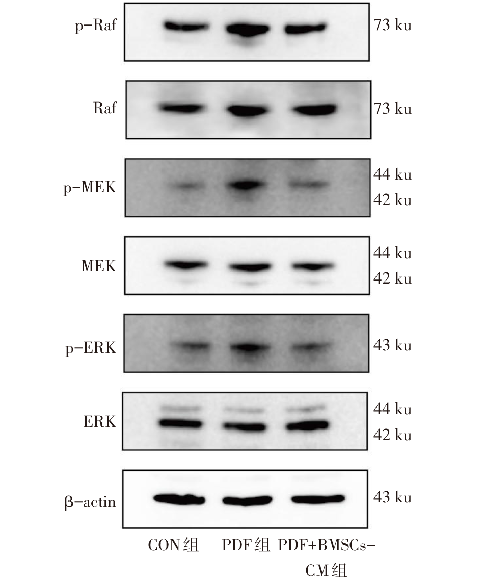
| [23] |
WU P K, BECKER A, PARK J I. Growth inhibitory signaling of the Raf/MEK/ERK pathway[J]. Int J Mol Sci, 2020, 21(15):5436. doi:10.3390/ijms21155436.
|
| [24] |
ZHONG Y, LI M Y, HAN L, et al. Galangin inhibits programmed cell death-ligand 1 expression by suppressing STAT3 and MYC and enhances T cell tumor-killing activity[J]. Phytomedicine, 2023, 116:154877. doi:10.1016/j.phymed.2023.154877.
|
| [25] |
KUMARI S, DHAPOLA R, REDDY D H. Apoptosis in Alzheimer's disease: insight into the signaling pathways and therapeutic avenues[J]. Apoptosis, 2023, 28(7/8):943-957. doi:10.1007/s10495-023-01848-y.
|
| [26] |
LIU Y, CHEN J, LIANG H, et al. Human umbilical cord-derived mesenchymal stem cells not only ameliorate blood glucose but also protect vascular endothelium from diabetic damage through a paracrine mechanism mediated by MAPK/ERK signaling[J]. Stem Cell Res Ther, 2022, 13(1):258. doi:10.1186/s13287-022-02927-8.
|
| [27] |
LANG J, YANG C, LIU L, et al. High glucose activates ERK1/2 to stabilize AP1 and increase MMP9 expression in diabetic foot ulcers[J]. Exp Cell Res, 2021, 403(1):112550. doi:10.1016/j.yexcr.2021.112550.
|
| [28] |
PAN L, ZHANG X, GAO Q. Histatin-1 alleviates high-glucose injury to skin keratinocytes through MAPK signaling pathway[J]. J Cosmet Dermatol, 2022, 21(11):6281-6291. doi:10.1111/jocd.15235.
|
 ), 代会博, 单云, 俞曼殊, 盛梅笑△(