| [1] |
中国心血管健康与疾病报告编写组. 中国心血管健康与疾病报告2020概要[J]. 中国循环杂志, 2021, 36(6):521-545.
|
|
The Writing Committee of the Report on Cardiovascular Health and Diseases in China. Report on cardiovascular health and diseases burden in China:An updated su-mmary of 2020[J]. Chinese Circulation Journal, 2021, 36(6):521-545. doi:10.3969/j.issn.1000-3614.2021.06.001.
|
| [2] |
储睿, 朱刚艳, 周兴宇. 衰老与心血管疾病关系的研究进展[J]. 医学研究杂志, 2018, 47(7):187-190,58.
|
|
CHU R, ZHU G Y, ZHOU X Y. Research progress on the relationship between aging and cardiovascular disease[J]. Journal of Medical Research, 2018, 47(7):187-190,58. doi:10.11969/j.issn.1673-548X.2018.07.044.
|
| [3] |
王曼怡, 朱家明, 孔昊. 基于Logistic模型对心血管疾病风险的预测[J]. 齐齐哈尔大学学报(自然科学版), 2017, 33(5):64-68.
|
|
WANG M Y, ZHU J M, KONG H. Prediction of cardiovascular disease risk based on Logistic model[J]. Journal of Qiqihar University(Natural Science Edition),2017, 33(5):64-68. doi:10.3969/j.issn.1007-984X.2017.05.015.
|
| [4] |
LIU X L, ZHAO Y C, ZHU H Y, et al. Taxifolin retards the D-galactose-induced aging process through inhibiting Nrf2-mediated oxidative stress and regulating the gut microbiota in mice[J]. Food Funct, 2021, 12(23):12142-12158. doi:10.1039/d1fo01349a.
|
| [5] |
于春艳, 于春荣, 敬舒, 等. 北五味子总木脂素对D-半乳糖诱导的小鼠脑衰老自噬和凋亡的影响[J]. 吉林大学学报(医学版), 2014, 40(6):1210-1215.
|
|
YU C Y, YU C R, JING S, et al. Effects of Schisandra total lignin on autophagy and apoptosis of mouse brain aging induced by D-galactose[J]. Journal of Jilin University (Medicine Edition), 2014, 40(6):1210-1215. doi:10.13481/j.1671-587x.20140618.
|
| [6] |
WANG D, WANG T, LI Z, et al. Green tea polyphenols upregulate the Nrf2 signaling pathway and suppress oxidative stress and inflammation markers in D-galactose-induced liver aging in mice[J]. Front Nutr, 2022, 9:836112. doi:10.3389/fnut.2022.836112.
|
| [7] |
CHEN X, LI D, SUN H Y, et al. Relieving ferroptosis may partially reverse neurodegeneration of the auditory cortex[J]. FEBS J, 2020, 287(21):4747-4766. doi:10.1111/febs.15266.
|
| [8] |
SUN C, PENG F, LI J, et al. Ferroptosis-specific inhibitor ferrostatin-1 relieves H2O2-induced redox imbalance in primary cardiomyocytes through the Nrf2/ARE pathway[J]. Dis Markers, 2022, 2022:4539932. doi:10.1155/2022/4539932.
|
| [9] |
HUANG F, YANG R, XIAO Z, et al. Targeting ferroptosis to treat cardiovascular diseases: A new continent to be explored[J]. Front Cell Dev Biol, 2021, 9:737971. doi:10.3389/fcell.2021.737971.
|
| [10] |
刘建亚, 冯文静, 牟婕, 等. hUCMSCs对D-半乳糖致衰老小鼠心脏保护作用[J]. 青岛大学学报(医学版), 2018, 54(2):189-192,196.
|
|
LIU J Y, FENG W J, MOU J, et al. Cardioprotective effect of human umbilical cord mesenchymal stem cells in D-galactose-induced aging mice[J]. Acta Aacademiae Medicinae Qingdao Universitatis, 2018, 54(2):189-192,196. doi:10.11712/jms201802015.
|
| [11] |
MAO H, ZHAO Y, LI H, et al. Ferroptosis as an emerging target in inflammatory diseases[J]. Prog Biophys Mol Biol, 2020, 155:20-28. doi:10.1016/j.pbiomolbio.2020.04.001.
|
| [12] |
TATARKOVÁ Z, KUKA S, RAČAY P, et al. Effects of aging on activities of mitochondrial electron transport chain complexes and oxidative damage in rat heart[J]. Physiol Res, 2011, 60(2):281-289. doi:10.33549/physiolres.932019.
|
| [13] |
TERMAN A, KURZ T, NAVRATIL M, et al. Mitochondrial turnover and aging of long-lived postmitotic cells:The mitochondrial-lysosomal axis theory of aging[J]. Antioxid Redox Signal, 2010, 12(4):503-535. doi:10.1089/ars.2009.2598.
|
| [14] |
LIANG W J, GUSTAFSSON Å B. The aging heart:Mitophagy at the center of rejuvenation[J]. Front Cardiovasc Med, 2020, 7:18. doi:10.3389/fcvm.2020.00018.
|
| [15] |
SHAKERI H, LEMMENS K, GEVAERT A B, et al. Cellular senescence links aging and diabetes in cardiovascular disease[J]. Am J Physiol Heart Circ Physiol, 2018, 315(3):H448-H462. doi:10.1152/ajpheart.00287.2018.
|
| [16] |
IZZO C, CARRIZZO A, ALFANO A, et al. The impact of aging on cardio and cerebrovascular diseases[J]. Int J Mol Sci, 2018, 19(2):481. doi:10.3390/ijms19020481.
|
| [17] |
SHIMIZU I, MINAMINO T. Cellular senescence in cardiac diseases[J]. J Cardiol, 2019, 74(4):313-319. doi:10.1016/j.jjcc.2019.05.002.
|
| [18] |
PIGNATELLI P, MENICHELLI D, PASTORI D, et al. Oxidative stress and cardiovascular disease:New insights[J]. Kardiol Pol, 2018, 76(4):713-722. doi:10.5603/KP.a2018.0071.
|
| [19] |
FENG W, LIU J, WANG S, et al. Alginate oligosaccharide alleviates D-galactose-induced cardiac ageing via regulating myocardial mitochondria function and integrity in mice[J]. J Cell Mol Med, 2021, 25(15):7157-7168. doi:10.1111/jcmm.16746.
|
| [20] |
SAHIN E, COLLA S, LIESA M, et al. Telomere dysfunction induces metabolic and mitochondrial compromise[J]. Nature, 2011, 470(7334):359-365. doi:10.1038/nature09787.
|
| [21] |
DIXON S J, LEMBERG K M, LAMPRECHT M R, et al. Ferroptosis:An iron-dependent form of nonapoptotic cell death[J]. Cell, 2012, 149(5):1060-1072. doi:10.1016/j.cell.2012.03.042.
|
| [22] |
STOCKWELL B R, FRIEDMANN ANGELI J P, BAYIR H, et al. Ferroptosis:A regulated cell death nexus linking metabolism,redox biology,and disease[J]. Cell, 2017, 171(2):273-285. doi:10.1016/j.cell.2017.09.021.
|
| [23] |
SUN Y, ZHENG Y, WANG C, et al. Glutathione depletion induces ferroptosis,autophagy, and premature cell senescence in retinal pigment epithelial cells[J]. Cell Death Dis, 2018, 9(7):753. doi:10.1038/s41419-018-0794-4.
|
| [24] |
FORCINA G C, DIXON S J. GPX4 at the crossroads of lipid homeostasis and ferroptosis[J]. Proteomics, 2019, 19(18):e1800311. doi:10.1002/pmic.201800311.
|
| [25] |
LIU P, FENG Y, LI H, et al. Ferrostatin-1 alleviates lipopolysaccharide-induced acute lung injury via inhibiting ferroptosis[J]. Cell Mol Biol Lett, 2020, 25:10. doi:10.1186/s11658-020-00205-0.
|
| [26] |
LARRICK J W, LARRICK J W, MENDELSOHN A R. Contribution of ferroptosis to aging and frailty[J]. Rejuvenation Res, 2020, 23(5):434-438. doi:10.1089/rej.2020.2390.
|
| [27] |
JENKINS N L, JAMES S A, SALIM A, et al. Changes in ferrous iron and glutathione promote ferroptosis and frailty in aging Caenorhabditis elegans[J]. Elife, 2020, 9:e56580. doi:10.7554/eLife.56580.
|
| [28] |
WU X, LI Y, ZHANG S, et al. Ferroptosis as a novel therapeutic target for cardiovascular disease[J]. Theranostics, 2021, 11(7):3052-3059. doi:10.7150/thno.54113.
|
| [29] |
RITZENTHALER J D, TORRES-GONZALEZ E, ZHENG Y, et al. The profibrotic and senescence phenotype of old lung fibroblasts is reversed or ameliorated by genetic and pharmacological manipulation of Slc7a11 expression[J]. Am J Physiol Lung Cell Mol Physiol, 2022, 322(3):L449-L461. doi:10.1152/ajplung.00593.2020.
|
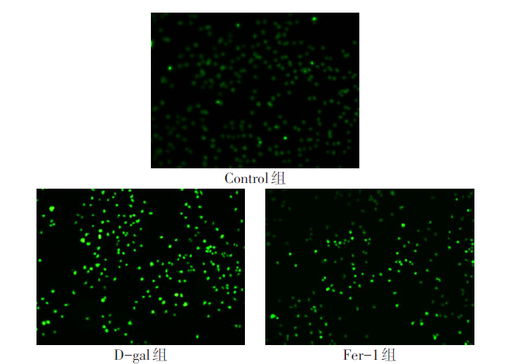
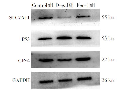
 ), 王一平1, 王刚1, 张甜1, 包雅丽1, 迪娜·艾尼瓦尔1, 孙湛1,2,∆(