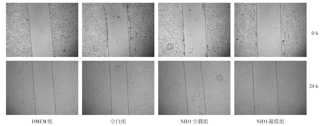
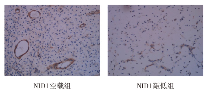
| [1] |
RIZZO M, CALIÒ A, BRUNELLI M, et al. Clinico-pathological implications of the 2022 WHO renal cell carcinoma classification[J]. Cancer Treat Rev, 2023, 116:102558. doi:10.1016/j.ctrv.2023.102558.
|
| [2] |
CHEN Y W, WANG L, PANIAN J, et al. Treatment landscape of renal cell carcinoma[J]. Curr Treat Options Oncol, 2023, 24(12):1889-1916. doi:10.1007/s11864-023-01161-5.
|
| [3] |
CHOUEIRI T K, KAELIN W G Jr. Targeting the HIF2-VEGF axis in renal cell carcinoma[J]. Nat Med, 2020, 26(10):1519-1530. doi:10.1038/s41591-020-1093-z.
|
| [4] |
KUAN M I, CARUSO L B, ZAVALA A G, et al. Human cytomegalovirus utilizes multiple viral proteins to regulate the basement membrane protein nidogen 1[J]. J Virol, 2022, 96(20):e0133622. doi:10.1128/jvi.01336-22.
|
| [5] |
MAO X, TEY S K, YEUNG C, et al. Nidogen 1-enriched extracellular vesicles facilitate extrahepatic metastasis of liver cancer by activating pulmonary fibroblasts to secrete tumor necrosis factor receptor 1[J]. Adv Sci(Weinh), 2020, 7(21):2002157. doi:10.1002/advs.202002157.
|
| [6] |
ALEČKOVIĆ M, WEI Y, LEROY G, et al. Identification of nidogen 1 as a lung metastasis protein through secretome analysis[J]. Genes Dev, 2017, 31(14):1439-1455. doi:10.1101/gad.301937.117.
|
| [7] |
ROKAVEC M, JAECKEL S, HERMEKING H. Nidogen-1/NID1 function and regulation during progression and metastasis of colorectal cancer[J]. Cancers(Basel), 2023, 15(22):5316. doi:10.3390/cancers15225316.
|
| [8] |
JONASCH E, DONSKOV F, ILIOPOULOS O, et al. Belzutifan for renal cell carcinoma in von hippel-lindau disease[J]. N Engl J Med, 2021, 385(22):2036-2046. doi:10.1056/NEJMoa2103425.
|
| [9] |
BERGQVIST C, EZZEDINE K. Vitiligo:a review[J]. Dermatology, 2020, 236(6):571-592. doi:10.1159/000506103.
|
| [10] |
KIM H, SHIM B Y, LEE S J, et al. Loss of Von hippel-lindau(VHL) tumor suppressor gene function: VHL-HIF pathway and advances in treatments for metastatic renal cell carcinoma(RCC)[J]. Int J Mol Sci, 2021, 22(18):9795. doi:10.3390/ijms22189795.
|
| [11] |
KAELIN W G Jr. Von Hippel-Lindau disease: insights into oxygen sensing, protein degradation, and cancer[J]. J Clin Invest, 2022, 132(18):e162480. doi:10.1172/JCI162480.
|
| [12] |
TELEANU R I, CHIRCOV C, GRUMEZESCU A M, et al. Tumor angiogenesis and anti-angiogenic strategies for cancer treatment[J]. J Clin Med, 2019, 9(1):84. doi:10.3390/jcm9010084.
|
| [13] |
JIANG X, WANG J, DENG X, et al. The role of microenvironment in tumor angiogenesis[J]. J Exp Clin Cancer Res, 2020, 39(1):204. doi:10.1186/s13046-020-01709-5.
|
| [14] |
YANG J, WANG K, YANG Z. Treatment strategies for clear cell renal cell carcinoma: Past, present and future[J]. Front Oncol, 2023, 13:1133832. doi:10.3389/fonc.2023.1133832.
|
| [15] |
ANGULO J C, SHAPIRO O. The changing therapeutic landscape of metastatic renal cancer[J]. Cancers(Basel), 2019, 11(9):1227. doi:10.3390/cancers11091227.
|
| [16] |
ASTORE S, BACIARELLO G, CERBONE L, et al. Primary and acquired resistance to first-line therapy for clear cell renal cell carcinoma[J]. Cancer Drug Resist, 2023, 6(3):517-546. doi:10.20517/cdr.2023.33.
|
| [17] |
ZHOU S, CHEN S, PEI Y A, et al. Nidogen: a matrix protein with potential roles in musculoskeletal tissue regeneration[J]. Genes Dis, 2022, 9(3):598-609. doi:10.1016/j.gendis.2021.03.004.
|
| [18] |
CHENG P, CAO T, ZHAO X, et al. Nidogen1-enriched extracellular vesicles accelerate angiogenesis and bone regeneration by targeting myosin-10 to regulate endothelial cell adhesion[J]. Bioact Mater, 2022, 12:185-197. doi:10.1016/j.bioactmat.2021.10.021.
|
| [19] |
HAN N, LI X, WANG Y, et al. HIF-1α induced NID1 expression promotes pulmonary metastases via the PI3K-AKT pathway in salivary gland adenoid cystic carcinoma[J]. Oral Oncol, 2022, 131:105940. doi:10.1016/j.oraloncology.2022.105940.
|
| [20] |
LEE H W, XU Y, HE L, et al. Role of venous endothelial cells in developmental and pathologic angiogenesis[J]. Circulation, 2021, 144(16):1308-1322. doi:10.1161/CIRCULATIONAHA.121.054071.
|
 ), 李刚△(