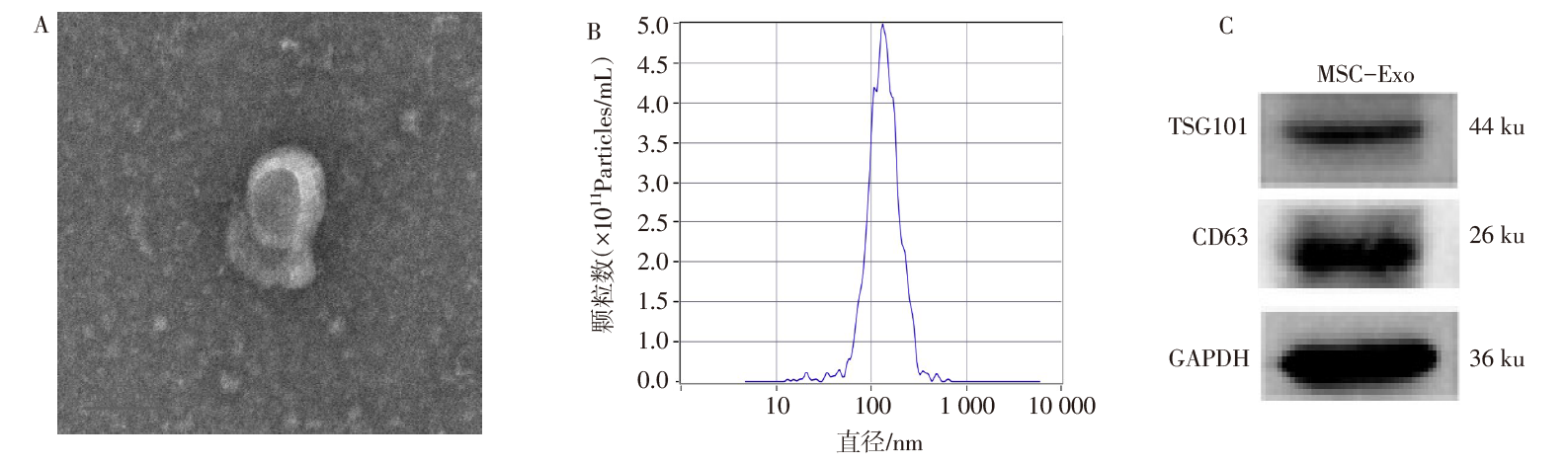
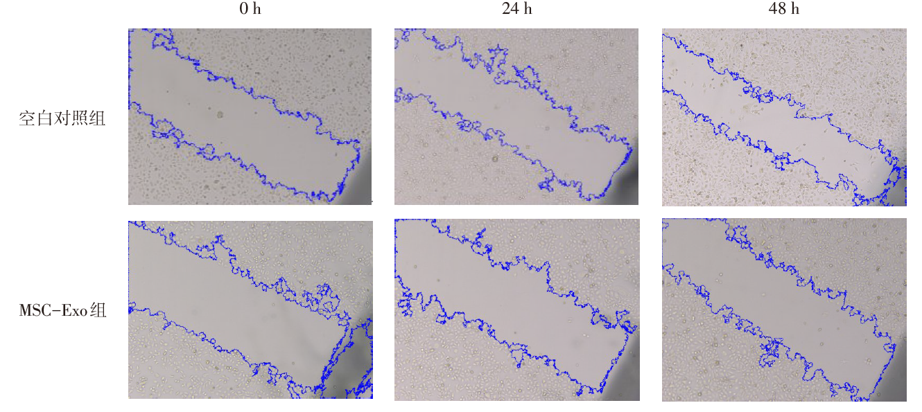
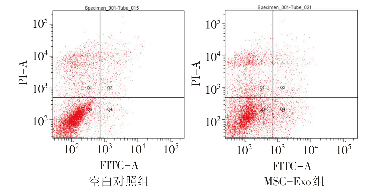
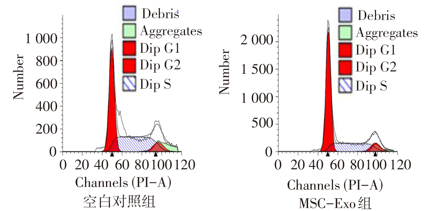
| [1] |
XIA C, DONG X, LI H, et al. Cancer statistics in China and United States[J]. Chin Med J (Engl), 2022, 135(5):584-590. doi:10.1097/CM9.0000000000002108.
|
| [2] |
ZHU H, MA X, YE T, et al. Esophageal cancer in China:Practice and research in the new era[J]. Int J Cancer, 2023, 152(9):1741-1751. doi:10.1002/ijc.34301.
|
| [3] |
MENSHIKOV M, ZUBKOVA E, STAFEEV I, et al. Autophagy,mesenchymal stem cell differentiation,and secretion[J]. Biomedicines, 2021, 9(9):1178. doi:10.3390/biomedicines9091178.
|
| [4] |
ARSHAD M, JALIL F, JALEELl H, et al. Bone marrow derived mesenchymal stem cells therapy for rheumatoid arthritis-a concise review of past ten years[J]. Mol Biol Rep, 2023, 50(5):4619-4629. doi:10.1007/s11033-023-08277-9.
|
| [5] |
VAN GRIENSVEN M, BALMAYOR E R. Extracellularvesicles are key players in mesenchymal stem cells dual potential to regenerate and modulate the immune system[J]. Adv Drug Deliv Rev, 2024, 207:115203. doi:10.1016/j.addr.2024.115203.
|
| [6] |
ZHU Y, CHEN X, LIAO Y. Mesenchymal stem cells derived apoptotic extracellular vesicles (ApoEVs):mechanism and application in tissue regeneration[J]. Stem Cells, 2023, 41(9):837-849. doi:10.1093/stmcls/sxad046.
|
| [7] |
褚小莉, 陈林, 朱栋炜. 脐带间充质干细胞来源的外泌体抑制胃癌细胞的增殖和侵袭[J]. 现代肿瘤医学, 32(12):2140-2145.
|
|
CHU X L, CHEN L, ZHU D W. Exosomes derived from umbilical cord mesenchymal stem cells inhibit proliferation and invasion of gastric cancer cells[J]. Modern Oncology, 2024, 32(12):2140-2145. doi:10.3969/j.issn.1672-4992.2024.12.002.
|
| [8] |
VAN V S, KOURTI A, AUSLOOS E, et al. ATP13A4 upregulation drives the elevated polyamine transport system in the breast cancer cell line MCF7[J]. Biomolecules, 2023, 13(6):918. doi:10.3390/biom13060918.
|
| [9] |
WANG S, SU X, XU M, et al. Exosomes secreted by mesenchymal stromal/stem cell-derived adipocytes promote breast cancer cell growth via activation of Hippo signaling pathway[J]. Stem Cell Res Ther, 2019, 10(1):117. doi:10.1186/s13287-019-1220-2.
|
| [10] |
WANG J, ZHANG A, HUANG F, et al. MSC-EXO and tempol ameliorate bronchopulmonary dysplasia in newborn rats by activating HIF-1α[J]. Pediatr Pulmonol, 2023, 58(5):1367-1379. doi:10.1002/ppul.26317.
|
| [11] |
HU Z, YUAN Y, ZHANG X, et al. Human umbilical cord mesenchymal stem cell-derived exosomes attenuate oxygen-glucose deprivation/reperfusion-induced microglial pyroptosis by promoting FOXO3a-dependent mitophagy[J]. Oxid Med Cell Longev, 2021, 2021:6219715. doi:10.1155/2021/6219715.
|
| [12] |
CAO M, ZHAO Y, CHEN T, et al. Adipose mesenchymal stem cell-derived exosomal microRNAs ameliorate polycystic ovary syndrome by protecting against metabolic disturbances[J]. Biomaterials, 2022, 288:121739. doi:10.1016/j.biomaterials.2022.121739.
|
| [13] |
QIU Y, SUN J, QIU J, et al. Antitumor activity of cabazitaxel and MSC-TRAIL derived extracellular vesicles in drug-resistant oral squamous cell carcinoma[J]. Cancer Manag Res, 2020, 12:10809-10820. doi:10.2147/CMAR.S277324.
|
| [14] |
SURAMAN M, KEDRACKA-KROK S, JANKOWSKA U, et al. Proteomic profiling of ectosomes derived from paired urothelial bladder cancer and normal cells reveals the presence of biologically-relevant molecules[J]. Int J Mol Sci, 2021, 22(13):6816. doi:10.3390/ijms22136816.
|
| [15] |
CHANG W, CERIONE R, ANTONYAK M. Extracellular vesicles and their roles in cancer progression[J]. Methods Mol Biol, 2021, 2174:143-170. doi:10.1007/978-1-0716-0759-6_10.
|
| [16] |
LIU J, XIAO Q, XIAO J, et al. Wnt/β-catenin signalling:function,biological mechanisms,and therapeutic opportunities[J]. Signal Transduct Target Ther, 2022, 7(1):3. doi:10.1038/s41392-021-00762-6.
|
| [17] |
ZHAO Q, BI Y, ZHONG J, et al. Pristimerin suppresses colorectal cancer through inhibiting inflammatory responses and Wnt/β-catenin signaling[J]. Toxicol Appl Pharmacol, 2020, 386:114813. doi:10.1016/j.taap.2019.114813.
|
| [18] |
WANG J, HU K, CAI X, et al. Targeting PI3K/AKT signaling for treatment of idiopathic pulmonary fibrosis[J]. Acta Pharm Sin B, 2022, 12(1):18-32. doi:10.1016/j.apsb.2021.07.023.
|
| [19] |
QIU L, WANG J, CHEN M, et al. Exosomal microRNA 146a derived from mesenchymal stem cells increases the sensitivity of ovarian cancer cells to docetaxel and taxane via a LAMC2 mediated PI3K/Akt axis[J]. Int J Mol Med, 2020, 46(2):609-620. doi:10.3892/ijmm.2020.4634.
|
 ), 李子沐2, 王亮1, 许彭3, 李秀梅3,△(