
天津医药 ›› 2026, Vol. 54 ›› Issue (7): 673-681.doi: 10.11958/20260112
• 细胞与分子生物学 • 下一篇
闫奇1( ), 陈晨2, 解维敏1,△()
收稿日期:2026-01-07
修回日期:2026-02-28
出版日期:2026-07-15
发布日期:2026-07-13
通讯作者:
△E-mail:312027454@qq.com
作者简介:闫奇(1985),男,主治医师,主要从事胰腺、胆道相关疾病方面研究。E-mail:基金资助:
YAN Qi1(), CHEN Chen2, XIE Weimin1,△()
Received:2026-01-07
Revised:2026-02-28
Published:2026-07-15
Online:2026-07-13
Contact:
△E-mail:312027454@qq.com
闫奇, 陈晨, 解维敏. 杠柳次苷调控STAT3抑制人胰腺癌细胞系增殖和侵袭[J]. 天津医药, 2026, 54(7): 673-681.
YAN Qi, CHEN Chen, XIE Weimin. Periplocymarin regulates STAT3 to inhibit proliferation and invasion of human pancreatic cancer cell lines[J]. Tianjin Medical Journal, 2026, 54(7): 673-681.
摘要:
目的 探究杠柳次苷(PPM)对人胰腺癌细胞系增殖和侵袭的影响,并分析其是否通过调控信号转导和转录激活因子3(STAT3)发挥抗癌作用。方法 (1)体外实验:将SW1990细胞分为对照组,0.05、0.1、0.2 PPM组(分别用0.05、0.1和0.2 μmol/L PPM培养48 h),NC-oe+0.2 PPM组(转染NC-oe,0.2 μmol/L PPM培养)和STAT3-oe+0.2 PPM组(转染STAT3-oe,0.2 μmol/L PPM培养)。采用MTT实验及EdU染色检测细胞增殖,TUNEL检测细胞凋亡,Transwell实验检测细胞侵袭。(2)体内实验:将裸鼠随机分为模型组、L-PPM组[1.0 mg/(kg·d) PPM]、H-PPM组[3.0 mg/(kg·d) PPM]、NC-oe+H-PPM组[转染NC-oe,3.0 mg/(kg·d) PPM]和STAT3-oe+H-PPM组[转染STAT3-oe,3.0 mg/(kg·d) PPM],每组6只,给药周期为28 d。采用HE染色观察肿瘤组织形态,免疫组化检测肿瘤组织p-STAT3、Ki-67和B细胞淋巴瘤(Bcl)-2表达,qRT-PCR检测细胞和肿瘤组织STAT3 mRNA水平。Western blot检测SW1990细胞和肿瘤组织p-STAT3(Tyr705)、STAT3、Bcl-2、Bcl-2相关X蛋白(Bax)、基质金属蛋白酶(MMP)-2和MMP-9蛋白表达。结果 (1)体外实验:与对照组比较,0.05、0.1和0.2 PPM组SW1990细胞的STAT3 mRNA和p-STAT3(Tyr705)蛋白水平均降低(P<0.05),相对细胞活力、EdU阳性率、侵袭细胞数量以及Bcl-2、MMP-2和MMP-9蛋白水平均降低(P<0.05),TUNEL阳性率和Bax蛋白水平均升高(P<0.05)。与NC-oe+0.2 PPM组比较,STAT3-oe+0.2 PPM组SW1990细胞的STAT3 mRNA和p-STAT3(Tyr705)蛋白水平均升高(P<0.05),相对细胞活力、EdU阳性率、侵袭细胞数量以及Bcl-2、MMP-2和MMP-9蛋白水平均升高(P<0.05),TUNEL阳性率和Bax蛋白水平均降低(P<0.05)。(2)体内实验:与模型组比较,L-PPM组和H-PPM组裸鼠肿瘤体积、STAT3 mRNA水平、p-STAT3(Tyr705)蛋白水平以及p-STAT3、Ki-67和Bcl-2的阳性率均降低(P<0.05)。与NC-oe+H-PPM组比较,STAT3-oe+H-PPM组裸鼠肿瘤体积、STAT3 mRNA水平、p-STAT3(Tyr705)蛋白水平以及p-STAT3、Ki-67和Bcl-2的阳性率均升高(P<0.05)。结论 PPM具有抗胰腺癌活性,其通过调控STAT3抑制胰腺癌细胞增殖和侵袭。
中图分类号:
| 组别 | STAT3 mRNA | p-STAT3(Tyr705)/STAT3 |
|---|---|---|
| 对照组 | 1.00±0.06 | 1.00±0.05 |
| 0.05 PPM组 | 0.70±0.06a | 0.65±0.08a |
| 0.1 PPM组 | 0.45±0.04ab | 0.43±0.02ab |
| 0.2 PPM组 | 0.24±0.02abc | 0.20±0.02abc |
| NC-oe+0.2 PPM组 | 0.23±0.02 | 0.19±0.02 |
| STAT3-oe+0.2 PPM组 | 2.25±0.13d | 1.66±0.16d |
| F | 762.529** | 342.966** |
表1 各组SW1990细胞的STAT3 mRNA和p-STAT3(Tyr705)蛋白表达水平比较
Tab.1 Comparison of STAT3 mRNA and p-STAT3 (Tyr705) protein expression levels between six groups of SW1990 cells (n=6,$\stackrel{-}{x}\pm s$)
| 组别 | STAT3 mRNA | p-STAT3(Tyr705)/STAT3 |
|---|---|---|
| 对照组 | 1.00±0.06 | 1.00±0.05 |
| 0.05 PPM组 | 0.70±0.06a | 0.65±0.08a |
| 0.1 PPM组 | 0.45±0.04ab | 0.43±0.02ab |
| 0.2 PPM组 | 0.24±0.02abc | 0.20±0.02abc |
| NC-oe+0.2 PPM组 | 0.23±0.02 | 0.19±0.02 |
| STAT3-oe+0.2 PPM组 | 2.25±0.13d | 1.66±0.16d |
| F | 762.529** | 342.966** |
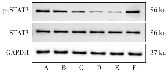
图1 各组SW1990细胞p-STAT3表达水平比较 A:对照组;B:0.05 PPM组;C:0.1 PPM组;D:0.2 PPM组;E:NC-oe+0.2 PPM组;F:STAT3-oe+0.2 PPM组。图4、6—7同。
Fig.1 p-STAT3 expression levels in each group of SW1990 cells
| 组别 | 相对细胞活力/% | EdU阳性率/% | ||||
|---|---|---|---|---|---|---|
| 对照组 | 100.00±3.23 | 100.00±5.11 | ||||
| 0.05 PPM组 | 77.81±6.51a | 82.70±6.47a | ||||
| 0.1 PPM组 | 60.35±3.62ab | 64.46±4.11ab | ||||
| 0.2 PPM组 | 38.46±3.49abc | 43.12±3.57abc | ||||
| NC-oe+0.2 PPM组 | 39.22±2.96 | 39.93±2.77 | ||||
| STAT3-oe+0.2 PPM组 | 69.91±4.79d | 68.63±11.78d | ||||
| F | 183.597** | 78.132** | ||||
| 组别 | TUNEL阳性率/% | Bcl-2/GAPDH | Bax/GAPDH | |||
| 对照组 | 4.82±0.32 | 1.00±0.06 | 1.00±0.07 | |||
| 0.05 PPM组 | 15.35±1.26a | 0.81±0.06a | 2.79±0.36a | |||
| 0.1 PPM组 | 26.91±2.56ab | 0.65±0.04ab | 4.25±0.39ab | |||
| 0.2 PPM组 | 34.58±4.22abc | 0.41±0.03abc | 5.11±0.66abc | |||
| NC-oe+0.2 PPM组 | 35.41±3.76 | 0.40±0.02 | 5.28±0.70 | |||
| STAT3-oe+0.2 PPM组 | 18.92±2.33d | 0.62±0.05d | 3.53±0.24d | |||
| F | 111.874** | 152.983** | 73.371** | |||
表2 各组SW1990细胞的相对细胞活力、EdU、TUNEL 阳性率和Bcl-2、Bax蛋白表达水平比较
Tab.2 Comparison of the relative cell viability, EdU positive rate, TUNEL positive rate, and protein expression levels of Bcl-2 and Bax between each group of SW1990 cells (n=6,$\stackrel{-}{x}\pm s$)
| 组别 | 相对细胞活力/% | EdU阳性率/% | ||||
|---|---|---|---|---|---|---|
| 对照组 | 100.00±3.23 | 100.00±5.11 | ||||
| 0.05 PPM组 | 77.81±6.51a | 82.70±6.47a | ||||
| 0.1 PPM组 | 60.35±3.62ab | 64.46±4.11ab | ||||
| 0.2 PPM组 | 38.46±3.49abc | 43.12±3.57abc | ||||
| NC-oe+0.2 PPM组 | 39.22±2.96 | 39.93±2.77 | ||||
| STAT3-oe+0.2 PPM组 | 69.91±4.79d | 68.63±11.78d | ||||
| F | 183.597** | 78.132** | ||||
| 组别 | TUNEL阳性率/% | Bcl-2/GAPDH | Bax/GAPDH | |||
| 对照组 | 4.82±0.32 | 1.00±0.06 | 1.00±0.07 | |||
| 0.05 PPM组 | 15.35±1.26a | 0.81±0.06a | 2.79±0.36a | |||
| 0.1 PPM组 | 26.91±2.56ab | 0.65±0.04ab | 4.25±0.39ab | |||
| 0.2 PPM组 | 34.58±4.22abc | 0.41±0.03abc | 5.11±0.66abc | |||
| NC-oe+0.2 PPM组 | 35.41±3.76 | 0.40±0.02 | 5.28±0.70 | |||
| STAT3-oe+0.2 PPM组 | 18.92±2.33d | 0.62±0.05d | 3.53±0.24d | |||
| F | 111.874** | 152.983** | 73.371** | |||
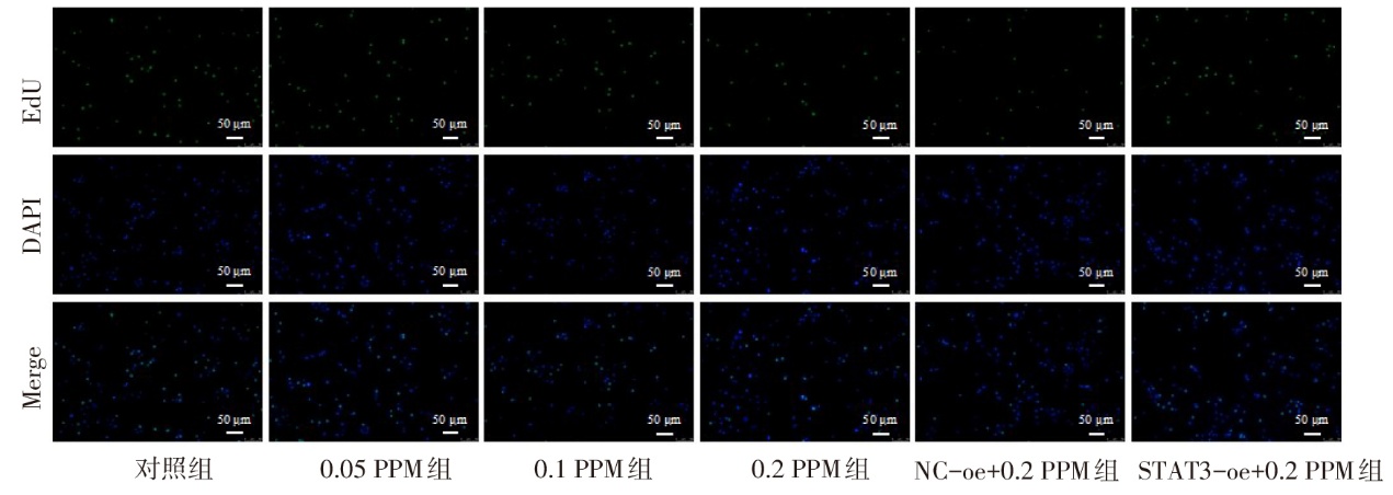
图2 EdU染色检测各组SW1990细胞增殖水平
Fig.2 The proliferation levels of SW1990 cells in each group detected by EdU staining
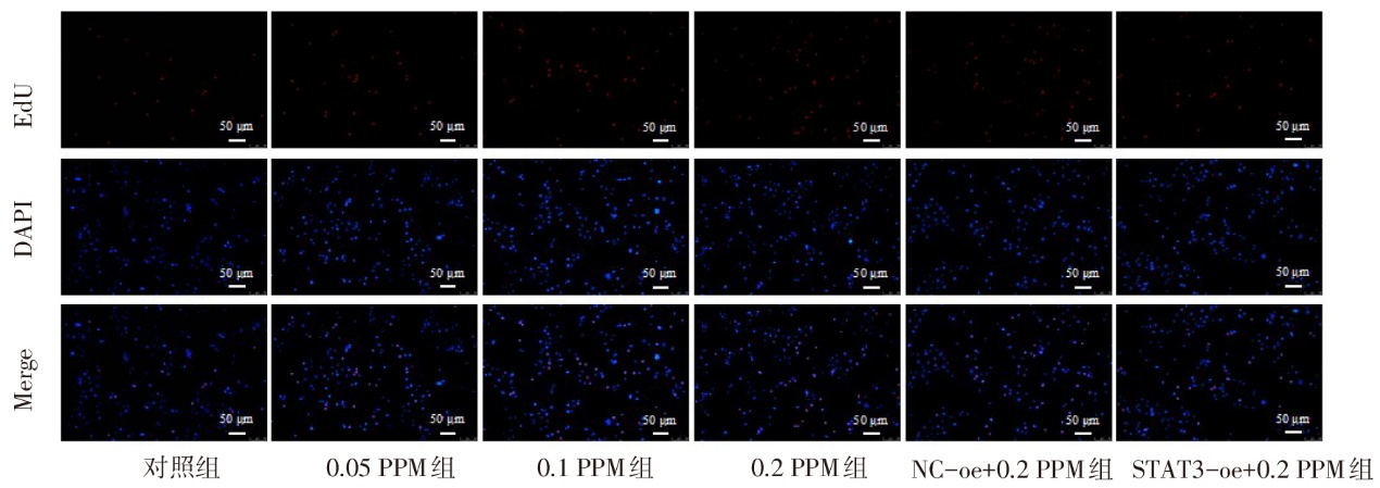
图3 TUNEL染色检测各组SW1990细胞凋亡水平
Fig.3 The apoptotic levels of SW1990 cells in each group detected by TUNEL staining
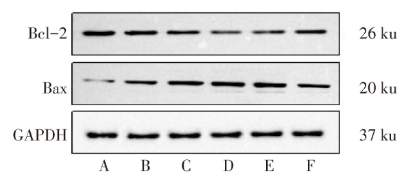
图4 各组SW1990细胞Bcl-2和Bax蛋白表达水平比较
Fig. 4 Comparison of Bcl-2 and Bax protein expression levels between each group of SW1990 cells
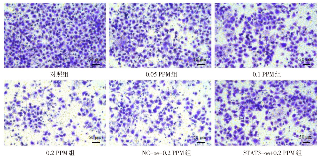
图5 Transwell实验中结晶紫染色检测各组SW1990细胞侵袭水平
Fig.5 The invasion levels of SW1990 cells in each group detected by crystal violet staining in Transwell assay
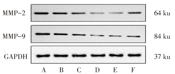
图6 各组SW1990细胞MMP-2和MMP-9蛋白表达水平比较
Fig. 6 Comparison of the expression levels of MMP-2 and MMP-9 proteins between each group of SW1990 cells
| 组别 | 侵袭细胞数/个 | MMP-2/GAPDH | MMP-9/GAPDH |
|---|---|---|---|
| 对照组 | 128.17±6.91 | 1.00±0.02 | 1.00±0.06 |
| 0.05 PPM组 | 107.17±12.02a | 0.79±0.06a | 0.69±0.05a |
| 0.1 PPM组 | 79.33±10.61ab | 0.52±0.03ab | 0.55±0.03ab |
| 0.2 PPM组 | 49.67±8.04abc | 0.25±0.02abc | 0.31±0.02abc |
| NC-oe+0.2 PPM组 | 53.50±3.02 | 0.24±0.02 | 0.29±0.02 |
| STAT3-oe+0.2 PPM组 | 88.67±3.14d | 0.62±0.03d | 0.59±0.04d |
| F | 85.943** | 487.081** | 243.020** |
表3 各组SW1990细胞的侵袭细胞数以及MMP-2和MMP-9蛋白相对水平
Tab.3 The number of invasive cells and the relative levels of MMP-2 and MMP-9 proteins in SW1990 cells of each group (n=6,$\stackrel{-}{x}\pm s$)
| 组别 | 侵袭细胞数/个 | MMP-2/GAPDH | MMP-9/GAPDH |
|---|---|---|---|
| 对照组 | 128.17±6.91 | 1.00±0.02 | 1.00±0.06 |
| 0.05 PPM组 | 107.17±12.02a | 0.79±0.06a | 0.69±0.05a |
| 0.1 PPM组 | 79.33±10.61ab | 0.52±0.03ab | 0.55±0.03ab |
| 0.2 PPM组 | 49.67±8.04abc | 0.25±0.02abc | 0.31±0.02abc |
| NC-oe+0.2 PPM组 | 53.50±3.02 | 0.24±0.02 | 0.29±0.02 |
| STAT3-oe+0.2 PPM组 | 88.67±3.14d | 0.62±0.03d | 0.59±0.04d |
| F | 85.943** | 487.081** | 243.020** |
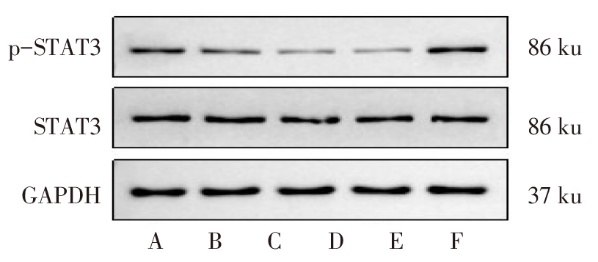
图7 各组小鼠肿瘤组织p-STAT3表达水平比较
Fig.7 p-STAT3 expression levels in tumor tissue of each group of mice
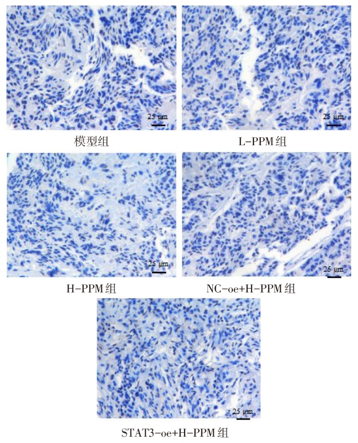
图8 各组小鼠肿瘤组织p-STAT3免疫组化染色
Fig.8 Immunohistochemical staining of p-STAT3 in tumor tissue of each group of mice
| 组别 | STAT3 mRNA | p-STAT3(Tyr705)/STAT3 | p-STAT3阳性率/% |
|---|---|---|---|
| 模型组 | 1.00±0.06 | 1.00±0.07 | 60.38±7.99 |
| L-PPM组 | 0.66±0.05a | 0.57±0.06a | 37.44±4.61a |
| H-PPM组 | 0.31±0.02ab | 0.26±0.02ab | 20.47±0.96ab |
| NC-oe+H-PPM组 | 0.30±0.03 | 0.25±0.02 | 20.42±0.85 |
| STAT3-oe+H-PPM组 | 2.61±0.19c | 2.17±0.24c | 78.08±5.90c |
| F | 622.450** | 290.289** | 159.239** |
表4 各组小鼠肿瘤组织STAT3 mRNA和p-STAT3(Tyr705)蛋白相对水平以及p-STAT3阳性率比较
Tab.4 Comparison of the relative levels of STAT3 mRNA, p-STAT3 (Tyr705) protein and the positive rate of p-STAT3 in tumor tissue between the six groups of mice (n=6,$\stackrel{-}{x}\pm s$)
| 组别 | STAT3 mRNA | p-STAT3(Tyr705)/STAT3 | p-STAT3阳性率/% |
|---|---|---|---|
| 模型组 | 1.00±0.06 | 1.00±0.07 | 60.38±7.99 |
| L-PPM组 | 0.66±0.05a | 0.57±0.06a | 37.44±4.61a |
| H-PPM组 | 0.31±0.02ab | 0.26±0.02ab | 20.47±0.96ab |
| NC-oe+H-PPM组 | 0.30±0.03 | 0.25±0.02 | 20.42±0.85 |
| STAT3-oe+H-PPM组 | 2.61±0.19c | 2.17±0.24c | 78.08±5.90c |
| F | 622.450** | 290.289** | 159.239** |
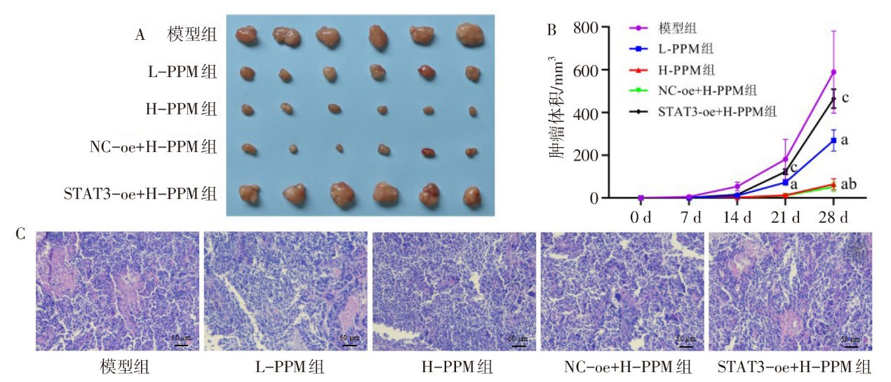
图9 各组小鼠肿瘤体积和病理学检测 A:瘤体图像;B:肿瘤生长曲线(0 d和7 d肿瘤尚未出现,14、21、28 d时F分别为67.706、164.498、208.867,均P<0.01),a与模型组比较,b与L-PPM组比较,c与NC-oe+H-PPM组比较,P<0.05;C:肿瘤组织HE染色。
Fig. 9 Tumor volume and pathological examination of each group of mice
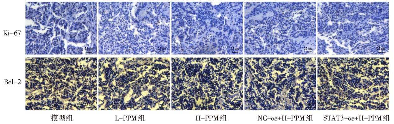
图10 各组小鼠肿瘤组织Ki-67和Bcl-2表达水平分析
Fig.10 Analysis of the expression levels of Ki-67 and Bcl-2 in tumor tissue of each group of mice
| 组别 | Ki-67阳性率/% | Bcl-2阳性率/% |
|---|---|---|
| 模型组 | 51.15±8.14 | 67.84±6.57 |
| L-PPM组 | 36.98±3.93a | 52.98±4.33a |
| H-PPM组 | 19.18±0.86ab | 30.78±1.72ab |
| NC-oe+H-PPM组 | 18.94±1.68 | 29.49±2.73 |
| STAT3-oe+H-PPM组 | 44.80±5.50c | 58.47±2.68c |
| F | 56.125** | 110.005** |
表5 各组小鼠肿瘤组织中Ki-67和Bcl-2的阳性率比较
Tab.5 Comparison of the positive rates of Ki-67 and Bcl-2 in mouse tumor tissue between different groups (n=6,$\stackrel{-}{x}\pm s$)
| 组别 | Ki-67阳性率/% | Bcl-2阳性率/% |
|---|---|---|
| 模型组 | 51.15±8.14 | 67.84±6.57 |
| L-PPM组 | 36.98±3.93a | 52.98±4.33a |
| H-PPM组 | 19.18±0.86ab | 30.78±1.72ab |
| NC-oe+H-PPM组 | 18.94±1.68 | 29.49±2.73 |
| STAT3-oe+H-PPM组 | 44.80±5.50c | 58.47±2.68c |
| F | 56.125** | 110.005** |
| [1] | 闫玲新, 李森, 郭改莉, 等. 异牡荆素通过miR-339-5p/HSPA8轴调节胰腺癌细胞的生物学行为[J]. 天津医药, 2025, 53(3):230-235. |
| Yan L X, Li S, Guo G L, et al. Isovitexin regulates proliferation,migration and invasion of pancreatic cancer cells via the miR-339-5p/HSPA8 axis[J]. Tianjin Med J, 2025, 53(3):230-235. doi:10.11958/20242056. | |
| [2] | Hamel Z, Sanchez S, Standing D, et al. Role of STAT3 in pancreatic cancer[J]. Explor Target Antitumor Ther, 2024, 5(5):20-34. doi:10.37349/etat.2024.00202. |
| [3] | Li X, Jiang W, Dong S, et al. STAT3 inhibitors:a novel insight for anticancer therapy of pancreatic cancer[J]. Biomolecules, 2022, 12(10):1450. doi:10.3390/biom12101450. |
| [4] | Petrocca F, Lieberman J. Promise and challenge of RNA interference-based therapy for cancer[J]. J Clin Oncol, 2011, 29(6):747-754. doi:10.1200/JCO.2009.27.6287. |
| [5] | Cheng Y, Wang G, Zhao L, et al. Periplocymarin induced colorectal cancer cells apoptosis via impairing PI3K/AKT pathway[J]. Front Oncol, 2021, 11:753598. doi:10.3389/fonc.2021.753598. |
| [6] | Hao Y, Song T, Wang M, et al. Dual targets of lethal apoptosis and protective autophagy in liver cancer with periplocymarin elicit a limited therapeutic effect[J]. Int J Oncol, 2023, 62(3):44. doi:10.3892/ijo.2023.5492. |
| [7] | Ke A, Yang W, Zhang W, et al. The cardiac glycoside periplocymarin sensitizes gastric cancer to ferroptosis via the ATP1A1-Src-YAP/TAZ-TFRC axis[J]. Phytomedicine, 2025, 142:156804. doi:10.1016/j.phymed.2025.156804. |
| [8] | Fan C, Liang S, Ye M, et al. Periplocymarin alleviates pathological cardiac hypertrophy via inhibiting the JAK2/STAT3 signalling pathway[J]. J Cell Mol Med, 2022, 26(9):2607-2619. doi:10.1111/jcmm.17267. |
| [9] | 邢雪, 郭晓, 任晓亮, 等. 杠柳毒苷、杠柳次苷及杠柳苷元生物药剂学分类研究[J]. 中草药, 2019, 50(5):1082-1087. |
| Xing X, Guo X, Ren X L, et al. Biopharmaceuticals classification system (BCS) of periplocin,periplocymarin and periplogenin[J]. Chinese Traditional and Herbal Drugs, 2019, 50(5):1082-1087. doi:10.7501/j.issn.0253-2670.2019.05.009. | |
| [10] | Martey O N K, He X, Xing H, et al. Periplocymarin is a potential natural compound for drug development:highly permeable with absence of P-glycoprotein efflux and cytochrome P450 inhibitions[J]. Biopharm Drug Dispos, 2014, 35(4):195-206. doi:10.1002/bdd.1884. |
| [11] | Chen H, Bian A, Yang L, et al. Targeting STAT3 by a small molecule suppresses pancreatic cancer progression[J]. Oncogene, 2021, 40(8):1440-1457. doi:10.1038/s41388-020-01626-z. |
| [12] | 董赟, 冯一中, 李峰. 应用组织芯片研究Pim-3、p-STAT3在胰腺癌中的表达及临床意义[J]. 临床与实验病理学杂志, 2008, 24(3):291-295. |
| Dong Y, Feng Y Z, Li F. Expression and clinical significance of pim-3 and p-STAT3 in pancreatic cancer with tissue microarray[J]. Chinese Journal of Clinical and Experimental Pathology, 2008, 24(3):291-295. doi:10.3969/j.issn.1001-7399.2008.03.010. | |
| [13] | Bai L, Zhou H, Xu R, et al. A potent and selective small-molecule degrader of STAT3 achieves complete tumor regression in vivo[J]. Cancer Cell, 2019, 36(5):498-511.e17. doi:10.1016/j.ccell.2019.10.002. |
| [14] | Qin A, Yu Q, Gao Y, et al. Inhibition of STAT3/cyclinD1 pathway promotes chemotherapeutic sensitivity of colorectal caner[J]. Biochem Biophys Res Commun, 2015, 457(4):681-687. doi:10.1016/j.bbrc.2015.01.048. |
| [15] | Zhang S, Li J, Xie P, et al. STAT3/c-Myc Axis-mediated metabolism alternations of inflammation-related glycolysis involve with colorectal carcinogenesis[J]. Rejuvenation Res, 2019, 22(2):138-145. doi:10.1089/rej.2018.2089. |
| [16] | Minus M B, Wang H, Munoz J O, et al. Targeting STAT3 anti-apoptosis pathways with organic and hybrid organic-inorganic inhibitors[J]. Org Biomol Chem, 2020, 18(17):3288-3296. doi:10.1039/c9ob02682g. |
| [17] | Xu Y, Feng S, Niu B. Silencing Stat3 inhibits viability and induces apoptosis in BGC-823 human gastric cancer cell line[J]. Biotech Histochem, 2021, 96(1):76-81. doi:10.1080/10520295.2020.1770332. |
| [18] | Bhattacharya S, Ray R M, Johnson L R. STAT3-mediated transcription of Bcl-2,Mcl-1 and c-IAP2 prevents apoptosis in polyamine-depleted cells[J]. Biochem J, 2005, 392(Pt 2):335-344. doi:10.1042/BJ20050465. |
| [19] | Guha P, Gardell J, Darpolor J, et al. STAT3 inhibition induces Bax-dependent apoptosis in liver tumor myeloid-derived suppressor cells[J]. Oncogene, 2019, 38(4):533-548. doi:10.1038/s41388-018-0449-z. |
| [20] | Xiang T, Xia X, Yan W. Expression of matrix metalloproteinases-2/-9 is associated with microvessel density in pancreatic cancer[J]. Am J Ther, 2017, 24(4):e431-e434. doi:10.1097/MJT.0000000000000424. |
| [21] | Zhang F, Wang Z, Fan Y, et al. Elevated STAT3 signaling-mediated upregulation of MMP-2/9 confers enhanced invasion ability in multidrug-resistant breast cancer cells[J]. Int J Mol Sci, 2015, 16(10):24772-24790. doi:10.3390/ijms161024772. |
| [1] | 杨立恒, 李毅, 刘爽, 孔楠, 张冬睿, 贾玮. 1例肺癌患者免疫检查点抑制剂治疗后肺结核潜伏感染激活的多学科诊疗分析[J]. 天津医药, 2026, 54(8): 874-880. |
| [2] | 胡南, 王捷夫. 新辅助化疗对肿瘤患者术后认知功能影响的研究进展[J]. 天津医药, 2026, 54(8): 886-890. |
| [3] | 张渤, 徐伟, 张静, 马肖. GFPT1通过调节PD-L1通路对非小细胞肺癌细胞的影响[J]. 天津医药, 2026, 54(7): 687-692. |
| [4] | 董倩, 陈淑华, 张飞. ACE2在结直肠癌耐药中的作用及其预测价值的研究[J]. 天津医药, 2026, 54(6): 561-569. |
| [5] | 张富允, 蒋龙超, 杨学燕. 食管癌患者血清TM4SF1、KLF5、SOX3水平与预后的关系[J]. 天津医药, 2026, 54(6): 623-626. |
| [6] | 康晓娜, 刘亚萍, 冯云, 罗玉梅, 贾皑. 基于超声内镜与增强CT构建胃肠道间质瘤恶性潜能的术前评估模型[J]. 天津医药, 2026, 54(6): 637-642. |
| [7] | 黄秀强, 刘倩, 刘鲁荣, 李慕然, 刘艳迪. 人工智能辅助结肠镜在结直肠癌早期及癌前病变诊断中的应用进展[J]. 天津医药, 2026, 54(6): 668-672. |
| [8] | 彭静, 余小倩. 结直肠癌术后患者肠内营养喂养不耐受的影响因素及预警模型的构建[J]. 天津医药, 2026, 54(5): 484-488. |
| [9] | 于波, 蒲肖琳, 刘乃婷, 李雅滨, 吴志茹. 消化道黏膜肿瘤患者EMR术后迟发性出血危险因素分析[J]. 天津医药, 2026, 54(5): 522-527. |
| [10] | 徐彤, 杨星旺, 刘永辉. Toll样受体激动剂及其在肿瘤疫苗中的应用研究进展[J]. 天津医药, 2026, 54(5): 550-555. |
| [11] | 钱永, 陈沐秀, 郑爽. COX-2在口腔鳞状细胞癌增殖与侵袭中的作用研究[J]. 天津医药, 2026, 54(4): 352-357. |
| [12] | 陈一铭, 戈超, 徐姝琪, 孙威, 戴颖, 徐选福. CYP1B1和PSMD14在结直肠癌中的表达及其对预后的影响[J]. 天津医药, 2026, 54(4): 412-416. |
| [13] | 孙广海, 刘春林, 韩鸿雁, 徐汉军, 曹春晖. 维迪西妥单抗联合特瑞普利单抗治疗肌层浸润性膀胱癌的疗效观察[J]. 天津医药, 2026, 54(4): 429-433. |
| [14] | 石岩, 张鹏, 张园, 王晨光, 续文栋, 陈渊. 胸壁巨大肉瘤切除伴碳纤维人工肋骨重建和背阔肌肌皮瓣移植1例[J]. 天津医药, 2026, 54(4): 434-437. |
| [15] | 刘家妮, 苏帅, 张爱婷, 徐昕, 王邦茂. 霍乱毒素抗肿瘤作用的研究进展[J]. 天津医药, 2026, 54(4): 438-443. |
| 阅读次数 | ||||||
|
全文 |
|
|||||
|
摘要 |
|
|||||
