天津医药 ›› 2024, Vol. 52 ›› Issue (11): 1164-1170.doi: 10.11958/20240758
郝凯凯1( ), 王晓敏2,△(), 刘峥1, 刘东洋3, 李静1
收稿日期:2024-06-11
修回日期:2024-07-19
出版日期:2024-11-15
发布日期:2024-11-12
通讯作者:
△E-mail:r61ejj@163.com
作者简介:郝凯凯(1988),男,主治医师,主要从事肿瘤内科学方面研究。E-mail:基金资助:
HAO Kaikai1(), WANG Xiaomin2,△(), LIU Zheng1, LIU Dongyang3, LI Jing1
Received:2024-06-11
Revised:2024-07-19
Published:2024-11-15
Online:2024-11-12
Contact:
△E-mail:r61ejj@163.com
郝凯凯, 王晓敏, 刘峥, 刘东洋, 李静. 藁本内酯调节RhoA/ROCK信号通路对食管癌细胞生物学行为的影响[J]. 天津医药, 2024, 52(11): 1164-1170.
HAO Kaikai, WANG Xiaomin, LIU Zheng, LIU Dongyang, LI Jing. Effects of ligustilide regulating RhoA/ROCK signaling pathway on biological behavior of esophageal cancer cells[J]. Tianjin Medical Journal, 2024, 52(11): 1164-1170.
摘要:
目的 探讨藁本内酯(LIG)对食管癌细胞增殖、凋亡、血管生成拟态及Ras同源基因家族蛋白A(Rho A)/Rho关联含卷曲螺旋结合蛋白激酶(ROCK)信号通路的影响。方法 用浓度为0、12.5、25、50、100、200 μmol/L LIG处理食管癌细胞EC-109,检测细胞活性,筛选适宜浓度进行后续实验。将EC-109细胞分为对照组(Control组),LIG低、中、高浓度组(LIG-L、LIG-M、LIG-H组),LIG高浓度+RhoA激活剂Naciclasine组(LIG-H+Naciclasine组)。Edu检测细胞增殖,流式细胞术检测细胞凋亡;观察血管生成拟态;Western blot检测细胞增殖、凋亡相关蛋白及RhoA、ROCK蛋白表达,裸鼠移植瘤实验验证LIG对食管癌肿瘤生长的影响,免疫组化检测移植瘤血管内皮生长因子(VEGF)、RhoA、ROCK表达水平。结果 与Control组相比,LIG-L、LIG-M、LIG-H组EC-109细胞血管拟态管状结构依次减少,Edu阳性率、细胞周期蛋白(Cyclin) D1、细胞增殖核抗原(Ki67)、B细胞淋巴瘤/白血病-2(Bcl-2)、RhoA、ROCK表达依次降低,P21、细胞凋亡率、Bcl-2相关蛋白(Bax)、胱天蛋白酶(Caspase)-3表达依次升高(P<0.05)。RhoA激活剂Naciclasine可部分逆转LIG对食管癌细胞增殖、凋亡和血管生成拟态的改善作用。裸鼠移植瘤实验显示,与Control组相比,LIG组裸鼠移植瘤生长减缓,肿瘤体积减小,RhoA、ROCK、VEGF表达水平降低(P<0.05)。结论 LIG通过抑制RhoA/ROCK信号通路抑制食管癌细胞的增殖及血管生成拟态,促进食管癌细胞凋亡。
中图分类号:
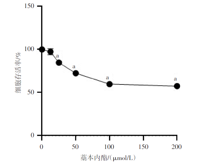
图1 LIG对EC-109细胞存活率的影响 n=6,F=470.147,P<0.01;a与Control组比较,P<0.05。
Fig.1 Effect of ligustilide on survival rate of EC-109 cells
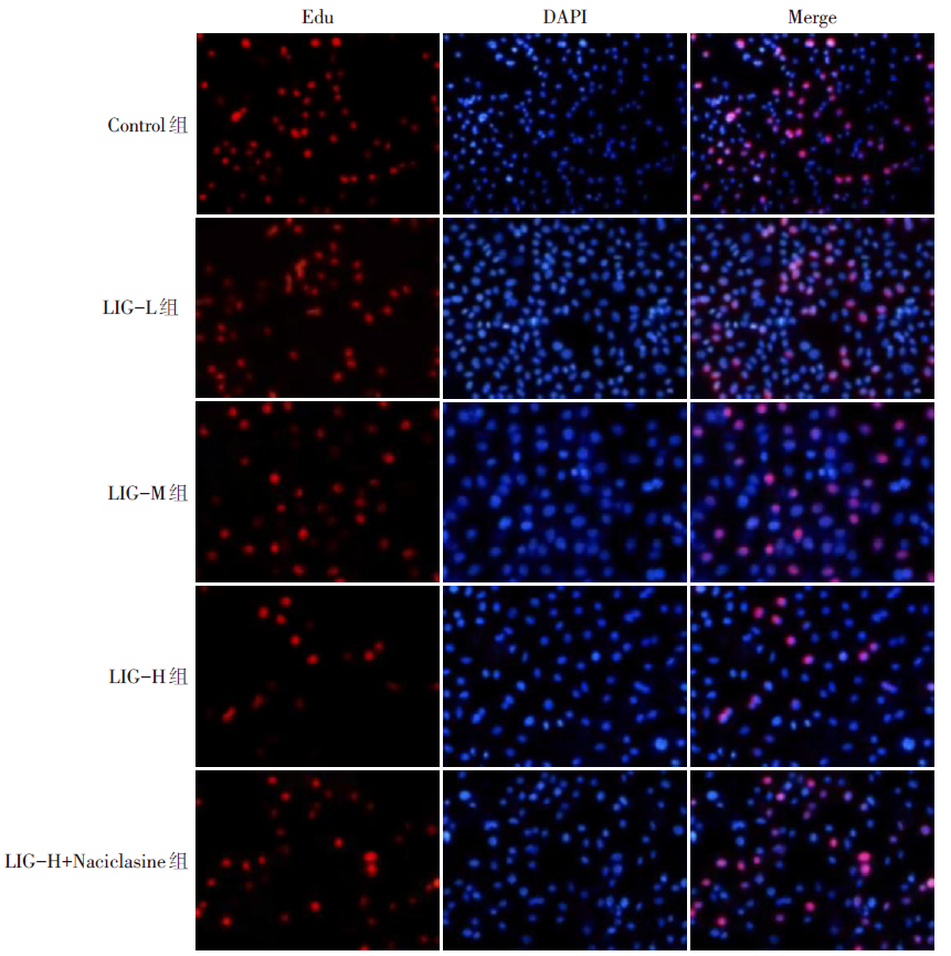
图2 Edu检测各组EC-109细胞增殖情况(×200)
Fig.2 Proliferation of EC-109 cells in each group tested by Edu (×200)
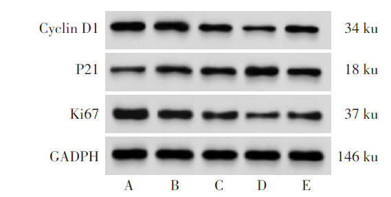
图3 Western blot检测Cyclin D1、P21、Ki67蛋白表达 A:Control组;B:LIG-L组;C:LIG-M组;D:LIG-H组;E:LIG-H+Naciclasine组
Fig.3 Western blot analysis of Cyclin D1, P21 and Ki67 protein expression
| 组别 | Edu阳性率/% | Cyclin D1 |
|---|---|---|
| Control组 | 42.36±2.14 | 1.03±0.08 |
| LIG-L组 | 36.79±1.85a | 0.71±0.07a |
| LIG-M组 | 28.52±1.43ab | 0.48±0.06ab |
| LIG-H组 | 19.48±0.97abc | 0.24±0.04abc |
| LIG-H+Naciclasine组 | 26.13±1.32d | 0.45±0.06d |
| F | 191.213** | 135.030** |
| 组别 | P21 | Ki67 |
| Control组 | 0.25±0.03 | 1.21±0.09 |
| LIG-L组 | 0.37±0.04a | 0.89±0.08a |
| LIG-M组 | 0.69±0.05ab | 0.47±0.07ab |
| LIG-H组 | 0.95±0.07abc | 0.31±0.05abc |
| LIG-H+Naciclasine组 | 0.72±0.08d | 0.48±0.06d |
| F | 147.387** | 160.494** |
表1 LIG对EC-109细胞增殖及增殖相关蛋白表达的影响(n=6,$\bar{x} \pm s$)
Tab.1 Effects of ligustilide on proliferation and proliferation-related protein expression in EC-109 cells
| 组别 | Edu阳性率/% | Cyclin D1 |
|---|---|---|
| Control组 | 42.36±2.14 | 1.03±0.08 |
| LIG-L组 | 36.79±1.85a | 0.71±0.07a |
| LIG-M组 | 28.52±1.43ab | 0.48±0.06ab |
| LIG-H组 | 19.48±0.97abc | 0.24±0.04abc |
| LIG-H+Naciclasine组 | 26.13±1.32d | 0.45±0.06d |
| F | 191.213** | 135.030** |
| 组别 | P21 | Ki67 |
| Control组 | 0.25±0.03 | 1.21±0.09 |
| LIG-L组 | 0.37±0.04a | 0.89±0.08a |
| LIG-M组 | 0.69±0.05ab | 0.47±0.07ab |
| LIG-H组 | 0.95±0.07abc | 0.31±0.05abc |
| LIG-H+Naciclasine组 | 0.72±0.08d | 0.48±0.06d |
| F | 147.387** | 160.494** |
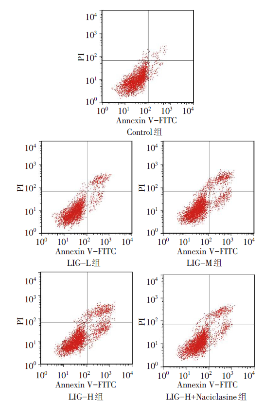
图4 流式细胞术检测各组EC-109细胞凋亡情况
Fig.4 The apoptosis of EC-109 cells in each group detected by flow cytometry
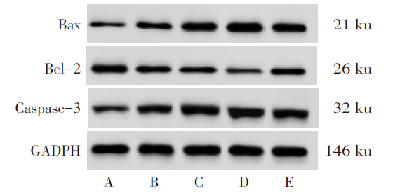
图5 Western blot检测Bax、Bcl-2、Caspase-3蛋白表达 A:Control组;B:LIG-L组;C:LIG-M组;D:LIG-H组;E:LIG-H+Naciclasine组
Fig.5 Western blot analysis of Bax, Bcl-2 and Caspase-3 protein expression
| 组别 | 细胞凋亡率/% | Bax | Bcl-2 | Caspase-3 |
|---|---|---|---|---|
| Control组 | 3.16±0.25 | 0.26±0.04 | 0.76±0.08 | 0.34±0.04 |
| LIG-L组 | 20.73±1.82a | 0.48±0.05a | 0.54±0.06a | 0.64±0.05a |
| LIG-M组 | 27.15±2.53ab | 0.80±0.06ab | 0.32±0.05ab | 0.97±0.07ab |
| LIG-H组 | 36.74±3.24abc | 1.15±0.08abc | 0.18±0.04abc | 1.22±0.11abc |
| LIG-H+Naciclasine组 | 25.38±2.36d | 0.83±0.06d | 0.39±0.05d | 0.99±0.06d |
| F | 176.966** | 199.712** | 88.952** | 143.769** |
表2 LIG对EC-109细胞凋亡及凋亡相关蛋白表达的影响 (n=6,$\bar{x} \pm s$)
Tab.2 Effects of ligustilide on apoptosis and apoptosis-related protein expression in EC-109 cells
| 组别 | 细胞凋亡率/% | Bax | Bcl-2 | Caspase-3 |
|---|---|---|---|---|
| Control组 | 3.16±0.25 | 0.26±0.04 | 0.76±0.08 | 0.34±0.04 |
| LIG-L组 | 20.73±1.82a | 0.48±0.05a | 0.54±0.06a | 0.64±0.05a |
| LIG-M组 | 27.15±2.53ab | 0.80±0.06ab | 0.32±0.05ab | 0.97±0.07ab |
| LIG-H组 | 36.74±3.24abc | 1.15±0.08abc | 0.18±0.04abc | 1.22±0.11abc |
| LIG-H+Naciclasine组 | 25.38±2.36d | 0.83±0.06d | 0.39±0.05d | 0.99±0.06d |
| F | 176.966** | 199.712** | 88.952** | 143.769** |
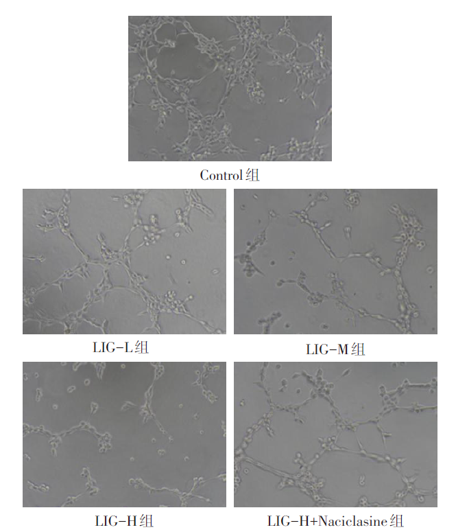
图6 各组EC-109细胞血管生成拟态情况(×100)
Fig. 6 Mimicry of angiogenesis of EC-109 cells in each group (×100)
| 组别 | 管状结构数 | RhoA | ROCK |
|---|---|---|---|
| Control组 | 34.50±3.27 | 1.03±0.09 | 1.14±0.12 |
| LIG-L组 | 27.16±2.54a | 0.80±0.08a | 0.91±0.07a |
| LIG-M组 | 20.33±1.83ab | 0.47±0.07ab | 0.58±0.06ab |
| LIG-H组 | 11.83±0.96abc | 0.32±0.05abc | 0.23±0.04abc |
| LIG-H+Naciclasine组 | 18.17±1.62d | 0.54±0.06d | 0.55±0.06d |
| F | 94.523** | 93.729** | 131.819** |
表3 LIG对EC-109细胞血管生成管状结构数及RhoA、ROCK蛋白表达的影响 (n=6,$\bar{x} \pm s$)
Tab.3 Effects of ligustilide on angiogenic tubular structure number and protein expression of RhoA and ROCK in EC-109 cells
| 组别 | 管状结构数 | RhoA | ROCK |
|---|---|---|---|
| Control组 | 34.50±3.27 | 1.03±0.09 | 1.14±0.12 |
| LIG-L组 | 27.16±2.54a | 0.80±0.08a | 0.91±0.07a |
| LIG-M组 | 20.33±1.83ab | 0.47±0.07ab | 0.58±0.06ab |
| LIG-H组 | 11.83±0.96abc | 0.32±0.05abc | 0.23±0.04abc |
| LIG-H+Naciclasine组 | 18.17±1.62d | 0.54±0.06d | 0.55±0.06d |
| F | 94.523** | 93.729** | 131.819** |
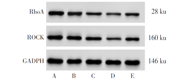
图7 Western blot检测RhoA、ROCK蛋白表达 A:Control组;B:LIG-L组;C:LIG-M组;D:LIG-H组;E:LIG-H+Naciclasine组。
Fig.7 Western blot analysis of RhoA and ROCK protein expression
| 组别 | 0 d | 2 d | 4 d | 6 d |
|---|---|---|---|---|
| Control组 | 0.00±0.00 | 0.06±0.01 | 0.20±0.02 | 0.40±0.04 |
| LIG组 | 0.00±0.00 | 0.03±0.01 | 0.06±0.01 | 0.12±0.02 |
| t | 0.000 | 6.708** | 19.799** | 19.799** |
| 组别 | 8 d | 10 d | 12 d | 14 d |
| Control组 | 0.65±0.06 | 0.87±0.08 | 1.06±0.10 | 1.22±0.12 |
| LIG组 | 0.20±0.02 | 0.32±0.03 | 0.44±0.04 | 0.53±0.05 |
| t | 22.500** | 20.356** | 18.204** | 16.784** |
表4 裸鼠移植瘤不同时间段体积比较 (n=10,cm3,$\bar{x} \pm s$)
Tab.4 Volume comparison of transplanted tumor in nude mice between different time periods
| 组别 | 0 d | 2 d | 4 d | 6 d |
|---|---|---|---|---|
| Control组 | 0.00±0.00 | 0.06±0.01 | 0.20±0.02 | 0.40±0.04 |
| LIG组 | 0.00±0.00 | 0.03±0.01 | 0.06±0.01 | 0.12±0.02 |
| t | 0.000 | 6.708** | 19.799** | 19.799** |
| 组别 | 8 d | 10 d | 12 d | 14 d |
| Control组 | 0.65±0.06 | 0.87±0.08 | 1.06±0.10 | 1.22±0.12 |
| LIG组 | 0.20±0.02 | 0.32±0.03 | 0.44±0.04 | 0.53±0.05 |
| t | 22.500** | 20.356** | 18.204** | 16.784** |
| 组别 | 移植瘤 质量/g | RhoA/OD值 | ROCK/OD值 | VEGF/OD值 |
|---|---|---|---|---|
| Control组 | 1.37±0.38 | 0.91±0.09 | 0.82±0.08 | 0.76±0.10 |
| LIG组 | 0.59±0.17 | 0.46±0.05 | 0.38±0.04 | 0.25±0.06 |
| t | 5.925** | 13.822** | 15.556** | 13.829** |
表5 LIG对14 d时裸鼠移植瘤质量及RhoA、ROCK、VEGF蛋白表达的影响 (n=10,$\bar{x} \pm s$)
Tab.5 Effects of ligustilide on tumor quality and protein expression of RhoA, ROCK and VEGF in nude mice at 14 days
| 组别 | 移植瘤 质量/g | RhoA/OD值 | ROCK/OD值 | VEGF/OD值 |
|---|---|---|---|---|
| Control组 | 1.37±0.38 | 0.91±0.09 | 0.82±0.08 | 0.76±0.10 |
| LIG组 | 0.59±0.17 | 0.46±0.05 | 0.38±0.04 | 0.25±0.06 |
| t | 5.925** | 13.822** | 15.556** | 13.829** |
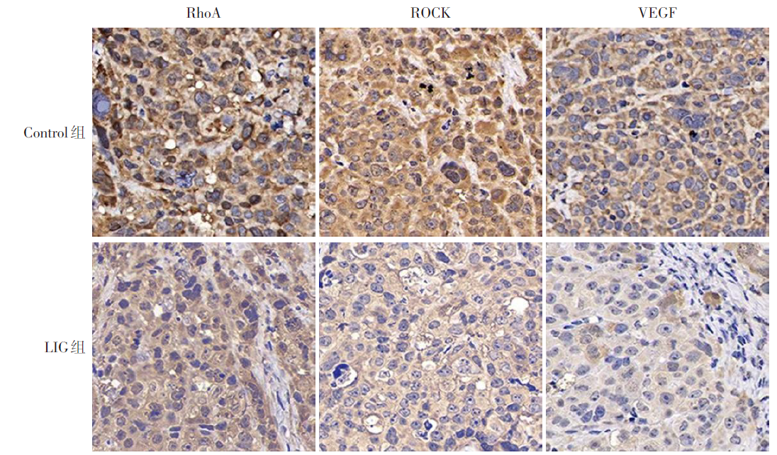
图8 免疫组化检测移植瘤组织中RhoA、ROCK、VEGF蛋白表达(×400)
Fig.8 Immunohistochemical detection of RhoA, ROCK and VEGF protein expression in transplanted tumor tissue (×400)
| [1] | SUNG H, FERLAY J, SIEGEL R L, et al. Global Cancer Statistics 2020:GLOBOCAN estimates of incidence and mortality worldwide for 36 cancers in 185 countries[J]. CA Cancer J Clin, 2021, 71(3):209-249. doi:10.3322/caac.21660. |
| [2] | WATANABE M, OTAKE R, KOZUKI R, et al. Recent progress in multidisciplinary treatment for patients with esophageal cancer[J]. Surg Today, 2020, 50(1):12-20. doi:10.1007/s00595-019-01878-7. |
| [3] | 杨慧超, 荀敬, 姜晓琳, 等. 中医药治疗食管癌的作用机制研究进展[J]. 中国中西医结合外科杂志, 2024, 30(3):419-423. |
| YANG H C, XUN J, JIANG X L, et al. Research progress on the mechanism of action of traditional Chinese medicine in the treatment of esophageal cancer[J]. Chinese Journal of Integrated Traditional and Western Medicine Surgery, 2024, 30(3):419-423. doi:10.3969/j.issn.1007-6948.2024.03.026. | |
| [4] | 李登辉, 蔡润茁, 朱丽明, 等. lncRNA HOTAIR靶向miR-126激活RhoA/ROCK通路促进喉癌Hep-2细胞增殖、侵袭与EMT[J]. 现代肿瘤医学, 2022, 30(3):365-370. |
| LI D H, CAI R Z, ZHU L M, et al. Activation of RhoA/ROCK pathway by lncRNA HOTAIR targeting miR-126 promotes proliferation,invasion and EMT of laryngeal carcinoma Hep-2 cells[J]. Modern Oncology Medicine, 2022, 30(3):365-370. doi:10.3969/j.issn.1672-4992.2022.03.001. | |
| [5] | HSU R J, PENG K Y, HSU W L, et al. Z-Ligustilide induces c-Myc-dependent apoptosis via activation of ER-stress signaling in hypoxic oral cancer cells[J]. Front Oncol, 2022,12:824043. doi:10.3389/fonc.2022.824043. |
| [6] | MA J, MEI J, LU J, et al. Ligustilide promotes apoptosis of cancer-associated fibroblasts via the TLR4 pathways[J]. Food Chem Toxicol, 2020,135:110991. doi:10.1016/j.fct.2019.110991. |
| [7] | 王红云, 王武亮. CHK1通过RhoA/ROCK信号通路对宫颈癌细胞放疗敏感性的影响及机制研究[J]. 医药论坛杂志, 2021, 42(10):76-80. |
| WANG H Y, WANG W L. Effect of CHK1 on radiotherapy sensitivity of cervical cancer cells through RhoA/ROCK signaling pathway and its mechanism[J]. Journal of Medical Forum,2021, 42(10): 76-80. doi:1672-3422(2021)10-0076-05. | |
| [8] | ALSHEHRI A F, KHODIER A E, AL-GAYYAR M M. Antitumor activity of ligustilide against ehrlich solid carcinoma in rats via inhibition of proliferation and activation of autophagy[J]. Cureus, 2023, 15(6):e40499. doi:10.7759/cureus.40499. |
| [9] | 林布雷, 肖轩. 西妥昔单抗联合放疗对食管癌患者血清肿瘤标志物及新生血管指标水平的影响[J]. 中外医学研究, 2023, 21(19):161-164. |
| LIN B L, XIAO X. Effects of cetuximab combined with radiotherapy on serum tumor markers and neovascularization in patients with esophageal cancer[J]. Chinese and Foreign Medical Research, 2023, 21(19):161-164. doi:10.14033/j.cnki.cfmr.2023.19.041. | |
| [10] | 何树苗, 陈元堃, 曾奥, 等. 藁本内酯药理作用及机制研究进展[J]. 广东药科大学学报, 2021, 37(2):152-156. |
| HE S M, CHEN Y K, ZENG A, et al. Pharmacological effects and mechanisms of ligustilide[J]. Journal of Guangdong Pharmaceutical University, 2021, 37(2):152-156. doi:10.16809/j.cnki.2096-3653.2020081202. | |
| [11] | 于冬冬. 藁本内酯对骨肉瘤细胞凋亡的影响及其作用机制[J]. 中国医科大学学报, 2023, 52(4):301-307. |
| YU D D. Effect of ligustilide on apoptosis of osteosarcoma cells and its mechanism of action[J]. Journal of China Medical University, 2023, 52(4):301-307. doi:10.12007/j.issn.0258-4646.2023.04.003. | |
| [12] | JIANG X, ZHAO W, ZHU F, et al. Ligustilide inhibits the proliferation of non-small cell lung cancer via glycolytic metabolism[J]. Toxicol Appl Pharmacol, 2021,410:115336. doi:10.1016/j.taap.2020.115336. |
| [13] | YIN L, YING L, GUO R, et al. Ligustilide induces apoptosis and reduces proliferation in human bladder cancer cells by NFκB1 and mitochondria pathway[J]. Chem Biol Drug Des, 2023, 101(6):1252-1261. doi:10.1111/cbdd.14207. |
| [14] | 张洁. 番茄碱通过AMPK/COX-2通路调控食管癌细胞凋亡的机制研究[J]. 中国临床药理学杂志, 2023, 39(16):2348-2352. |
| ZHANG J. The mechanism of tomato line regulating apoptosis of esophageal cancer cells through AMPK/COX-2 pathway[J]. Chinese Journal of Clinical Pharmacology, 2023, 39(16):2348-2352. doi:10.13699/j.cnki.1001-6821.2023.16.015. | |
| [15] | 孙振峰, 刘公哲, 朱应超, 等. 木犀草素调控食管癌细胞生物学行为及其机制[J]. 中华实验外科杂志, 2022, 39(2):287-290. |
| SUN Z F, LIU G Z, ZHU Y C, et al. Luteolin regulates biological behavior of esophageal carcinoma cells and its mechanism[J]. Chinese Journal of Experimental Surgery, 2022, 39(2):287-290. doi:10.3760/cma.j.cn421213-20210713-01210. | |
| [16] | MABETA P, STEENKAMP V. The VEGF/VEGFR axis revisited:implications for cancer therapy[J]. Int J Mol Sci, 2022, 23(24):15585. doi:10.3390/ijms232415585. |
| [17] | AHMAD A, NAWAZ M I. Molecular mechanism of VEGF and its role in pathological angiogenesis[J]. J Cell Biochem, 2022, 123(12):1938-1965. doi:10.1002/jcb.30344. |
| [18] | MA J, CHEN X, CHEN Y, et al. Ligustilide inhibits tumor angiogenesis by downregulating VEGFA secretion from cancer-associated fibroblasts in prostate cancer via TLR4[J]. Cancers(Basel), 2022, 14(10):2406. doi:10.3390/cancers14102406. |
| [19] | JOSHI R, QIN L, CAO X, et al. DLC1 SAM domain-binding peptides inhibit cancer cell growth and migration by inactivating RhoA[J]. J Biol Chem, 2020, 295(2):645-656. doi:10.1074/jbc.RA119.011929. |
| [20] | 严铃铃, 冯晓云, 马庆霞. VEGF联合TRAP1、CEA在食管癌患者肿瘤转移、分化、临床分期中的临床价值[J]. 国际检验医学杂志, 2023, 44(18):2301-2304. |
| YAN L L, FENG X Y, MA Q X. The clinical value of VEGF combined with TRAP1 and CEA in tumor metastasis,differentiation and clinical staging in patients with esophageal cancer[J]. International Journal of Laboratory Medicine, 2019, 44(18):2301-2304. doi:10.3969/j.issn.1673-4130.2023.18.026. | |
| [21] | LIU D, XIA A D, WU L P, et al. IGF2BP2 promotes gastric cancer progression by regulating the IGF1R-RhoA-ROCK signaling pathway[J]. Cell Signal, 2022,94:110313. doi:10.1016/j.cellsig.2022.110313. |
| [22] | 廖山婴, 刘超, 王蓓蓓, 等. 复方斑蝥胶囊调控RhoA/ROCK信号通路抑制MNNG诱导的大鼠胃癌发生[J]. 中国中西医结合杂志, 2019, 39(6):728-732. |
| LIAO S Y, LIU C, WANG B B, et al. Compound cantharides capsules regulate RhoA/ROCK signaling pathway to inhibit the occurrence of MNNG-induced gastric cancer in rats[J]. Chinese Journal of Integrated Traditional and Western Medicine, 2019, 39(6):728-732. doi:10.7661/j.cjim.20181031.321. |
| [1] | 杨晓芳, 贾新燕, 丰文君. miR-181a-5p通过HMGB1/NF-κB信号通路调控狼疮性肾炎小鼠肾小球系膜细胞增殖和凋亡[J]. 天津医药, 2026, 54(3): 232-237. |
| [2] | 张婧, 魏玉英, 宁海虹, 韦红梅, 王嘉玮, 曹薇, 吴宾. DUSP9在2型糖尿病心肌病小鼠心肌损伤中的保护作用及其机制[J]. 天津医药, 2026, 54(3): 238-244. |
| [3] | 王喆, 邱林, 马贲. 番茄来源胞外囊泡样颗粒对口腔鳞状细胞癌的作用效果研究[J]. 天津医药, 2026, 54(2): 145-150. |
| [4] | 李志伟, 张会超, 杨凤鸣, 曾垂义. 基于miR-144-3p/MAPK1通路探讨红参总皂苷对扩张型心肌病小鼠心肌细胞凋亡的影响[J]. 天津医药, 2026, 54(1): 23-29. |
| [5] | 赵兰君, 李良惠, 马馨, 巩娇娇, 郑臣辉, 石琳. 穿心莲内酯调控STAT3/GPX4通路对骨髓瘤细胞增殖和凋亡的影响[J]. 天津医药, 2026, 54(1): 8-13. |
| [6] | 孔翠文, 路延双, 孙丽萍, 于芬芬. LncRNA SNHG14靶向miR-30a-5p对高糖诱导的足细胞损伤的影响[J]. 天津医药, 2025, 53(9): 903-909. |
| [7] | 刘虹, 张玥玥, 王一琳, 王彩丽, 王晓敏, 毛敏, 李燕. MicroRNA-34a通过调控Wnt途径影响慢性淋巴细胞白血病进展的机制探讨[J]. 天津医药, 2025, 53(8): 785-790. |
| [8] | 万艳波, 刘明, 王勇. 秦皮甲素调节HMGB1/RAGE信号通路对缺氧/复氧诱导的心肌细胞损伤的影响[J]. 天津医药, 2025, 53(8): 796-801. |
| [9] | 刘海威, 杨洁, 王力, 蒙诗波, 唐旭松, 刘成仁, 王永旺. 木犀草素通过NFE2L2/x-CT/GPX4信号轴调控ROS水平抑制胶质母细胞瘤[J]. 天津医药, 2025, 53(7): 673-678. |
| [10] | 韩建存, 周谊. 川陈皮素调节FAK/AKT信号通路对喉鳞状细胞癌细胞增殖和凋亡的影响[J]. 天津医药, 2025, 53(6): 561-565. |
| [11] | 余朝霞, 马贲, 邱林, 高倩, 尼娜. 基于网络药理学和实验验证探究鲍式层孔菌多酚的抗头颈鳞癌机制[J]. 天津医药, 2025, 53(5): 456-461. |
| [12] | 李晨, 李占恩, 苏宏伟, 侯彩云, 董少文. KRT17调节Wnt/β-catenin信号通路对膀胱癌细胞增殖、凋亡及上皮间质转化的影响[J]. 天津医药, 2025, 53(5): 462-467. |
| [13] | 苏红见, 张春艳, 张卫东, 韩利, 乔亚红. 鸢尾素调控EBF3/ALOX15通路影响肺腺癌细胞增殖和迁移[J]. 天津医药, 2025, 53(4): 337-342. |
| [14] | 祁卫华, 黄广磊, 张媛媛, 班宏英, 毛诏旭. 连翘脂素调节cAMP/EPAC1/RAP1信号通路对肺癌细胞恶性进展的影响[J]. 天津医药, 2025, 53(4): 343-348. |
| [15] | 闫玲新, 李森, 郭改莉, 孟婉秋, 徐超. 异牡荆素通过miR-339-5p/HSPA8轴调节胰腺癌细胞的生物学行为[J]. 天津医药, 2025, 53(3): 230-235. |
| 阅读次数 | ||||||
|
全文 |
|
|||||
|
摘要 |
|
|||||