天津医药 ›› 2024, Vol. 52 ›› Issue (8): 785-790.doi: 10.11958/20240086
• 细胞与分子生物学 • 下一篇
方杰1( ), 黄芮1, 郑红慧1,2, 贾倩倩1, 鲍静2,△()
收稿日期:2024-01-12
修回日期:2024-04-11
出版日期:2024-08-15
发布日期:2024-08-16
通讯作者:
E-mail:作者简介:方杰(1986),男,主治医师,主要从事血液肿瘤方面研究。E-mail:基金资助:
FANG Jie1(), HUANG Rui1, ZHENG Honghui1,2, JIA Qianqian1, BAO Jing2,△()
Received:2024-01-12
Revised:2024-04-11
Published:2024-08-15
Online:2024-08-16
Contact:
E-mail:方杰, 黄芮, 郑红慧, 贾倩倩, 鲍静. miR-9-5p靶向TIMP2诱导多发性骨髓瘤细胞自噬和凋亡的机制[J]. 天津医药, 2024, 52(8): 785-790.
FANG Jie, HUANG Rui, ZHENG Honghui, JIA Qianqian, BAO Jing. miR-9-5p-induced autophagy and apoptosis in multiple myeloma cells by targeting TIMP2[J]. Tianjin Medical Journal, 2024, 52(8): 785-790.
摘要:
目的 探究miR-9-5p和组织金属蛋白酶抑制因子2(TIMP2)相互作用对多发性骨髓瘤(MM)细胞自噬和凋亡的影响机制。方法 采用实时荧光定量PCR(qRT-PCR)检测初诊MM和复发MM各9例患者骨髓样本中miR-9-5p和TIMP2的表达水平,分析两者表达水平的相关性。U266细胞分为miR-对照组、miR-9-5p组、pcDNA3.1组、pcDNA3.1-TIMP2组、miR-9-5p+pcDNA3.1组、miR-9-5p+pcDNA3.1-TIMP2组。采用流式细胞术、免疫荧光染色、蛋白质印迹实验检测过表达miR-9-5p和TIMP2对U266细胞自噬和凋亡的影响;双萤光素酶报告实验验证miR-9-5p和TIMP2的靶向关系。结果 与初诊MM患者相比,复发MM患者miR-9-5p表达水平升高,TIMP2表达降低;miR-9-5p和TIMP2表达水平呈负相关(P<0.05)。与miR-对照组相比,miR-9-5p组MAP1LC3B-Ⅱ的表达水平降低,MAP1LC3B-Ⅰ和SQSTM1的表达水平增加,细胞凋亡率降低(P<0.05)。与pcDNA3.1组相比,pcDNA3.1-TIMP2组MAP1LC3B-Ⅱ的表达水平升高,MAP1LC3B-Ⅰ和SQSTM1的表达水平降低,细胞凋亡率增加(P<0.05)。生物信息学和双萤光素酶报告实验证实TIMP2是miR-9-5p的靶基因。结论 miR-9-5p靶向TIMP2抑制MM细胞的自噬和凋亡,从而促进MM的发生发展。
中图分类号:
| 基因名称 | 引物序列(5'→3') | 产物大小/bp |
|---|---|---|
| miR-9-5p | 上游:GCCCGCTCTTTGGTTATCTAG | 95 |
| 下游:CCAGTGCAGGGTCCGAGGT | ||
| TIMP2 | 上游:AGCACCACCCAGAAGAAGAG | 183 |
| 下游:GTGACCCAGTCCATCCAGAG | ||
| U6 | 上游:GCTTCGGCAGCACATATACTAAAAT | 113 |
| 下游:CGCTTCACGAATTTGCGTGTCAT | ||
| GAPDH | 上游:TGGAGAAACCTGCCAAGTATG | 120 |
| 下游:GGAGACAACCTGGTCCTCAG |
表1 引物序列
Tab.1 Primer sequence
| 基因名称 | 引物序列(5'→3') | 产物大小/bp |
|---|---|---|
| miR-9-5p | 上游:GCCCGCTCTTTGGTTATCTAG | 95 |
| 下游:CCAGTGCAGGGTCCGAGGT | ||
| TIMP2 | 上游:AGCACCACCCAGAAGAAGAG | 183 |
| 下游:GTGACCCAGTCCATCCAGAG | ||
| U6 | 上游:GCTTCGGCAGCACATATACTAAAAT | 113 |
| 下游:CGCTTCACGAATTTGCGTGTCAT | ||
| GAPDH | 上游:TGGAGAAACCTGCCAAGTATG | 120 |
| 下游:GGAGACAACCTGGTCCTCAG |
| 细胞系 | n | miR-9-5p | TIMP2 |
|---|---|---|---|
| GM50113 | 3 | 1.00±0.12 | 1.00±0.03 |
| U266 | 3 | 3.58±0.24a | 0.50±0.05a |
| NCI-H929 | 3 | 2.67±0.24a | 0.80±0.03a |
| RPMI-8226 | 3 | 2.25±0.31a | 0.65±0.06a |
| F | 61.060** | 69.304** |
表2 miR-9-5p和TIMP2在多发性骨髓瘤不同细胞系中的表达水平比较 ($\bar{x} \pm s$)
Tab.2 Comparison of expression levels of miR-9-5p and TIMP 2 between different cell lines of multiple myeloma
| 细胞系 | n | miR-9-5p | TIMP2 |
|---|---|---|---|
| GM50113 | 3 | 1.00±0.12 | 1.00±0.03 |
| U266 | 3 | 3.58±0.24a | 0.50±0.05a |
| NCI-H929 | 3 | 2.67±0.24a | 0.80±0.03a |
| RPMI-8226 | 3 | 2.25±0.31a | 0.65±0.06a |
| F | 61.060** | 69.304** |
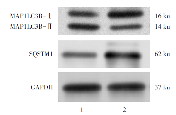
图1 过表达miR-9-5p对U266细胞自噬相关蛋白表达的影响 1:miR-对照组;2:miR-9-5p组。
Fig.1 Effects of overexpressed miR-9-5p on the expression of autophagy-related proteins in U266 cells
图2 过表达miR-9-5p对U266细胞凋亡的影响
Fig.2 Effect of overexpressed miR-9-5p on apoptosis in U266 cells
| 组别 | MAP1LC3B-Ⅰ | MAP1LC3B-Ⅱ | SQSTM1 | 细胞凋亡率/% |
|---|---|---|---|---|
| miR-对照组 | 0.66±0.01 | 0.42±0.02 | 0.50±0.01 | 8.95±1.33 |
| miR-9-5p组 | 0.86±0.03 | 0.35±0.03 | 0.67±0.01 | 5.91±0.33 |
| t | 10.955** | 3.363* | 20.821** | 3.843* |
表3 U266细胞miR-对照组和miR-9-5p组中3种自噬相关蛋白的表达水平及凋亡率比较 (n=6, $\bar{x} \pm s$)
Tab.3 Comparison of protein expression levels of three autophagy-related proteins and apoptosis ratio between the miR-control group and the miR-9-5p group of U266 cells
| 组别 | MAP1LC3B-Ⅰ | MAP1LC3B-Ⅱ | SQSTM1 | 细胞凋亡率/% |
|---|---|---|---|---|
| miR-对照组 | 0.66±0.01 | 0.42±0.02 | 0.50±0.01 | 8.95±1.33 |
| miR-9-5p组 | 0.86±0.03 | 0.35±0.03 | 0.67±0.01 | 5.91±0.33 |
| t | 10.955** | 3.363* | 20.821** | 3.843* |
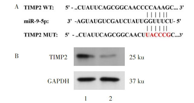
图3 miR-9-5p与TIMP2之间的靶向结合位点及调控关系 1:miR-对照组;2:miR-9-5p组。
Fig.3 Targeted binding sites and regulatory relationships between miR-9-5p and TIMP2
| 组别 | n | TIMP2 mRNA | TIMP2 蛋白 |
|---|---|---|---|
| miR-对照组 | 3 | 1.01±0.01 | 1.01±0.03 |
| miR-9-5p组 | 3 | 0.46±0.02 | 0.58±0.01 |
| t | 46.603** | 23.552** |
表4 U266细胞miR-对照组和miR-9-5p组中TIMP2的mRNA和蛋白表达水平比较 ($\bar{x} \pm s$)
Tab.4 Comparison of protein expression levels of TIMP2 between the miR-control group and the miR-9-5p group in U266 cells
| 组别 | n | TIMP2 mRNA | TIMP2 蛋白 |
|---|---|---|---|
| miR-对照组 | 3 | 1.01±0.01 | 1.01±0.03 |
| miR-9-5p组 | 3 | 0.46±0.02 | 0.58±0.01 |
| t | 46.603** | 23.552** |
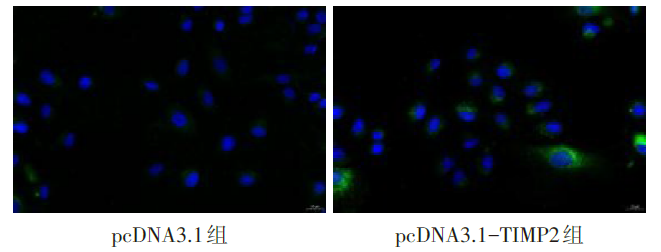
图4 过表达TIMP2后GFP的荧光表达情况 绿色为GFP染色,蓝色为DAPI染色。
Fig.4 The fluorescence expression of GFP after overexpressed TIMP2
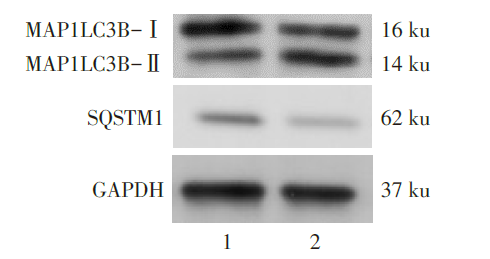
图5 U266细胞中过表达TIMP2后,MAP1LC3B-Ⅰ、MAP1LC3B-Ⅱ、SQSTM1蛋白表达电泳图 1:pcDNA3.1组;2:pcDNA3.1-TIMP2组。
Fig.5 Protein electrophoretogram of MAP1LC3B-I, MAP1LC3B-II and SQSTM1 proteins after overexpressed TIMP2 in U266 cells
| 组别 | MAP1LC3B-Ⅰ | MAP1LC3B-Ⅱ |
|---|---|---|
| pcDNA3.1组 | 0.51±0.01 | 0.44±0.02 |
| pcDNA3.1-TIMP2组 | 0.31±0.02 | 0.65±0.01 |
| t | 15.492** | 16.267** |
| 组别 | SQSTM1 | 细胞凋亡率/% |
| pcDNA3.1组 | 0.33±0.03 | 4.97±0.52 |
| pcDNA3.1-TIMP2组 | 0.12±0.03 | 19.61±3.23 |
| t | 8.573** | 7.751** |
表5 U266细胞中过表达TIMP2后,3种自噬相关蛋白的表达水平以及凋亡率比较 (n=6,$\bar{x} \pm s$)
Tab.5 Comparison of protein expression levels of three autophagy-related proteins, and apoptosis ratio after overexpressed TIMP2 in U266 cells
| 组别 | MAP1LC3B-Ⅰ | MAP1LC3B-Ⅱ |
|---|---|---|
| pcDNA3.1组 | 0.51±0.01 | 0.44±0.02 |
| pcDNA3.1-TIMP2组 | 0.31±0.02 | 0.65±0.01 |
| t | 15.492** | 16.267** |
| 组别 | SQSTM1 | 细胞凋亡率/% |
| pcDNA3.1组 | 0.33±0.03 | 4.97±0.52 |
| pcDNA3.1-TIMP2组 | 0.12±0.03 | 19.61±3.23 |
| t | 8.573** | 7.751** |
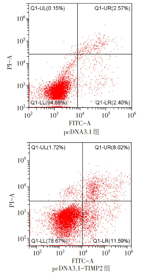
图6 过表达TIMP2细胞凋亡的流式图
Fig.6 Flow diagram of cell apoptosis after overexpressed TIMP2
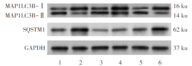
图7 U266细胞中过表达miR-9-5p和TIMP2,MAP1LC3B-Ⅰ、MAP1LC3B-Ⅱ、SQSTM1蛋白表达电泳图 1:miR-对照组;2:miR-9-5p组;3:pcDNA3.1-TIMP2组;4:pcDNA3.1组;5:miR-9-5p+pcDNA3.1-TIMP2组;6:miR-9-5p+pcDNA3.1组。
Fig.7 Protein electrophoretogram of MAP1LC3B-I, MAP1LC3B-II and SQSTM1 after overexpressed miR-9-5p and TIMP2 in U266 cells
| 组别 | MAP1LC3B-Ⅰ | MAP1LC3B-Ⅱ | ||
|---|---|---|---|---|
| miR-对照组 | 0.32±0.02 | 0.56±0.04 | ||
| miR-9-5p组 | 0.53±0.01a | 0.39±0.02a | ||
| pcDNA3.1组 | 0.59±0.01 | 0.36±0.03 | ||
| pcDNA3.1-TIMP2组 | 0.35±0.03b | 0.60±0.05b | ||
| miR-9-5p+pcDNA3.1组 | 0.58±0.04 | 0.39±0.02 | ||
| miR-9-5p+pcDNA3.1-TIMP2组 | 0.36±0.04c | 0.60±0.04c | ||
| F | 59.550** | 31.978** | ||
| 组别 | SQSTM1 | 细胞凋亡率/% | ||
| miR-对照组 | 0.22±0.02 | 8.40±0.98 | ||
| miR-9-5p组 | 0.37±0.03a | 5.78±0.55a | ||
| pcDNA3.1组 | 0.41±0.05 | 5.82±0.45 | ||
| pcDNA3.1-TIMP2组 | 0.28±0.01b | 24.03±1.59b | ||
| miR-9-5p+pcDNA3.1组 | 0.45±0.05 | 5.39±0.48 | ||
| miR-9-5p+pcDNA3.1-TIMP2组 | 0.30±0.02c | 20.07±1.35c | ||
| F | 19.844** | 203.821** | ||
表6 U266细胞中过表达miR-9-5p和TIMP2后3种自噬相关蛋白的表达水平以及凋亡率比较 (n=3,$\bar{x} \pm s$)
Tab.6 Comparison of protein expression levels of three autophagy-related proteins and apoptosis ratio after overexpressed miR-9-5p and TIMP2
| 组别 | MAP1LC3B-Ⅰ | MAP1LC3B-Ⅱ | ||
|---|---|---|---|---|
| miR-对照组 | 0.32±0.02 | 0.56±0.04 | ||
| miR-9-5p组 | 0.53±0.01a | 0.39±0.02a | ||
| pcDNA3.1组 | 0.59±0.01 | 0.36±0.03 | ||
| pcDNA3.1-TIMP2组 | 0.35±0.03b | 0.60±0.05b | ||
| miR-9-5p+pcDNA3.1组 | 0.58±0.04 | 0.39±0.02 | ||
| miR-9-5p+pcDNA3.1-TIMP2组 | 0.36±0.04c | 0.60±0.04c | ||
| F | 59.550** | 31.978** | ||
| 组别 | SQSTM1 | 细胞凋亡率/% | ||
| miR-对照组 | 0.22±0.02 | 8.40±0.98 | ||
| miR-9-5p组 | 0.37±0.03a | 5.78±0.55a | ||
| pcDNA3.1组 | 0.41±0.05 | 5.82±0.45 | ||
| pcDNA3.1-TIMP2组 | 0.28±0.01b | 24.03±1.59b | ||
| miR-9-5p+pcDNA3.1组 | 0.45±0.05 | 5.39±0.48 | ||
| miR-9-5p+pcDNA3.1-TIMP2组 | 0.30±0.02c | 20.07±1.35c | ||
| F | 19.844** | 203.821** | ||
| [1] | 付庆华, 夏冰, 杨洪亮, 等. 初诊伴髓外病变的多发性骨髓瘤患者的临床特征及预后分析[J]. 天津医药, 2020, 48(5):415-420. |
| FU Q H, XIA B, YANG H L, et al. Clinical features and prognostic analysis of newly diagnosed multiple myeloma with extramedullary disease[J]. Tianjin Med J, 2020, 48(5):415-420. doi:10.11958/20193032. | |
| [2] | WANG Z, ZHANG S, ZHAO Y, et al. MicroRNA-140-3p alleviates intervertebral disc degeneration via KLF5/N-cadherin/MDM2/Slug axis[J]. RNA Biol, 2021, 18(12):2247-2260. doi:10.1080/15476286.2021.1898176. |
| [3] | SALIMINEJAD K, KHORRAM KHORSHID H R, SOLEYMANI FARD S, et al. An overview of microRNAs:biology,functions,therapeutics,and analysis methods[J]. J Cell Physiol, 2019, 234(5):5451-5465. doi:10.1002/jcp.27486. |
| [4] | HUANG G, LIU X, ZHAO X, et al. MiR-9 promotes multiple myeloma progression by regulating TRIM56/NF-κB pathway[J]. Cell Biol Int, 2019, 43(11):1223-1233. doi:10.1002/cbin.11104. |
| [5] | GOBIN E, BAGWELL K, WAGNER J, et al. A pan-cancer perspective of matrix metalloproteases(MMP)gene expression profile and their diagnostic/prognostic potential[J]. BMC Cancer, 2019, 19(1):581. doi:10.1186/s12885-019-5768-0. |
| [6] | WANG Z, HE J, BACH D H, et al. Induction of m(6)A methylation in adipocyte exosomal LncRNAs mediates myeloma drug resistance[J]. J Exp Clin Cancer Res, 2022, 41(1):4. doi:10.1186/s13046-021-02209-w. |
| [7] | WANG J, LIU L. MiR-149-3p promotes the cisplatin resistance and EMT in ovarian cancer through downregulating TIMP2 and CDKN1A[J]. J Ovarian Res, 2021, 14(1):165. doi:10.1186/s13048-021-00919-5. |
| [8] | 中国医师协会血液科医师分会, 中华医学会血液学分会,中国医师协会多发性骨髓瘤专业委员会. 中国多发性骨髓瘤诊治指南(2022年修订)[J]. 中华内科杂志, 2022, 54(12):1066-1070. |
| Hematology Branch of Chinese Medical Doctor Association,Hematology Branch of Chinese Medical Association, Multiple Myeloma Professional Committee of Chinese Medical Doctor Association. Guidelines for the diagnosis and treatment of multiple myeloma in China (revised in 2015)[J]. Chin J Intern Med, 2022, 54(12):1066-1070. doi:10.3760/cma.j.issn.0578-1426.2022.12.020. | |
| [9] | WANG Y, DONG L, WAN F, et al. MiR-9-3p regulates the biological functions and drug resistance of gemcitabine-treated breast cancer cells and affects tumor growth through targeting MTDH[J]. Cell Death Dis, 2021, 12(10):861. doi:10.1038/s41419-021-04145-1. |
| [10] | 刘艺, 席振芳. miR-9-5p通过靶向FOXO1抑制急性淋巴细胞白血病进展[J]. 免疫学杂志, 2023, 39(9):809-820. |
| LIU Y, XI Z F. MiR-9-5p inhibits the progression of acute lymphocytic leukemia by targeting FOXO1[J]. Journal of Immunology, 2023, 39(9):809-820. doi:10.13431/j.cnki.immunol.j.20230106. | |
| [11] | CHIRON D, JEGO G, PELLAT-DEUCEUNYNCK C. Toll-like receptors: expression and involvement in multiple myeloma[J]. Leuk Res, 2010, 34(12):1545-1550. doi:10.1016/j.leukres.2010.06.002. |
| [12] | LUDDI A, MARROCCO C, GOVERNINI L, et al. Expression of matrix metalloproteinases and their inhibitors in endometrium:high levels in endometriotic lesions[J]. Int J Mol Sci, 2020, 21(8):2840. doi:10.3390/ijms21082840. |
| [13] | WANG J, ZHANG Q, WANG D, et al. Microenvironment-induced TIMP2 loss by cancer-secreted exosomal miR-4443 promotes liver metastasis of breast cancer[J]. J Cell Physiol, 2020, 235(7/8):5722-5735. doi:10.1002/jcp.29507. |
| [14] | FANG J, JIAO Y H, WEI L M, et al. TIMP2 is associated with prognosis and immune infiltrates of gastric and colon cancer[J]. Int Immunopharmacol, 2022, 110:109008. doi:10.1016/j.intimp.2022.109008. |
| [15] | LIU W, LI Z, CAI Z, et al. LncRNA-mRNA expression profiles and functional networks in osteoclast differentiation[J]. J Cell Mol Med, 2020, 24(17):9786-9797. doi:10.1111/jcmm.15560. |
| [16] | SUN H, CHEN X. MiR-106b-5p promotes malignant behaviors of cervical squamous cell carcinoma cells by targeting TIMP2[J]. Reprod Sci, 2022, 29(1):203-211. doi:10.1007/s43032-021-00788-9. |
| [17] | WU H, LIU C, YANG Q, et al. MIR145-3p promotes autophagy and enhances bortezomib sensitivity in multiple myeloma by targeting HDAC4[J]. Autophagy, 2020, 16(4):683-697. doi:10.1080/15548627.2019.1635380. |
| [1] | 杨晓芳, 贾新燕, 丰文君. miR-181a-5p通过HMGB1/NF-κB信号通路调控狼疮性肾炎小鼠肾小球系膜细胞增殖和凋亡[J]. 天津医药, 2026, 54(3): 232-237. |
| [2] | 张婧, 魏玉英, 宁海虹, 韦红梅, 王嘉玮, 曹薇, 吴宾. DUSP9在2型糖尿病心肌病小鼠心肌损伤中的保护作用及其机制[J]. 天津医药, 2026, 54(3): 238-244. |
| [3] | 王喆, 邱林, 马贲. 番茄来源胞外囊泡样颗粒对口腔鳞状细胞癌的作用效果研究[J]. 天津医药, 2026, 54(2): 145-150. |
| [4] | 李志伟, 张会超, 杨凤鸣, 曾垂义. 基于miR-144-3p/MAPK1通路探讨红参总皂苷对扩张型心肌病小鼠心肌细胞凋亡的影响[J]. 天津医药, 2026, 54(1): 23-29. |
| [5] | 赵兰君, 李良惠, 马馨, 巩娇娇, 郑臣辉, 石琳. 穿心莲内酯调控STAT3/GPX4通路对骨髓瘤细胞增殖和凋亡的影响[J]. 天津医药, 2026, 54(1): 8-13. |
| [6] | 黄慧琦, 伍秋苑, 张昆, 李佩贤, 熊亚明, 叶国麟, 周丹. 川楝素联合奥拉帕尼在三阴性乳腺癌中的抗肿瘤机制研究[J]. 天津医药, 2025, 53(9): 897-902. |
| [7] | 孔翠文, 路延双, 孙丽萍, 于芬芬. LncRNA SNHG14靶向miR-30a-5p对高糖诱导的足细胞损伤的影响[J]. 天津医药, 2025, 53(9): 903-909. |
| [8] | 林义伟, 魏谭军, 陈飞, 肖成, 袁烈, 王毅. 圣草酚调控UBA52表达对代谢相关脂肪性肝病的体内和体外作用[J]. 天津医药, 2025, 53(9): 916-922. |
| [9] | 万艳波, 刘明, 王勇. 秦皮甲素调节HMGB1/RAGE信号通路对缺氧/复氧诱导的心肌细胞损伤的影响[J]. 天津医药, 2025, 53(8): 796-801. |
| [10] | 刘海威, 杨洁, 王力, 蒙诗波, 唐旭松, 刘成仁, 王永旺. 木犀草素通过NFE2L2/x-CT/GPX4信号轴调控ROS水平抑制胶质母细胞瘤[J]. 天津医药, 2025, 53(7): 673-678. |
| [11] | 韩建存, 周谊. 川陈皮素调节FAK/AKT信号通路对喉鳞状细胞癌细胞增殖和凋亡的影响[J]. 天津医药, 2025, 53(6): 561-565. |
| [12] | 向立丽, 王倩, 蒙延娜, 付杰, 张璞. 来那度胺联合硼替佐米和地塞米松治疗多发性骨髓瘤的疗效[J]. 天津医药, 2025, 53(6): 665-669. |
| [13] | 马春梅, 于鹏, 张其程, 杨磊, 李棣华, 谭建, 孟召伟. 异硫氰酸苄酯联合索拉非尼治疗未分化甲状腺癌机制探讨[J]. 天津医药, 2025, 53(5): 449-455. |
| [14] | 祁卫华, 黄广磊, 张媛媛, 班宏英, 毛诏旭. 连翘脂素调节cAMP/EPAC1/RAP1信号通路对肺癌细胞恶性进展的影响[J]. 天津医药, 2025, 53(4): 343-348. |
| [15] | 李冰心, 许军英, 张雅茹, 周小兵. 冬虫夏草通过调控AMPK/mTOR通路保护高糖诱导的足细胞损伤[J]. 天津医药, 2025, 53(3): 225-229. |
| 阅读次数 | ||||||
|
全文 |
|
|||||
|
摘要 |
|
|||||