Tianjin Medical Journal ›› 2023, Vol. 51 ›› Issue (11): 1205-1210.doi: 10.11958/20230140
• Experimental Research • Previous Articles Next Articles
LEI Hui1( ), BAO Yaling1,△(), BAO Xijing2, MA Jun2
Received:2023-02-20
Revised:2023-05-03
Published:2023-11-15
Online:2023-11-07
Contact:
△E-mail:LEI Hui, BAO Yaling, BAO Xijing, MA Jun. Effects of hyperin on inflammatory reaction, wound healing and AMPK/SIRT1 signaling pathway in diabetic foot ulcer model rats[J]. Tianjin Medical Journal, 2023, 51(11): 1205-1210.
CLC Number:
| 基因名称 | 引物序列(5′→3′) | 产物大小/bp |
|---|---|---|
| AMPK | 上游:TCGATCGATCGATCGATGGC | 191 |
| 下游:AGCTAGCTAGCTAGCATTCT | ||
| SIRT1 | 上游:TAGCTAGCTAGTATTCCGA | 164 |
| 下游:GCTAGCTAGCTATCAAGTT | ||
| GAPDH | 上游:AGCTAGCTAGCTGTCGGA | 176 |
| 下游:GCGAGCTAGCTGACTAAC |
Tab.1 Primer sequence for qPCR
| 基因名称 | 引物序列(5′→3′) | 产物大小/bp |
|---|---|---|
| AMPK | 上游:TCGATCGATCGATCGATGGC | 191 |
| 下游:AGCTAGCTAGCTAGCATTCT | ||
| SIRT1 | 上游:TAGCTAGCTAGTATTCCGA | 164 |
| 下游:GCTAGCTAGCTATCAAGTT | ||
| GAPDH | 上游:AGCTAGCTAGCTGTCGGA | 176 |
| 下游:GCGAGCTAGCTGACTAAC |
| 组别 | 给药前 | 给药后 |
|---|---|---|
| 对照组 | 5.03±0.71 | 5.11±0.63 |
| 模型组 | 22.35±4.19a | 21.86±4.05a |
| 金丝桃苷低剂量组 | 21.97±4.02a | 15.97±3.11b |
| 金丝桃苷高剂量组 | 22.58±3.98a | 9.69±1.93bc |
| 二甲双胍组 | 21.86±3.67a | 9.85±2.06bc |
| F | 55.700** | 73.685** |
Tab.2 Comparison of FBG levels of rats between five groups
| 组别 | 给药前 | 给药后 |
|---|---|---|
| 对照组 | 5.03±0.71 | 5.11±0.63 |
| 模型组 | 22.35±4.19a | 21.86±4.05a |
| 金丝桃苷低剂量组 | 21.97±4.02a | 15.97±3.11b |
| 金丝桃苷高剂量组 | 22.58±3.98a | 9.69±1.93bc |
| 二甲双胍组 | 21.86±3.67a | 9.85±2.06bc |
| F | 55.700** | 73.685** |
| 组别 | TNF-α | IL-6 |
|---|---|---|
| 对照组 | 12.24±1.55 | 15.59±1.24 |
| 模型组 | 25.46±2.68a | 35.64±3.36a |
| 金丝桃苷低剂量组 | 20.49±2.12b | 27.24±2.93b |
| 金丝桃苷高剂量组 | 15.27±2.04bc | 19.88±2.05bc |
| 二甲双胍组 | 15.42±1.97bc | 20.03±2.31bc |
| F | 73.833** | 120.640** |
Tab.3 Comparison of serum levels of TNF-α and IL-6 between five groups
| 组别 | TNF-α | IL-6 |
|---|---|---|
| 对照组 | 12.24±1.55 | 15.59±1.24 |
| 模型组 | 25.46±2.68a | 35.64±3.36a |
| 金丝桃苷低剂量组 | 20.49±2.12b | 27.24±2.93b |
| 金丝桃苷高剂量组 | 15.27±2.04bc | 19.88±2.05bc |
| 二甲双胍组 | 15.42±1.97bc | 20.03±2.31bc |
| F | 73.833** | 120.640** |
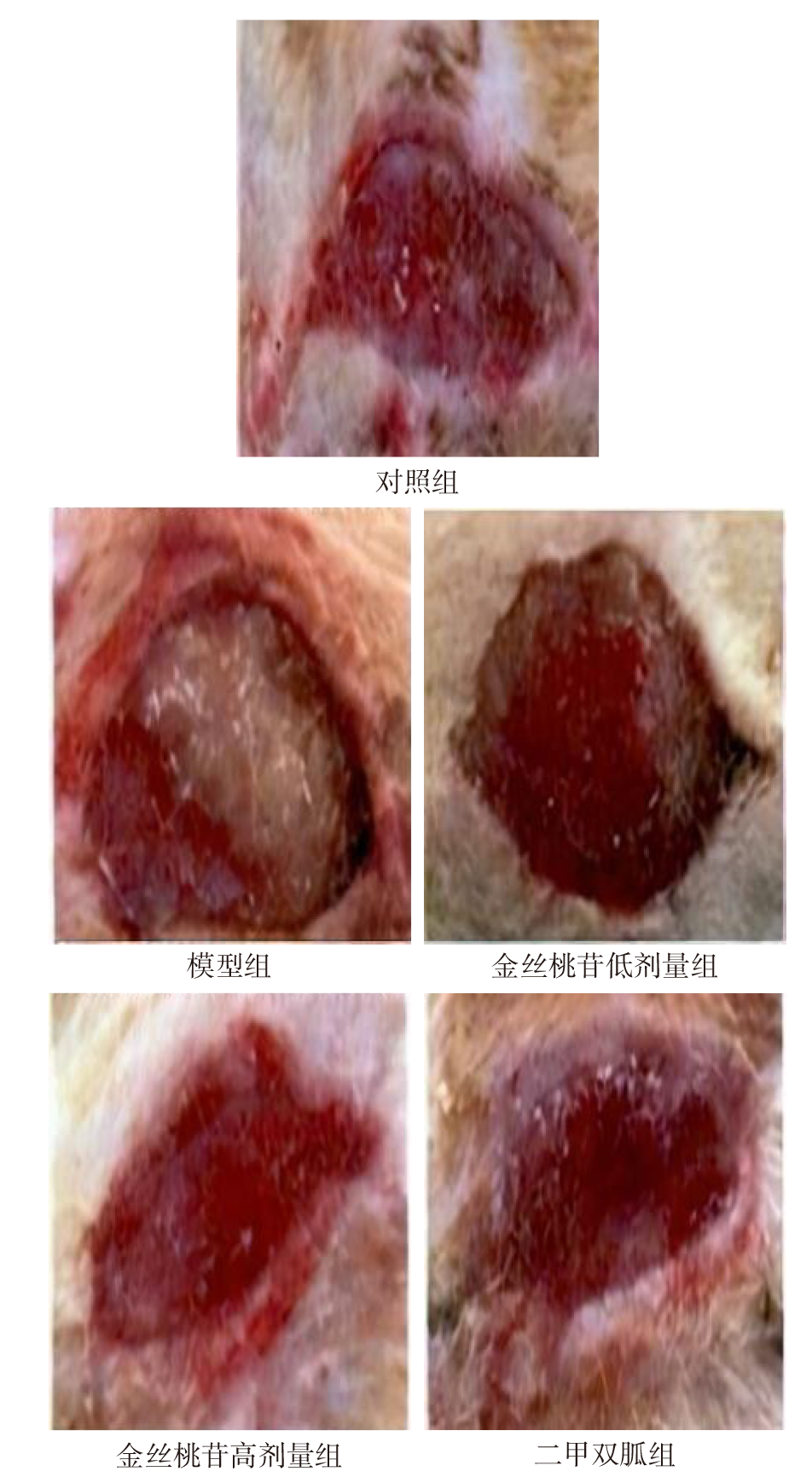
Fig.1 Wound healing of rats in each group after administration
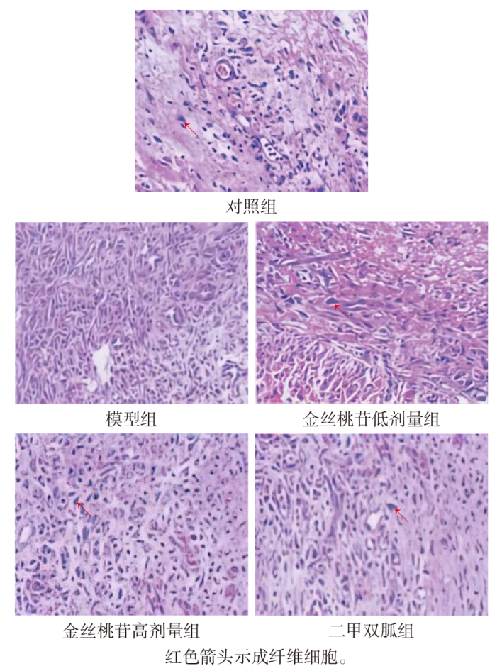
Fig.2 Histopathological changes of wound granulation tissue of rats in each group (HE staining, ×400)
| 组别 | AMPK mRNA | SIRT1 mRNA |
|---|---|---|
| 对照组 | 1.00±0.00 | 1.00±0.00 |
| 模型组 | 0.31±0.02a | 0.34±0.03a |
| 金丝桃苷低剂量组 | 0.52±0.04b | 0.55±0.06b |
| 金丝桃苷高剂量组 | 0.71±0.06bc | 0.85±0.08bc |
| 二甲双胍组 | 0.72±0.08bc | 0.87±0.10bc |
| F | 329.350** | 208.909** |
Tab.4 Comparison of mRNA expression levels of AMPK and SIRT1 in wound granulation tissue of rats between five groups
| 组别 | AMPK mRNA | SIRT1 mRNA |
|---|---|---|
| 对照组 | 1.00±0.00 | 1.00±0.00 |
| 模型组 | 0.31±0.02a | 0.34±0.03a |
| 金丝桃苷低剂量组 | 0.52±0.04b | 0.55±0.06b |
| 金丝桃苷高剂量组 | 0.71±0.06bc | 0.85±0.08bc |
| 二甲双胍组 | 0.72±0.08bc | 0.87±0.10bc |
| F | 329.350** | 208.909** |
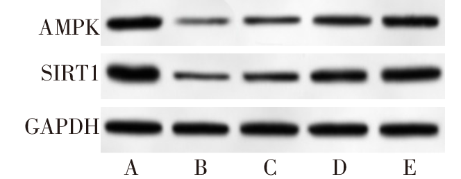
Fig.3 Western blot assay of AMPK and SIRT1 in wound granulation tissue of rats in each group
| 组别 | AMPK | SIRT1 |
|---|---|---|
| 对照组 | 0.95±0.15 | 1.34±0.17 |
| 模型组 | 0.18±0.03a | 0.31±0.04a |
| 金丝桃苷低剂量组 | 0.39±0.05b | 0.57±0.07b |
| 金丝桃苷高剂量组 | 0.72±0.09bc | 0.98±0.11bc |
| 二甲双胍组 | 0.75±0.09bc | 1.03±0.10bc |
| F | 135.349** | 171.997** |
Tab.5 Comparison of protein expression levels of AMPK and SIRT1 in wound granulation tissue of rats between five groups
| 组别 | AMPK | SIRT1 |
|---|---|---|
| 对照组 | 0.95±0.15 | 1.34±0.17 |
| 模型组 | 0.18±0.03a | 0.31±0.04a |
| 金丝桃苷低剂量组 | 0.39±0.05b | 0.57±0.07b |
| 金丝桃苷高剂量组 | 0.72±0.09bc | 0.98±0.11bc |
| 二甲双胍组 | 0.75±0.09bc | 1.03±0.10bc |
| F | 135.349** | 171.997** |
| [1] | BOYKO E J, ZELNICK L R, BRAFFETT B H, et al. Risk of foot ulcer and lower-extremity amputation among participants in the diabetes control and complications trial/epidemiology of diabetes interventions and complications study[J]. Diabetes Care, 2022, 45(2):357-364. doi:10.2337/dc21-1816. |
| [2] | AWASTHI A, SINGH S K, KUMAR B, et al. Treatment strategies against diabetic foot ulcer: success so far and the road ahead[J]. Curr Diabetes Rev, 2021, 17(4):421-436. doi:10.2174/1573399816999201102125537. |
| [3] | WAN R, WEISSMAN J P, GRUNDMAN K, et al. Diabetic wound healing:The impact of diabetes on myofibroblast activity and its potential therapeutic treatments[J]. Wound Repair Regen, 2021, 29(4):573-581. doi:10.1111/wrr.12954. |
| [4] | 曹明明, 车琳琳, 朱路文. 金丝桃苷药理作用及机制研究进展[J]. 辽宁中医药大学学报, 2022, 24(6):150-155. |
| CAO M M, CHE L L, ZHU L W. Research progress on the pharmacological action and mechanism of hyperoside[J]. Journal of Liaoning University of Traditional Chinese Medicine, 2022, 24(6):150-155. doi:10.13194/j.issn.1673-842x.2022.06.033. | |
| [5] | 夏骏, 姚晓丽, 刘潭, 等. 金丝桃苷改善脓毒症模型大鼠心肌损伤作用机制研究[J]. 中国药业, 2021, 30(5):21-25. |
| XIA J, YAO X L, LIU T, et al. Mechanism of hyperoside improving myocardial injury in model rats with sepsis[J]. China Pharmaceuticals, 2021, 30(5):21-25. doi:10.3969/j.issn.1006-4931.2021.05.006. | |
| [6] | 张元丽, 王素利, 张宇, 等. 金丝桃苷调控胰岛素受体底物1/磷脂酰肌醇3-激酶/蛋白激酶B信号通路对糖尿病肾病大鼠肾组织损伤的保护作用[J]. 中国临床药理学杂志, 2023, 39(2):241-245. |
| ZHANG Y L, WANG S L, ZHANG Y, et al. Protective effect of hyperoside on renal tissue damage in rats with diabetic nephropathy by regulating the insulin receptor substrate 1 /phosphatidylinositol 3 -kinase /protein kinase B signaling pathway[J]. Chin J Clin Pharmacol, 2023, 39(2):241-245. doi:10.13699/j.cnki.1001-6821.2023.02.019. | |
| [7] | 邓九红, 郑超, 王声遥, 等. 醋酸泼尼松对糖尿病肾病模型大鼠肾功能、肾脏炎性反应及AMPK/SIRT1信号通路的影响[J]. 基础医学与临床, 2022, 42(2):243-248. |
| DENG J H, ZHENG C, WANG S Y, et al. Effects of prednisone acetate on renal function,renal inflammation and AMPK/SIRT1 signaling pathway in diabetic nephropathy rats[J]. Basic and Clinical Medicine, 2022, 42(2):243-248. doi:10.3969/j.issn.1001-6325.2022.02.007. | |
| [8] | JIA W, BAI T, ZENG J, et al. Combined administration of metformin and atorvastatin attenuates diabetic cardiomyopathy by inhibiting inflammation, apoptosis, and oxidative stress in type 2 diabetic mice[J]. Front Cell Dev Biol, 2021, 9:634900-634913. doi:10.3389/fcell.2021.634900. |
| [9] | 景亮, 祁永章. 红景天苷对糖尿病足溃疡大鼠Nrf2/Keap1信号通路及伤口愈合的影响[J]. 中国比较医学杂志, 2021, 31(8):48-54. |
| JING L, QI Y Z. Effects of salidroside on the Nrf2/Keap1 signaling pathway and wound healing in rats with diabetic foot ulcer[J]. Chin J Comp Med, 2021, 31(8):48-54. doi:10.3969/j.issn.1671-7856.2021.08.007. | |
| [10] | 郭晓, 曲凤霞, 辛越, 等. 金丝桃苷对心力衰竭大鼠肝纤维化的影响及其分子机制[J]. 山东医药, 2021, 61(2):40-45. |
| GUO X, QU F X, XIN Y, et al. Effect and mechanism of hyperin on liver fibrosis in rats with heart failure[J]. Shandong Medical Journal, 2021, 61(2):40-45. doi:10.3969/j.issn.1002-266X.2021.02.009. | |
| [11] | 于泽洋, 李天博, 王江宁. 复方芪参提取物对糖尿病足溃疡模型大鼠创面愈合及HIF-1α/VEGF/VEGFR2通路的影响[J]. 河北医学, 2021, 27(3):374-379. |
| YU Z Y, LI T B, WANG J N. Effects of compound Qishen extract on wound healing and HIF-1α/VEGF/VEGFR2 pathway in diabetic foot ulcer model rats[J]. Hebei Medicine, 2021, 27(3):374-379. doi:10.3969/j.issn.1006-6233.2021.03.005. | |
| [12] | LAUWERS P, DIRINCK E, VaN BOUWEL S, et al. Malnutrition and its relation with diabetic foot ulcer severity and outcome: a review[J]. Acta Clin Belg, 2022, 77(1):79-85. doi:10.1080/17843286.2020.1800315. |
| [13] | DÖRR S, FREIER F, SCHLECHT M, et al. Bacterial diversity and inflammatory response at first-time visit in younger and older individuals with diabetic foot infection(DFI)[J]. Acta Diabetol, 2021, 58(2):181-189. doi:10.1007/s00592-020-01587-5. |
| [14] | MARÔNEK M, MARAFINI I, GARDLÍK R, et al. Metalloproteinases in inflammatory bowel diseases[J]. J Inflamm Res, 2021, 14(1):1029-1041. doi:10.2147/JIR.S288280. |
| [15] | WANG Q, WEI H C, ZHOU S J, et al. Hyperoside:A review on its sources,biological activities, and molecular mechanisms[J]. Phytother Res, 2022, 36(7):2779-2802. doi:10.1002/ptr.7478. |
| [16] | 朱妍妍, 王桐生, 戴宁, 等. 金丝桃苷通过激活Keap1/Nrf2/HO-1通路保护小鼠GC-2细胞的氧化损伤[J]. 南方医科大学学报, 2022, 42(5):673-680. |
| ZHU Y Y, WANG T S, DAI N, et al. Hyperoside protects mouse spermatocytes GC-2 cells from oxidative damage by activating the Keap1/Nrf2/HO-1 pathway[J]. J South Med Univ, 2022, 42(5):673-680. doi:10.12122/j.issn.1673-4254.2022.05.07. | |
| [17] | 申玲君, 张海瑞, 邵伟, 等. 金丝桃苷下调微小RNA-199a对脂多糖诱导的人肺泡上皮细胞的影响[J]. 安徽医药, 2022, 26(2):217-221. |
| SHEN L J, ZHANG H R, SHAO W, et al. Effect of hypericin on LPS-induced HPAEpiC cells by down-regulating miR-199a[J]. Anhui Medical and Pharmaceutical Journal, 2022, 26(2):217-221. doi:10.3969/j.issn.1009-6469.2022.02.002. | |
| [18] | 王旭东, 赵玉良, 史为涛, 等. 黄芪多糖对脓毒症急性肾损伤大鼠AMPK/SIRT1信号通路介导的肾上皮细胞能量代谢的影响[J]. 中国医院药学杂志, 2021, 41(21):2181-2185. |
| WANG X D, ZHAO Y L, SHI W T, et al. Effects of astragalus polysaccharides on energy metabolism of renal epithelial cells mediated by AMPK/SIRT1 signaling pathway in rats with septic acute kidney injury[J]. Chin J Hosp Pharm, 2021, 41(21):2181-2185. doi:10.13286/j.1001-5213.2021.21.05. | |
| [19] | 张蕴, 韩新生, 张洪阳, 等. N-乙酰半胱氨酸通过AMPK/SIRT1途径抑制缺氧诱导的大鼠脑血管内皮细胞损伤[J]. 中国动脉硬化杂志, 2020, 28(2):107-112. |
| ZHANG Y, HAN X S, ZHANG H Y, et al. N-acetylcysteine inhibits hypoxia-induced cerobro-vascular endothelial cell injury via AMPK/SIRT1 pathway[J]. Chin J Arterioscler, 2020, 28(2):107-112. doi:10.3969/j.issn.1007-3949.2020.02.004. | |
| [20] | LI L, CHEN J, ZHOU Y, et al. Artesunate alleviates diabetic retinopathy by activating autophagy via the regulation of AMPK/SIRT1 pathway[J]. Arch Physiol Biochem, 2023, 129(4):943-950. doi:10.1080/13813455.2021.1887266. |
| [21] | XUE W, MAO J, CHEN Q, et al. Mogroside ⅢE alleviates high glucose-induced inflammation, oxidative stress and apoptosis of podocytes by the activation of AMPK/SIRT1 signaling pathway[J]. Diabetes Metab Syndr Obes, 2020, 13:3821-3830. doi:10.2147/DMSO.S276184. |
| Viewed | ||||||
|
Full text |
|
|||||
|
Abstract |
|
|||||