Tianjin Medical Journal ›› 2023, Vol. 51 ›› Issue (12): 1326-1331.doi: 10.11958/20230708
• Experimental Research • Previous Articles Next Articles
NIE Jin1( ), LIU Daishun2, ZHANG Jianyong1, LIU Chu1, ZHOU Liang1,△()
Received:2023-05-16
Revised:2023-06-15
Published:2023-12-15
Online:2023-12-22
Contact:
△ E-mail:NIE Jin, LIU Daishun, ZHANG Jianyong, LIU Chu, ZHOU Liang. The effect and mechanism of exosomes from umbilical cord mesenchymal stem cells on pulmonary inflammation in chronic obstructive pulmonary disease rats[J]. Tianjin Medical Journal, 2023, 51(12): 1326-1331.
CLC Number:
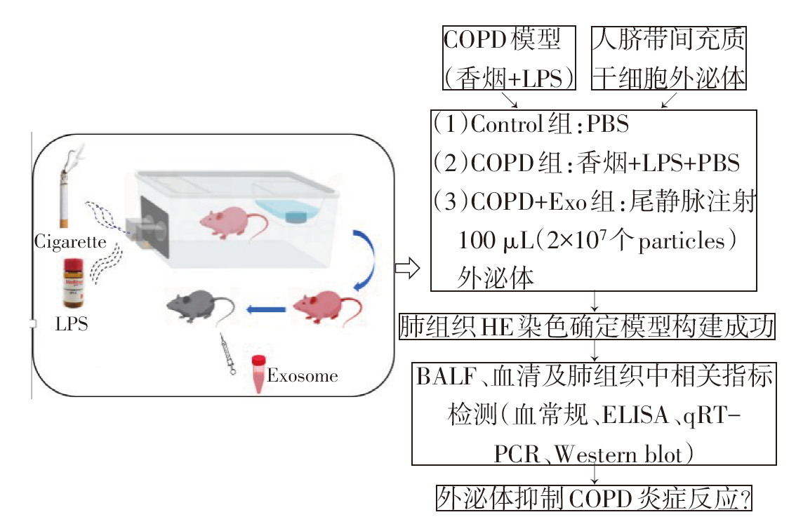
Fig.1 Flow chart of experimental study on inhibition of lung inflammation by human umbilical mesenchymal stem cell exosomes in COPD rats
| 基因名称 | 引物(5′→3′) | 产物大小/bp |
|---|---|---|
| IL-1β | 上游:GCACAGTTCCCCAACTGGTA | 109 |
| 下游:ACACGGGTTCCATGGTGAAG | ||
| IL-6 | 上游:CTCTCCGCAAGAGACTTCCA | 92 |
| 下游:TCTCCTCTCCGGACTTGTGAA | ||
| TNF-α | 上游:CCACGCTCTTCTGTCTACTG | 145 |
| 下游:GCTACGGGCTTGTCACTC | ||
| GAPDH | 上游:CAGGAGGCATTGCTGATGAT | 138 |
| 下游:GAAGGCTGGGGCTCATTT |
Tab.1 Primer sequence for qRT-PCR
| 基因名称 | 引物(5′→3′) | 产物大小/bp |
|---|---|---|
| IL-1β | 上游:GCACAGTTCCCCAACTGGTA | 109 |
| 下游:ACACGGGTTCCATGGTGAAG | ||
| IL-6 | 上游:CTCTCCGCAAGAGACTTCCA | 92 |
| 下游:TCTCCTCTCCGGACTTGTGAA | ||
| TNF-α | 上游:CCACGCTCTTCTGTCTACTG | 145 |
| 下游:GCTACGGGCTTGTCACTC | ||
| GAPDH | 上游:CAGGAGGCATTGCTGATGAT | 138 |
| 下游:GAAGGCTGGGGCTCATTT |
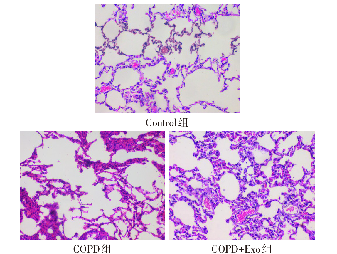
Fig.2 Representative images of HE staining of lung tissue in each group of rats (×200)
| 组别 | WBC | NEU | MON | LYM |
|---|---|---|---|---|
| Control组 | 0.48±0.07 | 0.77±0.24 | 0.03±0.00 | 0.25±0.00 |
| COPD组 | 3.10±0.27a | 2.01±0.20a | 0.15±0.01a | 2.63±0.19a |
| COPD+Exo组 | 1.59±0.55ab | 0.35±0.15b | 0.06±0.02b | 1.20±0.47ab |
| F | 27.197* | 39.738* | 51.100* | 33.116* |
Tab.2 Number of inflammatory cells in peripheral blood of rats in each group (n=3,×109/L,$\bar{x}±s$)
| 组别 | WBC | NEU | MON | LYM |
|---|---|---|---|---|
| Control组 | 0.48±0.07 | 0.77±0.24 | 0.03±0.00 | 0.25±0.00 |
| COPD组 | 3.10±0.27a | 2.01±0.20a | 0.15±0.01a | 2.63±0.19a |
| COPD+Exo组 | 1.59±0.55ab | 0.35±0.15b | 0.06±0.02b | 1.20±0.47ab |
| F | 27.197* | 39.738* | 51.100* | 33.116* |
| 组别 | BALF | |||||
|---|---|---|---|---|---|---|
| IL-1β | IL-6 | TNF-α | ||||
| Control组 | 55.05±11.49 | 71.58±8.77 | 290.51±28.28 | |||
| COPD组 | 85.23±13.44a | 104.16±7.92a | 513.71±59.00a | |||
| COPD+Exo组 | 61.02±6.42b | 84.66±12.84b | 406.74±52.32ab | |||
| F | 10.827* | 13.234* | 26.634* | |||
| 组别 | 血清 | |||||
| IL-1β | IL-6 | TNF-α | ||||
| Control组 | 64.02±6.84 | 72.00±4.45 | 309.18±5.25 | |||
| COPD组 | 88.54±8.44a | 90.99±0.97a | 483.74±9.74a | |||
| COPD+Exo组 | 73.22±4.92b | 73.41±7.93b | 364.64±2.40b | |||
| F | 9.708* | 8.036* | 11.869* | |||
Tab.3 Expression levels of IL-1β, IL-6 and TNF-α in BALF and serum of rats in each group (n=6,ng/L,$\bar{x}±s$)
| 组别 | BALF | |||||
|---|---|---|---|---|---|---|
| IL-1β | IL-6 | TNF-α | ||||
| Control组 | 55.05±11.49 | 71.58±8.77 | 290.51±28.28 | |||
| COPD组 | 85.23±13.44a | 104.16±7.92a | 513.71±59.00a | |||
| COPD+Exo组 | 61.02±6.42b | 84.66±12.84b | 406.74±52.32ab | |||
| F | 10.827* | 13.234* | 26.634* | |||
| 组别 | 血清 | |||||
| IL-1β | IL-6 | TNF-α | ||||
| Control组 | 64.02±6.84 | 72.00±4.45 | 309.18±5.25 | |||
| COPD组 | 88.54±8.44a | 90.99±0.97a | 483.74±9.74a | |||
| COPD+Exo组 | 73.22±4.92b | 73.41±7.93b | 364.64±2.40b | |||
| F | 9.708* | 8.036* | 11.869* | |||
| 组别 | IL-1β | IL-6 | TNF-α |
|---|---|---|---|
| Control组 | 1.00±0.02 | 1.00±0.10 | 1.02±0.18 |
| COPD组 | 3.10±0.31a | 6.58±1.03a | 5.17±0.35a |
| COPD+Exo组 | 1.56±0.12ab | 1.65±0.02ab | 2.52±0.31ab |
| F | 62.411* | 51.588* | 104.294* |
Tab.4 Expression levels of IL-1β, IL-6 and TNF-α in lung tissue of rats in each group (n=6, $\bar{x}±s$)
| 组别 | IL-1β | IL-6 | TNF-α |
|---|---|---|---|
| Control组 | 1.00±0.02 | 1.00±0.10 | 1.02±0.18 |
| COPD组 | 3.10±0.31a | 6.58±1.03a | 5.17±0.35a |
| COPD+Exo组 | 1.56±0.12ab | 1.65±0.02ab | 2.52±0.31ab |
| F | 62.411* | 51.588* | 104.294* |
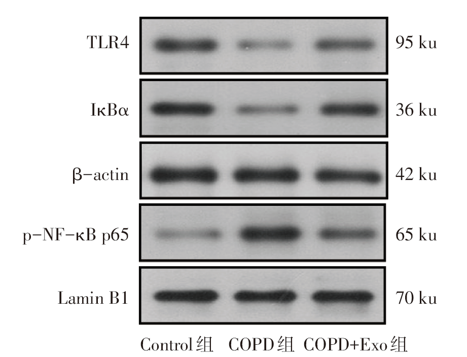
Fig.3 Expression levels of TLR4, IκBα and p-NF-κB p65 protein in lung tissue of rats in each group detected by Western blot assay
| 组别 | TLR4 | IκBα | p-NF-κB p65 |
|---|---|---|---|
| Control组 | 0.73±0.01 | 0.50±0.03 | 0.31±0.02 |
| COPD组 | 0.30±0.00a | 0.23±0.01a | 0.92±0.10a |
| COPD+Exo组 | 0.56±0.02ab | 0.46±0.02b | 0.58±0.06ab |
| F | 824.637* | 130.246* | 59.341* |
Tab.5 Comparison of TLR4, IκBα and p-NF-κB p65 protein levels in lung tissue between the three groups of rats (n=4,$\bar{x}±s$)
| 组别 | TLR4 | IκBα | p-NF-κB p65 |
|---|---|---|---|
| Control组 | 0.73±0.01 | 0.50±0.03 | 0.31±0.02 |
| COPD组 | 0.30±0.00a | 0.23±0.01a | 0.92±0.10a |
| COPD+Exo组 | 0.56±0.02ab | 0.46±0.02b | 0.58±0.06ab |
| F | 824.637* | 130.246* | 59.341* |
| [1] | CHRISTENSON S A, SMITH B M, BAFADHEL M, et al. Chronic obstructive pulmonary disease[J]. Lancet, 2022, 399(10342):2227-2242. doi:10.1016/S0140-6736(22)00470-6. |
| [2] | SONG Q, CHEN P, LIU X M. The role of cigarette smoke-induced pulmonary vascular endothelial cell apoptosis in COPD[J]. Respir Res, 2021, 22(1):39. doi:10.1186/s12931-021-01630-1. |
| [3] | 中华医学会呼吸病学分会慢性阻塞性肺疾病学组, 中国医师协会呼吸医师分会慢性阻塞性肺疾病工作委员会, 陈荣昌, 等. 慢性阻塞性肺疾病诊治指南(2021年修订版)[J]. 中华结核和呼吸杂志, 2021, 44(3):170-205. |
| Group of Chronic Obstructive Pulmonary Disease, Respiratory Branch of Chinese Medical Association, Working Committee of Chronic Obstructive Pulmonary Disease, Respiratory Branch of Chinese Medical Association, CHEN R C, et al. Guidelines for the diagnosis and treatment of chronic obstructive pulmonary disease (2021 revision)[J]. Chinese Journal of Tuberculosis and Respiration, 2021, 44(3):170-205. doi:10.3760/cma.j.cn112147-20210109-00031. | |
| [4] | LIU J, GAO J, LIANG Z, et al. Mesenchymal stem cells and their microenvironment[J]. Stem Cell Res Ther, 2022, 13(1):429. doi:10.1186/s13287-022-02985-y. |
| [5] | ZOU J X, YANG W N, CUI W S, et al. Therapeutic potential and mechanisms of mesenchymal stem cell-derived exosomes as bioactive materials in tendon-bone healing[J]. J Nanobiotechnology, 2023, 21(1):14. doi:10.1186/s12951-023-01778-6. |
| [6] | MOHAN A, AGARWAL S, CLAUSS M, et al. Extracellular vesicles:novel communicators in lung diseases[J]. Respir Res, 2020, 21(1):175. doi:10.1186/s12931-020-01423-y. |
| [7] | HARRELL C R, JOVICIC N, DJONOV V, et al. Mesenchymal stem cell-derived exosomes and other extracellular vesicles as new remedies in the therapy of inflammatory diseases[J]. Cells, 2019, 8(12):1605. doi:10.3390/cells8121605. |
| [8] | SHEN Z W, HUANG W, LIU J, et al. Effects of mesenchymal stem cell-derived exosomes on autoimmune diseases[J]. Front Immunol, 2021, 12:749192. doi:10.3389/fimmu.2021.749192. |
| [9] | LIN Z J, WU Y L, XU Y T, et al. Mesenchymal stem cell-derived exosomes in cancer therapy resistance:recent advances and therapeutic potential[J]. Mol Cancer, 2022, 21(1):179. doi:10.1186/s12943-022-01650-5. |
| [10] | ZHOU L, LUO H, LEE J W. Role of extracellular vesicles in lung diseases[J]. Chin Med J, 2022, 135(15):1765-1780. doi:10.1097/CM9.0000000000002118. |
| [11] | ABBASZADEH H, GHORBANI F, ABBASPOUR-AGHDAM S, et al. Chronic obstructive pulmonary disease and asthma:mesenchymal stem cells and their extracellular vesicles as potential therapeutic tools[J]. Stem Cell Res Ther, 2022, 13(1):262. doi:10.1186/s13287-022-02938-5. |
| [12] | SHU J Z, LI D F, OUYANG H P, et al. Comparison and evaluation of two different methods to establish the cigarette smoke exposure mouse model of COPD[J]. Sci Rep, 2017, 7(1):15454. doi:10.1038/s41598-017-15685-y. |
| [13] | KORSGREN M, LINDEN M, ENTWISTLE N, et al. Inhalation of LPS induces inflammatory airway responses mimicking characteristics of chronic obstructive pulmonary disease[J]. Clin Physiol Funct Imaging, 2012, 32(1):71-79. doi:10.1111/j.1475-097X.2011.01058.x. |
| [14] | SHI M M, YANG Q Y, MONSEL A, et al. Preclinical efficacy and clinical safety of clinical-grade nebulized allogenic adipose mesenchymal stromal cells-derived extracellular vesicles[J]. J Extracell Vesicles, 2021, 10(10):e12134. doi:10.1002/jev2.12134. |
| [15] | CHURG A, COSIO M, WRIGHT J L. Mechanisms of cigarette smoke-induced COPD:insights from animal models[J]. Am J Physiol Lung Cell Mol Physiol, 2008, 294(4):L612-L631. doi:10.1152/ajplung.00390.2007. |
| [16] | AGHAPOUR M, RAEE P, MOGHADDAM S J, et al. Airway epithelial barrier dysfunction in chronic obstructive pulmonary disease:role of cigarette smoke exposure[J]. Am J Respir Cell Mol Biol, 2018, 58(2):157-169. doi:10.1165/rcmb.2017-0200TR. |
| [17] | JONES B, DONOVAN C, LIU G, et al. Animal models of COPD:what do they tell us?[J]. Respirology, 2017, 22(1):21-32. doi:10.1111/resp.12908. |
| [18] | LIANG G B, HE Z H. Animal models of emphysema[J]. Chin Med J(Engl), 2019, 132(20):2465-2475. doi:10.1097/CM9.0000000000000469. |
| [19] | SMITH K R, LEONARD D, MCDONALD J D, et al. Inflammation,mucous cell metaplasia,and Bcl-2 expression in response to inhaled lipopolysaccharide aerosol and effect of rolipram[J]. Toxicol Appl Pharmacol, 2011, 253(3):253-260. doi:10.1016/j.taap.2011.04.001. |
| [20] | DE SOUZA XAVIER COSTA N, RIBEIRO JÚNIOR G,DOS SANTOS ALEMANY A A, et al. Early and late pulmonary effects of nebulized LPS in mice:An acute lung injury model[J]. PLoS One, 2017, 12(9):e0185474. doi:10.1371/journal.pone.0185474. |
| [21] | FONCECA A M, ZOSKY G R, BOZANICH E M, et al. Accumulation mode particles and LPS exposure induce TLR-4 dependent and independent inflammatory responses in the lung[J]. Respir Res, 2018, 19(1):15. doi:10.1186/s12931-017-0701-z. |
| [22] | 何永鸿, 强丽, 王宋平. 阿托伐他汀钙对COPD模型大鼠肺血管重塑的影响及机制探讨[J]. 天津医药, 2021, 49(6):598-602. |
| HE Y H, QIANG L, WANG S P. The effect of atorvastatin calcium on pulmonary vascular remodeling in chronic obstructive pulmonary disease rats[J]. Tianjin Med J, 2021, 49(6):598-602. doi:10.11958/202013011. | |
| [23] | ZHU W T, LI C H, DAI T T, et al. Effect of allyl isothiocyanate on oxidative stress in COPD via the AhR/CYP1A1 and Nrf2/NQO1 pathways and the underlying mechanism[J]. Phytomedicine, 2023, 114:154774. doi:10.1016/j.phymed.2023.154774. |
| [24] | JU J, LI Z, SHI Q. Baicalin inhibits inflammation in rats with chronic obstructive pulmonary disease by the TLR2/MYD88/NF-κBp65 signaling pathway[J]. Evid Based Complement Alternat Med, 2022, 2022:7273387. doi:10.1155/2022/7273387. |
| [25] | ARMITAGE J, TAN D, MOODLEY Y, et al. Mesenchymal stem cell infusion modulates systemic inflammation in patients with chronic obstructive pulmonary disease(COPD)[J]. Respirology, 2016, 21(Suppl 2):133. |
| [26] | GAO J, LIANG Y, CHEN J, et al. CXCR4 enhances the inhibitory effects of bone mesenchymal stem cells on lung cell apoptosis in a rat model of smoking-induced COPD[J]. Apoptosis, 2023, 28(3/4):639-652. doi:10.1007/s10495-022-01800-6. |
| [27] | WEISS D J, SEGAL K, CASABURI R, et al. Effect of mesenchymal stromal cell infusions on lung function in COPD patients with high CRP levels[J]. Respir Res, 2021, 22(1):142. doi:10.1186/s12931-021-01734-8. |
| [28] | ALVARENGA-NASCIMENTO C R, AADB LEIA, SANTOS T G, et al. Immunotherapeutic strategy with mesenchymal stem cells modulating inflammation in an experimental model of COPD[J]. European Respiratory Journal, 2020, 56(Suppl 64):314. |
| [29] | VOLAREVIC V, MARKOVIC B S, GAZDIC M, et al. Ethical and safety issues of stem cell-based therapy[J]. Int J Med Sci, 2018, 15(1):36-45. doi:10.7150/ijms.21666. |
| [30] | TANG Y Y, ZHOU Y, LI H J. Advances in mesenchymal stem cell exosomes:a review[J]. Stem Cell Res Ther, 2021, 12(1):71. doi:10.1186/s13287-021-02138-7. |
| [31] | CHAN A M L, SAMPASIVAM Y, LOKANATHAN Y. Biodistribution of mesenchymal stem cells (MSCs) in animal models and implied role of exosomes following systemic delivery of MSCs:a systematic review[J]. Am J Transl Res, 2022, 14(4):2147-2161. |
| [32] | XIA L J, ZHANG C L, LV N Y, et al. AdMSC-derived exosomes alleviate acute lung injury via transferring mitochondrial component to improve homeostasis of alveolar macrophages[J]. Theranostics, 2022, 12(6):2928-2947. doi:10.7150/thno.69533. |
| [33] | WAN X, CHEN S, FANG Y, et al. Mesenchymal stem cell-derived extracellular vesicles suppress the fibroblast proliferation by downregulating FZD6 expression in fibroblasts via micrRNA-29b-3p in idiopathic pulmonary fibrosis[J]. J Cell Physiol, 2020, 235(11):8613-8625. doi:10.1002/jcp.29706. |
| [34] | CHEN Q, LIN J, DENG Z Q, et al. Exosomes derived from human umbilical cord mesenchymal stem cells protect against papain-induced emphysema by preventing apoptosis through activating VEGF-VEGFR2-mediated AKT and MEK/ERK pathways in rats[J]. Regen Ther, 2022, 21:216-224. doi:10.1016/j.reth.2022.07.002. |
| [35] | YANG L, WEN M, LIU X, et al. Feikang granules ameliorate pulmonary inflammation in the rat model of chronic obstructive pulmonary disease via TLR2/4-mediated NF-κB pathway[J]. BMC Complement Med Ther, 2020, 20(1):170. doi:10.1186/s12906-020-02964-x. |
| [36] | ROY A, SRIVASTAVA M, SAQIB U, et al. Potential therapeutic targets for inflammation in toll-like receptor 4 (TLR4)-mediated signaling pathways[J]. Int Immunopharmacol, 2016, 40:79-89. doi:10.1016/j.intimp.2016.08.026. |
| [37] | LI Y, ZHAO J, SHAO H, et al. Preventive effect of total flavonoids of Trollius altaicus on a chronic obstructive pulmonary disease rat model based on the TLR4/NF-κB pathway[J]. Ann Transl Med, 2022, 10(4):222. doi:10.21037/atm-22-331. |
| Viewed | ||||||
|
Full text |
|
|||||
|
Abstract |
|
|||||