天津医药 ›› 2025, Vol. 53 ›› Issue (2): 170-175.doi: 10.11958/20241377
张小庆( ), 赵慧霞(), 曹鄂洪, 张连霞, 宫秀娟
收稿日期:2024-09-20
修回日期:2024-11-20
出版日期:2025-02-15
发布日期:2025-02-26
通讯作者:
△ E-mail:作者简介:张小庆(1992),女,主治医师,主要从事慢性阻塞性肺疾病、肺部感染方面研究。E-mail:
ZHANG Xiaoqing(), ZHAO Huixia(), CAO Ehong, ZHANG Lianxia, GONG Xiujuan
Received:2024-09-20
Revised:2024-11-20
Published:2025-02-15
Online:2025-02-26
Contact:
△ E-mail:张小庆, 赵慧霞, 曹鄂洪, 张连霞, 宫秀娟. PIV、HCAR、PCT/PLT在老年COPD肺部感染中的应用价值[J]. 天津医药, 2025, 53(2): 170-175.
ZHANG Xiaoqing, ZHAO Huixia, CAO Ehong, ZHANG Lianxia, GONG Xiujuan. Application value of PIV, HCAR and PCT/PLT in pulmonary infection in elderly patients with COPD[J]. Tianjin Medical Journal, 2025, 53(2): 170-175.
摘要:
目的 探究泛免疫炎症值(PIV)、超敏C反应蛋白(hs-CRP)/白蛋白(ALB)比值(HCAR)、降钙素原/血小板计数比值(PCT/PLT)在老年慢性阻塞性肺疾病(COPD)肺部感染患者中的应用价值。方法 选取143例老年COPD伴肺部细菌感染患者为感染组,另选取同期就诊的143例老年COPD不伴肺部细菌感染患者作为未感染组。另依据感染程度将感染组分为轻度组47例、中度组51例和重度组45例;依预后情况分为预后良好组112例和预后不佳组31例。比较各组血生化指标、PIV、HCAR及PCT/PLT水平,分析上述指标与肺部感染、感染程度及预后的关系。结果 感染组中性粒细胞(NEU)、单核细胞(MON)、hs-CRP、PCT水平高于未感染组,淋巴细胞(LYM)、ALB水平低于未感染组(P<0.05);感染组、预后不佳组PIV、HCAR、PCT/PLT水平分别高于未感染组、预后良好组(P<0.05);轻度组、中度组、重度组PIV、HCAR及PCT/PLT水平依次升高(P<0.05)。Spearman相关性分析显示,PIV、HCAR及PCT/PLT均与肺部感染程度呈正相关(P<0.05)。多因素Logistic回归分析显示,高水平PIV、HCAR及PCT/PLT是老年COPD患者发生肺部感染的独立危险因素(P<0.05),亦是肺部感染患者预后不佳的独立危险因素(P<0.05)。PIV、HCAR及PCT/PLT三指标联合诊断老年COPD肺部感染及患者预后不佳的曲线下面积(AUC)分别为0.980和0.910(P<0.05)。结论 PIV、HCAR及PCT/PLT与老年COPD伴肺部感染有关,有助于识别肺部感染、判断肺部感染病情及评估患者预后。
中图分类号:
| 组别 | n | 男性 | 年龄/岁 | 病程/年 | |||||
|---|---|---|---|---|---|---|---|---|---|
| 未感染组 | 143 | 112(78.3) | 66.66±3.50 | 8.37±2.27 | |||||
| 感染组 | 143 | 115(80.4) | 67.01±3.61 | 8.65±2.50 | |||||
| χ2或t | 0.192 | 0.815 | 0.990 | ||||||
| 组别 | BMI/ (kg/m2) | 高血压 | 糖尿病 | Gold分级 (1/2/3/4级) | |||||
| 未感染组 | 23.10±2.26 | 18(12.6) | 62(43.4) | 28/36/46/33 | |||||
| 感染组 | 23.44±2.05 | 24(16.8) | 71(49.7) | 23/34/45/41 | |||||
| χ2或t | 1.347 | 1.005 | 1.138 | 1.131 | |||||
表1 感染组与未感染组基线资料比较
Tab.1 Comparison of baseline information between the infected group and the uninfected group
| 组别 | n | 男性 | 年龄/岁 | 病程/年 | |||||
|---|---|---|---|---|---|---|---|---|---|
| 未感染组 | 143 | 112(78.3) | 66.66±3.50 | 8.37±2.27 | |||||
| 感染组 | 143 | 115(80.4) | 67.01±3.61 | 8.65±2.50 | |||||
| χ2或t | 0.192 | 0.815 | 0.990 | ||||||
| 组别 | BMI/ (kg/m2) | 高血压 | 糖尿病 | Gold分级 (1/2/3/4级) | |||||
| 未感染组 | 23.10±2.26 | 18(12.6) | 62(43.4) | 28/36/46/33 | |||||
| 感染组 | 23.44±2.05 | 24(16.8) | 71(49.7) | 23/34/45/41 | |||||
| χ2或t | 1.347 | 1.005 | 1.138 | 1.131 | |||||
| 组别 | NEU/(×109/L) | PLT/(×109/L) | MON/(×109/L) | LYM/(×109/L) | |||||||
|---|---|---|---|---|---|---|---|---|---|---|---|
| 未感染组 | 7.02±1.22 | 193.29±33.80 | 0.73±0.22 | 1.37±0.35 | |||||||
| 感染组 | 8.42±1.22 | 189.32±34.65 | 0.93±0.20 | 1.01±0.21 | |||||||
| t | 9.692** | 0.980 | 7.765** | 10.496** | |||||||
| 组别 | hs-CRP(mg/L) | ALB/(g/L) | PCT/(μg/L) | ||||||||
| 未感染组 | 86.46±19.21 | 49.09±1.34 | 2.02(1.38,2.48) | ||||||||
| 感染组 | 126.32±22.31 | 39.45±4.91 | 3.60(2.66,4.26) | ||||||||
| t或Z | 16.187** | 22.680** | 10.730** | ||||||||
| 组别 | PIV | HCAR | PCT/PLT | ||||||||
| 未感染组 | 706.20(496.04,1007.36) | 1.76±0.39 | 0.01(0.01,0.01) | ||||||||
| 感染组 | 1 367.26(1 051.93,1 856.15) | 3.27±0.81 | 0.02(0.01,0.02) | ||||||||
| Z或t | 10.555** | 19.986** | 10.675** | ||||||||
表2 感染组与未感染组患者的实验室各指标比较 (n=143,$\bar{x}±s$)
Tab.2 Comparison of laboratory indexes between the infected group and the uninfected group
| 组别 | NEU/(×109/L) | PLT/(×109/L) | MON/(×109/L) | LYM/(×109/L) | |||||||
|---|---|---|---|---|---|---|---|---|---|---|---|
| 未感染组 | 7.02±1.22 | 193.29±33.80 | 0.73±0.22 | 1.37±0.35 | |||||||
| 感染组 | 8.42±1.22 | 189.32±34.65 | 0.93±0.20 | 1.01±0.21 | |||||||
| t | 9.692** | 0.980 | 7.765** | 10.496** | |||||||
| 组别 | hs-CRP(mg/L) | ALB/(g/L) | PCT/(μg/L) | ||||||||
| 未感染组 | 86.46±19.21 | 49.09±1.34 | 2.02(1.38,2.48) | ||||||||
| 感染组 | 126.32±22.31 | 39.45±4.91 | 3.60(2.66,4.26) | ||||||||
| t或Z | 16.187** | 22.680** | 10.730** | ||||||||
| 组别 | PIV | HCAR | PCT/PLT | ||||||||
| 未感染组 | 706.20(496.04,1007.36) | 1.76±0.39 | 0.01(0.01,0.01) | ||||||||
| 感染组 | 1 367.26(1 051.93,1 856.15) | 3.27±0.81 | 0.02(0.01,0.02) | ||||||||
| Z或t | 10.555** | 19.986** | 10.675** | ||||||||
| 变量 | β | SE | Waldχ2 | P | OR(95%CI) |
|---|---|---|---|---|---|
| PIV | 0.001 | 0.001 | 6.516 | 0.011 | 1.001(1.000~1.003) |
| HCAR | 4.840 | 0.730 | 43.928 | <0.001 | 126.422(30.219~528.896) |
| PCT/PLT | 0.087 | 0.042 | 4.295 | 0.038 | 1.091(1.005~1.185) |
| 常数项 | -14.110 | 1.900 | 55.160 | <0.001 | 0.000 |
表3 老年COPD患者肺部感染的影响因素分析
Tab.3 Analysis of the influencing factors of pulmonary infection in elderly patients with COPD
| 变量 | β | SE | Waldχ2 | P | OR(95%CI) |
|---|---|---|---|---|---|
| PIV | 0.001 | 0.001 | 6.516 | 0.011 | 1.001(1.000~1.003) |
| HCAR | 4.840 | 0.730 | 43.928 | <0.001 | 126.422(30.219~528.896) |
| PCT/PLT | 0.087 | 0.042 | 4.295 | 0.038 | 1.091(1.005~1.185) |
| 常数项 | -14.110 | 1.900 | 55.160 | <0.001 | 0.000 |
| 指标 | AUC(95%CI) | 截断值 | 敏感度/% | 特异度/% | 约登指数 |
|---|---|---|---|---|---|
| PIV | 0.861(0.815~0.899) | 895.56 | 90.21 | 68.53 | 0.587 |
| HCAR | 0.975(0.950~0.990) | 2.40 | 92.31 | 94.41 | 0.867 |
| PCT/PLT | 0.816(0.767~0.860) | 0.01 | 72.73 | 86.71 | 0.594 |
| 指标联合 | 0.980(0.956~0.993) | - | 95.10 | 92.31 | 0.874 |
表4 PIV、HCAR、PCT/PLT及联合对老年COPD伴肺部感染的诊断价值分析
Tab.4 The diagnostic value of PIV, HCAR, PCT/PLT and their combination for pulmonary infection in elderly patients with COPD
| 指标 | AUC(95%CI) | 截断值 | 敏感度/% | 特异度/% | 约登指数 |
|---|---|---|---|---|---|
| PIV | 0.861(0.815~0.899) | 895.56 | 90.21 | 68.53 | 0.587 |
| HCAR | 0.975(0.950~0.990) | 2.40 | 92.31 | 94.41 | 0.867 |
| PCT/PLT | 0.816(0.767~0.860) | 0.01 | 72.73 | 86.71 | 0.594 |
| 指标联合 | 0.980(0.956~0.993) | - | 95.10 | 92.31 | 0.874 |
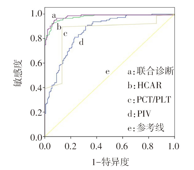
图1 PIV、HCAR、PCT/PLT及联合诊断老年COPD伴肺部感染的ROC曲线
Fig.1 ROC curves of PIV, HCAR, PCT/PLT and their combination for diagnosing pulmonary infection in elderly patients with COPD
| 组别 | n | PIV | HCAR | PCT/PLT |
|---|---|---|---|---|
| 轻度组 | 47 | 947.94±192.18 | 2.64±0.44 | 0.01(0.01,0.02) |
| 中度组 | 51 | 1 384.79±157.67a | 3.37±0.32a | 0.02(0.02,0.02)a |
| 重度组 | 45 | 2 437.00±671.45ab | 3.81±1.03ab | 0.03(0.02,0.04)ab |
| F或H | 165.208** | 37.271** | 49.006** |
表5 不同肺部感染程度的3组间PIV、HCAR及PCT/PLT水平比较
Tab.5 Comparison of PIV, HCAR and PCT/PLT between subgroups with different degrees of pulmonary infection
| 组别 | n | PIV | HCAR | PCT/PLT |
|---|---|---|---|---|
| 轻度组 | 47 | 947.94±192.18 | 2.64±0.44 | 0.01(0.01,0.02) |
| 中度组 | 51 | 1 384.79±157.67a | 3.37±0.32a | 0.02(0.02,0.02)a |
| 重度组 | 45 | 2 437.00±671.45ab | 3.81±1.03ab | 0.03(0.02,0.04)ab |
| F或H | 165.208** | 37.271** | 49.006** |
| 组别 | n | PIV | HCAR | PCT/PLT |
|---|---|---|---|---|
| 预后良好组 | 112 | 1 354.19±547.07 | 3.08±0.64 | 0.02(0.01,0.02) |
| 预后不佳组 | 31 | 2 360.44±790.27 | 3.96±1.23 | 0.02(0.02,0.04) |
| t或Z | 6.661** | 3.810** | 3.781** |
表6 不同预后亚组间PIV、HCAR及PCT/PLT水平比较 ($\bar{x}±s$)
Tab.6 Comparison of PIV, HCAR and PCT/PLT between subgroups with different prognoses
| 组别 | n | PIV | HCAR | PCT/PLT |
|---|---|---|---|---|
| 预后良好组 | 112 | 1 354.19±547.07 | 3.08±0.64 | 0.02(0.01,0.02) |
| 预后不佳组 | 31 | 2 360.44±790.27 | 3.96±1.23 | 0.02(0.02,0.04) |
| t或Z | 6.661** | 3.810** | 3.781** |
| 变量 | β | SE | Wald χ2 | P | OR(95%CI) |
|---|---|---|---|---|---|
| PIV | 0.001 | 0.000 | 6.326 | 0.012 | 1.001(1.000~1.002) |
| HCAR | 1.106 | 0.354 | 9.760 | 0.002 | 3.022(1.510~6.048) |
| PCT/PLT | 0.487 | 0.243 | 4.004 | 0.045 | 1.627(1.010~2.620) |
| 常数项 | -8.576 | 1.444 | 35.259 | <0.001 | 0.000 |
表7 老年COPD伴肺部感染患者预后不佳的影响因素分析
Tab.7 Analysis of the influencing factors of poor prognosis in elderly patients with COPD and pulmonary infection
| 变量 | β | SE | Wald χ2 | P | OR(95%CI) |
|---|---|---|---|---|---|
| PIV | 0.001 | 0.000 | 6.326 | 0.012 | 1.001(1.000~1.002) |
| HCAR | 1.106 | 0.354 | 9.760 | 0.002 | 3.022(1.510~6.048) |
| PCT/PLT | 0.487 | 0.243 | 4.004 | 0.045 | 1.627(1.010~2.620) |
| 常数项 | -8.576 | 1.444 | 35.259 | <0.001 | 0.000 |
| 指标 | AUC(95%CI) | 截断值 | 敏感度/ % | 特异度/ % | 约登 指数 | ||
|---|---|---|---|---|---|---|---|
| PIV | 0.889(0.826~0.935) | 1 648.82 | 90.32 | 84.82 | 0.751 | ||
| HCAR | 0.739(0.659~0.808) | 4.02 | 45.16 | 91.96 | 0.371 | ||
| PCT/PLT | 0.722(0.641~0.794) | 0.02 | 67.74 | 67.86 | 0.356 | ||
| 指标联合 | 0.910(0.850~0.951) | - | 90.32 | 81.25 | 0.716 | ||
表8 PIV、HCAR、PCT/PLT及联合对老年COPD伴肺部感染预后不佳的预测价值
Tab.8 Predictive value of PIV, HCAR, PCT/PLT and their combination for poor prognosis in elderly patients with COPD and pulmonary infection
| 指标 | AUC(95%CI) | 截断值 | 敏感度/ % | 特异度/ % | 约登 指数 | ||
|---|---|---|---|---|---|---|---|
| PIV | 0.889(0.826~0.935) | 1 648.82 | 90.32 | 84.82 | 0.751 | ||
| HCAR | 0.739(0.659~0.808) | 4.02 | 45.16 | 91.96 | 0.371 | ||
| PCT/PLT | 0.722(0.641~0.794) | 0.02 | 67.74 | 67.86 | 0.356 | ||
| 指标联合 | 0.910(0.850~0.951) | - | 90.32 | 81.25 | 0.716 | ||
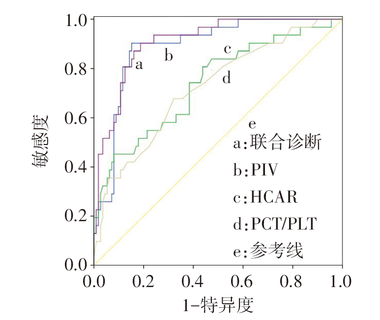
图2 PIV、HCAR、PCT/PLT及联合预测老年COPD伴肺部感染患者预后不佳的ROC曲线
Fig.2 ROC curves of PIV, HCAR, PCT/PLT and their combination for predicting poor prognosis in elderly patients with COPD and pulmonary infection
| [1] | SULAIMAN I, WU B G, CHUNG M, et al. Lower airway dysbiosis augments lung inflammatory injury in mild-to-moderate chronic obstructive pulmonary disease[J]. Am J Respir Crit Care Med, 2023, 208(10):1101-1114. doi:10.1164/rccm.202210-1865OC. |
| [2] | RITCHIE A I, WEDZICHA J A. Definition,causes,pathogenesis,and consequences of chronic obstructive pulmonary disease exacerbations[J]. Clin Chest Med, 2020, 41(3):421-438. doi:10.1016/j.ccm.2020.06.007. |
| [3] | DEMIRÖZ TAŞOLAR S, ÇIFTÇI N. Role of pan immune inflammatory value in the evaluation of hepatosteatosis in children and adolescents with obesity[J]. J Pediatr Endocrinol Metab, 2022, 35(12):1481-1486. doi:10.1515/jpem-2022-0494. |
| [4] | 付志彬, 李赵忠, 强仲惪, 等. HCAR与临床指标对COPD并发呼吸衰竭患者预后的预测作用研究[J]. 国际呼吸杂志, 2022, 42(12):922-927. |
| FU Z B, LI Z Z, QIANG Z D, et al. Study on the predictive effeet of high-sensitivity C-reactive protein/albumin ratio and clinical indexes on the prognosis of chronic obstructive pulmonary disease patients with respiratory failure[J]. Int J Respir, 2022, 42(12):922-927. doi:10.3760/cma.j.cn131368-20220302-00152. | |
| [5] | 刘梦婷, 钟文宏, 文茵, 等. PCT/PLT比值对脓毒症心肌损伤的预警价值[J]. 中华急诊医学杂志, 2022, 31(8):1071-1076. |
| LIU M T, ZHONG W H, WEN Y, et al. Early-warning value of PCT/PLT ratio on sepsis-induced myocardial injury[J]. Chin J Emerg Med, 2022, 31(8):1071-1076. doi:10.3760/cma.j.issn.1671-0282.2022.08.010. | |
| [6] | 徐慧, 赵登峰, 王淼, 等. 肺部感染对慢性阻塞性肺疾病患者呼吸功能状态与细胞因子表达的影响[J]. 中华医院感染学杂志, 2019, 29(7):1011-1014. |
| XU H, ZHAO D F, WANG M, et al. Effects of pulmonary infection on respiratory function and cytokine expression in patients with chronic obstructive pulmonary disease[J]. Chin J Nosocomiol, 2019, 29(7):1011-1014. | |
| [7] | REHMAN A U, SHAH S, ABBAS G, et al. Assessment of risk factors responsible for rapid deterioration of lung function over a period of one year in patients with chronic obstructive pulmonary disease[J]. Sci Rep, 2021, 11(1):13578. doi:10.1038/s41598-021-92968-5. |
| [8] | LEE S H, KIM K U, LEE H, et al. Factors associated with low-level physical activity in elderly patients with chronic obstructive pulmonary disease[J]. Korean J Intern Med, 2018, 33(1):130-137. doi:10.3904/kjim.2016.090. |
| [9] | 王远珍, 魏红艳, 常丽仙, 等. 原发性肝癌干预前并发肺部感染风险预测模型的建立与验证[J]. 天津医药, 2024, 52(9):940-945. |
| WANG Y Z, WEI H Y, CHANG L X, et al. Establishment and validation of a risk prediction model for primary liver cancer complicated with pulmonary infection before intervention[J]. Tianjin Med J, 2024, 52(9):940-945. doi:10.11958/20240147. | |
| [10] | 孙皎琳, 曹霞, 李婷婷, 等. 感染对慢性阻塞性肺疾病系统性炎症反应的影响[J]. 武汉大学学报(医学版), 2022, 43(5):768-773. |
| SUN J L, CAO X, LI T T, et al. Effects of infection on systemic inflammatory response in patients with chronic obstructive pulmonary disease[J]. Med J Wuhan Univ, 2022, 43(5):768-773. doi:10.14188/j.1671-8852.2021.0277. | |
| [11] | 陈学梅, 祁蕙燕, 成志飞. 老年COPD合并肺部感染患者呼出气一氧化氮水平与EOS、PCT、CRP水平相关性分析[J]. 中国急救复苏与灾害医学杂志, 2022, 17(4):482-485. |
| CHEN X M, QI H Y, CHENG Z F. Correlation analysis of exhaled nitric oxide level and EOS,PCT,CRP levels in elderly patients with COPD complicated with pulmonary infection[J]. Chin J Emerg Resusc Disaster Med, 2022, 17(4):482-485. doi:10.3969/j.issn.1673-6966.2022.04.015. | |
| [12] | 余莹, 罗琳琳, 李洁, 等. 老年COPD患者下呼吸道微生物定植与肺部感染的炎症指标及TGFβ1/Smads信号通路[J]. 中华医院感染学杂志, 2023, 33(12):1804-1808. |
| YU Y, LUO L L, LI J, et al. Inflammatory indicators of microbial colonization of the lower respiratory tract and lung infection in elderly patients with COPD and TGFβ1/Smads signaling pathway[J]. Chin J Nosocomiol, 2023, 33(12):1804-1808. doi:10.11816/cn.ni.2023-221939. | |
| [13] | 康李蕊, 肖漓. 慢性阻塞性肺疾病急性加重相关的免疫细胞及因子研究进展[J]. 解放军医学杂志, 2023, 48(11):1353-1358. |
| KANG L R, XIAO L. Research progress of immunocytes and immunologic factors related to acute exacerbation of chronic obstructive pulmonary disease[J]. Med J Chin PLA, 2023, 48(11):1353-1358. doi:10.11855/j.issn.0577-7402.1493.2023.0413. | |
| [14] | WANG M D, DUAN F F, HUA X, et al. A novel albumin-related nutrition biomarker predicts breast cancer prognosis in neoadjuvant chemotherapy:a two-center cohort study[J]. Nutrients, 2023, 15(19):4292. doi:10.3390/nu15194292. |
| [15] | ECKART A, STRUJA T, KUTZ A, et al. Relationship of nutritional status,inflammation,and serum albumin levels during acute illness:a prospective study[J]. Am J Med, 2020, 133(6):713-722. doi:10.1016/j.amjmed.2019.10.031. |
| [16] | 张辉, 伍冬冬, 马东波, 等. 血小板平均体积与血小板计数比值预测老年人慢性阻塞性肺疾病急性加重期近期预后的临床价值[J]. 中华老年医学杂志, 2020, 39(6):627-631. |
| ZHANG H, WU D D, MA D B, et al. Clinical value of mean platelet volume/platelet count ratio in predicting short-term prognosis of elderly patients with acute exacerbation of chronic obstructive pulmonary disease[J]. Chin J Geriatr, 2020, 39(6):627-631. doi:10.3760/cma.j.issn.0254-9026.2020.06.005. | |
| [17] | 和军辉, 万大国, 董静, 等. 泛免疫炎症值、全身免疫炎症指数与急性冠脉综合征患者易损斑块的相关性研究[J]. 中国全科医学, 2024, 27(24):2976-2981. |
| HE J H, WAN D G, DONG J, et al. Correlation analysis between pan-immune inflammatory value,systemic immune-inflammatory index,and vulnerable plaques in patients with acute coronary syndrome[J]. Chin Gen Prac, 2024, 27(24):2976-2981. doi:10.12114/j.issn.1007-9572.2023.0679. | |
| [18] | INAN D, ERDOGAN A, PAY L, et al. The prognostic impact of inflammation in patients with decompensated acute heart failure,as assessed using the pan-immune inflammation value (PIV)[J]. Scand J Clin Lab Invest, 2023, 83(6):371-378. doi:10.1080/00365513.2023.2233890. |
| [19] | HAO X, WANG X, WEI H, et al. Development and validation of the prediction model of sepsis in patients after percutaneous nephrolithotomy and sepsis progresses to septic shock[J]. J Endourol, 2023, 37(4):377-386. doi:10.1089/end.2022.0384. |
| [20] | 郭红玲, 欧阳艳红, 王圣, 等. 急性ST段抬高型心肌梗死患者的hs-CRP/ALB变化对预后的预测价值[J]. 中国急救医学, 2020, 40(2):102-107. |
| GUO H L, OUYANG Y H, WANG S, et al. Prognostic value of hs-CRP/ALB changes in the patients with acute ST-segment elevation myocardial infarction[J]. Chin J Crit Care Med, 2020, 40(2):102-107. doi:10.3969/j.issn.1002-1949.2020.02.003. | |
| [21] | 杨金强, 张仁敏. 降钙素原与血小板比值评估发热伴血小板减少综合征预后的价值[J]. 临床荟萃, 2023, 38(4):346-351. |
| YANG J Q, ZHANG R M. Prognostic value of procalcitonin to platelet ratio in patients with fever with thrombocytopenia syndrome[J]. Clinical Focus, 2023, 38(4):346-351. doi:10.3969/j.issn.1004-583X.2023.04.010. | |
| [22] | 王勇飞, 朱勤贤, 曹旭, 等. 加味瓜蒌枳实汤联合哌拉西林钠他唑巴坦钠对痰热蕴肺型卒中相关性肺炎患者的免疫功能和血清炎症因子水平的影响[J]. 中国实验方剂学杂志, 2023, 29(21):140-146. |
| WANG Y F, ZHU Q X, CAO X, et al. Effect of modified Gualou Zhishitang combined with piperacillin sodium and tazobactam sodium on immune function and serum levels of inflammatory cytokines in patients with stroke-associated pneumonia with syndrome of phlegm-heat accumulation in lung[J]. Chin J Exp Tradit Med Formulae, 2023, 29(21):140-146. doi:10.13422/j.cnki.syfjx. |
| [1] | 陈丽, 陈楠. COPD进展为Ⅱ型呼吸衰竭预警模型的建立与验证[J]. 天津医药, 2026, 54(2): 189-195. |
| [2] | 付莎莎, 贾月, 邵红霞, 郭禹, 马龙艳, 韩同, 孙浩, 于洪志. 1例老年抑郁患者有机磷和抗凝血鼠药中毒的MDT救治策略[J]. 天津医药, 2025, 53(9): 1000-1004. |
| [3] | 焦爱菊, 朱若兰, 张春花, 李文瑞, 孙霞, 赵玮婧, 任宝龙. 血清Lp-PLA2、Hcy联合WMH对脑小血管病患者认知障碍的预测价值[J]. 天津医药, 2025, 53(8): 846-849. |
| [4] | 封金花, 黄晗, 许丽萍, 孙爽爽, 王艳琼. 血清PCT、外周血补体及淋巴细胞亚群对难治性肺炎支原体肺炎患儿发生塑型性支气管炎的预测价值[J]. 天津医药, 2025, 53(7): 714-718. |
| [5] | 沙亚娟, 赵莹琰, 李海燕. 那不勒斯预后评分和预后营养指数对子宫内膜癌患者术后复发的预测价值[J]. 天津医药, 2025, 53(6): 634-639. |
| [6] | 周美娟, 缪小祥. 帕金森病患者血清miR-214-3p、miR-124-3p表达水平与病情严重程度的关系及其早期诊断价值[J]. 天津医药, 2025, 53(5): 498-502. |
| [7] | 王欣欣, 许慧, 吴晓. 妊娠期肝内胆汁淤积症合并GDM孕妇不良妊娠结局的影响因素及预测模型构建[J]. 天津医药, 2025, 53(5): 503-508. |
| [8] | 胡卓, 谢松波, 游诗伟. 序贯器官衰竭评分联合Clara细胞蛋白和血管生成素-2对脓毒症所致ARDS的预测价值[J]. 天津医药, 2025, 53(5): 519-522. |
| [9] | 宋志新, 叶建军, 杨道武. 交通创伤患者继发创伤性凝血功能障碍的危险因素分析[J]. 天津医药, 2025, 53(4): 416-419. |
| [10] | 高丹青, 马笑风, 钱英杰, 裴小妹, 王恺文. 血清SACE、MMP-9和HPT与精神分裂症患者肺部感染的相关性研究[J]. 天津医药, 2025, 53(2): 176-179. |
| [11] | 李晓宁, 李妮. 血清PCT、CXCL10、IFN-γ对ICU多重耐药菌感染患者疗效的预测价值[J]. 天津医药, 2025, 53(2): 180-184. |
| [12] | 吴静静, 张福森, 陈皓, 赵亿, 刘泉, 李冬梅. 血清Syndecan-1、endocan-1联合qSOFA评分在脓毒症诊断及预后评估中的应用价值[J]. 天津医药, 2025, 53(2): 185-188. |
| [13] | 陈璐, 张瑞家, 陈国庆. 妊娠期糖尿病孕妇分娩期宫内感染的相关因素及其预测价值[J]. 天津医药, 2025, 53(12): 1258-1262. |
| [14] | 徐淑光, 管淑红, 张云凤. 集束化管理在慢性阻塞性肺疾病急性加重期患者围出院期中的应用效果[J]. 天津医药, 2025, 53(11): 1170-1175. |
| [15] | 廖洪柳, 胡艳, 李艳, 邓才望. 血清CRP、SAA、PCT联合脑脊液LDH、ADA水平测定在老年中枢神经系统感染中的应用[J]. 天津医药, 2025, 53(10): 1048-1052. |
| 阅读次数 | ||||||
|
全文 |
|
|||||
|
摘要 |
|
|||||