
天津医药 ›› 2026, Vol. 54 ›› Issue (10): 1087-1092.doi: 10.11958/20260051
董文鹏( ), 崔培贺△(), 赵师岳, 崔岩
收稿日期:2026-01-06
修回日期:2026-03-06
出版日期:2026-10-15
发布日期:2026-10-10
通讯作者:
△E-mail:534679177@qq.com
作者简介:董文鹏(1981),男,副主任医师,主要从事肾脏病临床及血液净化治疗的研究。E-mail:9951010513@163.com
DONG Wenpeng(), CUI Peihe△(), ZHAO Shiyue, CUI Yan
Received:2026-01-06
Revised:2026-03-06
Published:2026-10-15
Online:2026-10-10
Contact:
△E-mail:534679177@qq.com
董文鹏, 崔培贺, 赵师岳, 崔岩. 膜增生性肾小球肾炎伴单克隆免疫球蛋白沉积的临床特征观察[J]. 天津医药, 2026, 54(10): 1087-1092.
DONG Wenpeng, CUI Peihe, ZHAO Shiyue, CUI Yan. Observation of clinical characteristics of membranoproliferative glomerulonephritis with monoclonal immunoglobulin deposition[J]. Tianjin Medical Journal, 2026, 54(10): 1087-1092.
摘要:
目的 分析膜增生性肾小球肾炎伴单克隆免疫球蛋白沉积(PGNMID)的临床及病理特征、治疗方法与转归。方法 回顾性分析我院经肾穿刺活检确诊的3例PGNMID患者的临床资料、病理资料、治疗方案及预后特点,探讨发病机制并评价不同治疗方法的疗效。结果 3例PGNMID患者均为女性,平均年龄(68.00±12.29)岁,均表现为肾小球源性血尿、大量蛋白尿及不同程度肾功能损伤。肾活检病理光镜下均以膜增生性肾炎为特征,免疫荧光可见IgG亚型免疫球蛋白及轻链沉积于毛细血管襻与系膜区,均伴补体C3沉积,电镜下均可见足突弥漫性融合(>95%),电子致密物沉积于内皮下及系膜区,其中2例基底膜内亦可见电子致密物沉积。3例患者均采用肾素-血管紧张素转化酶抑制剂联合针对致病B细胞或浆细胞克隆实施靶向治疗,1个月后24 h尿蛋白定量均有不同程度下降。2例患者在治疗2~3个月时达到部分缓解,在第6~7个月达完全缓解,1例患者经治疗后随访至6个月仍为部分缓解。结论 PGNMID患者常表现为肾病综合征并伴有肾功能减退,病理上常以IgG3沉积的膜增生性肾小球肾炎为特征,早期诊治可获得较好临床治疗效果。
中图分类号:
| 病例 | 年龄/岁 | 基本病情 | 血尿/HPF | 24 h UTP/ (g/d) | ALB/ (g/L) | SCr/ (μmol/L) | eGFR/ [mL/(min·1.73m2)] | ||||||||||
|---|---|---|---|---|---|---|---|---|---|---|---|---|---|---|---|---|---|
| 1 | 54 | 颜面部水肿1年,加重1个月 | 满视野 | 5.07 | 35.00 | 85.00 | 70.17 | ||||||||||
| 2 | 73 | 发现Scr升高3个月,双下肢水肿2个月 | 57.00 | 6.30 | 26.80 | 108.00 | 46.78 | ||||||||||
| 3 | 77 | 确诊膜增生性肾炎15年,蛋白尿加重3个月 | 10.20 | 11.24 | 27.20 | 262.00 | 15.75 | ||||||||||
| 病例 | Hb/ (g/L) | 血κ轻链/(g/L) | 血λ轻链/(g/L) | κ/λ | 抗核抗体谱 | 免疫固定电泳 | 血清IgG/(g/L) | 补体C3/(g/L) | 补体C4/(g/L) | 骨髓活检浆 细胞数/个 | |||||||
| 1 | 99 | 5.44 | 2.58 | 2.11 | 抗SSA/Ro(+++)、抗Ro-52抗体(+++) | 阴性 | 23.80 | 0.60 | 0.09 | 1 | |||||||
| 2 | 108 | 1.87 | 0.68 | 2.75 | 抗SSA/Ro(+++)、抗Ro-52抗体(+++) | 阴性 | 7.70 | 0.93 | 0.30 | 2 | |||||||
| 3 | 108 | 1.35 | 1.03 | 1.31 | 抗SSA/Ro(+++)、抗Ro-52抗体(++) | 阴性 | 6.83 | 0.94 | 0.34 | 1 | |||||||
表1 3例PGNMID患者的临床资料
Tab.1 Clinical data of 3 patients with PGNMID
| 病例 | 年龄/岁 | 基本病情 | 血尿/HPF | 24 h UTP/ (g/d) | ALB/ (g/L) | SCr/ (μmol/L) | eGFR/ [mL/(min·1.73m2)] | ||||||||||
|---|---|---|---|---|---|---|---|---|---|---|---|---|---|---|---|---|---|
| 1 | 54 | 颜面部水肿1年,加重1个月 | 满视野 | 5.07 | 35.00 | 85.00 | 70.17 | ||||||||||
| 2 | 73 | 发现Scr升高3个月,双下肢水肿2个月 | 57.00 | 6.30 | 26.80 | 108.00 | 46.78 | ||||||||||
| 3 | 77 | 确诊膜增生性肾炎15年,蛋白尿加重3个月 | 10.20 | 11.24 | 27.20 | 262.00 | 15.75 | ||||||||||
| 病例 | Hb/ (g/L) | 血κ轻链/(g/L) | 血λ轻链/(g/L) | κ/λ | 抗核抗体谱 | 免疫固定电泳 | 血清IgG/(g/L) | 补体C3/(g/L) | 补体C4/(g/L) | 骨髓活检浆 细胞数/个 | |||||||
| 1 | 99 | 5.44 | 2.58 | 2.11 | 抗SSA/Ro(+++)、抗Ro-52抗体(+++) | 阴性 | 23.80 | 0.60 | 0.09 | 1 | |||||||
| 2 | 108 | 1.87 | 0.68 | 2.75 | 抗SSA/Ro(+++)、抗Ro-52抗体(+++) | 阴性 | 7.70 | 0.93 | 0.30 | 2 | |||||||
| 3 | 108 | 1.35 | 1.03 | 1.31 | 抗SSA/Ro(+++)、抗Ro-52抗体(++) | 阴性 | 6.83 | 0.94 | 0.34 | 1 | |||||||
| 病例 | 免疫荧光 | 超微结构 电子致密物位 | 光镜 | ||
|---|---|---|---|---|---|
| 优势蛋白类型及位置 | 补体类型 | 肾小球病变类型 | 嗜复红物质沉积位置 | ||
| 1 | IgG2-Lambda;团块状/毛细血管襻、系膜区 | C3(+)、C4(+/-)、C1q(阴性) | 系膜区、内皮下 | 膜增生性肾炎 | 系膜区、内皮下沉积 |
| 2 | IgG3-Kappa;团块状、颗粒状/毛细血管襻、系膜区 | C3(+++)、C4(阴性)、C1q(阴性) | 系膜区、内皮下,基底膜内偶见 | 膜增生性肾炎 | 系膜区、内皮下沉积 |
| 3 | IgG3-Kappa;花瓣状/毛细血管襻、系膜区 | C3(+++)、C4(阴性)、C1q(+/-) | 系膜区、内皮下,个别基底膜内 | 膜增生性肾炎 | 系膜区、内皮下沉积 |
表2 3例PGNMID患者肾活检病理组织检查结果
Tab.2 Pathological examination results of renal biopsies from 3 patients
| 病例 | 免疫荧光 | 超微结构 电子致密物位 | 光镜 | ||
|---|---|---|---|---|---|
| 优势蛋白类型及位置 | 补体类型 | 肾小球病变类型 | 嗜复红物质沉积位置 | ||
| 1 | IgG2-Lambda;团块状/毛细血管襻、系膜区 | C3(+)、C4(+/-)、C1q(阴性) | 系膜区、内皮下 | 膜增生性肾炎 | 系膜区、内皮下沉积 |
| 2 | IgG3-Kappa;团块状、颗粒状/毛细血管襻、系膜区 | C3(+++)、C4(阴性)、C1q(阴性) | 系膜区、内皮下,基底膜内偶见 | 膜增生性肾炎 | 系膜区、内皮下沉积 |
| 3 | IgG3-Kappa;花瓣状/毛细血管襻、系膜区 | C3(+++)、C4(阴性)、C1q(+/-) | 系膜区、内皮下,个别基底膜内 | 膜增生性肾炎 | 系膜区、内皮下沉积 |

图1 PGNMID患者肾活检病理光镜下改变 A:蓝色箭头可见肾间质水肿、炎性细胞浸润(HE,×100);B:黑色箭头可见肾小球系膜细胞和基质中度至重度弥漫性增生,伴分叶状毛细血管袢;绿色箭头可见毛细血管管腔压缩(PAS,×400);C:灰色箭头可见基底膜增厚,系膜基质插入,双轨征形成,系膜区、内皮下嗜复红蛋白沉积,绿色箭头可见1个小细胞纤维性新月体形成(PASM,×400);D:可见黑色箭头系膜区及灰色箭头内皮下存在嗜复红物质沉积(Masson,×400)。
Fig.1 Pathological changes under light microscopy in renal biopsy of PGNMID patients
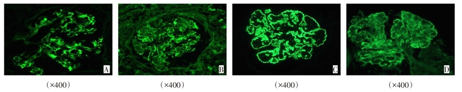
图2 PGNMID患者肾活检病理免疫荧光改变 A、B:IgG2(+++)、Lambda(++)呈弥漫性、团块状沉积于毛细血管襻及系膜区;C、D:IgG3(+++)、Kappa(+++)呈弥漫性、花瓣状沉积于毛细血管襻及系膜区。
Fig.2 Pathological immunofluorescence changes in renal biopsy of PGNMID patients
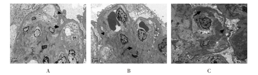
图3 PGNMID患者肾活检病理电镜下改变 A:肾小球基底膜节段性增厚(×5 000);B:足突弥漫融合(>95%),可见系膜插入(×5 000);C:内皮下、系膜区、基底膜内可见电子致密物沉积(×6 000)。
Fig.3 Pathological changes under electron microscopy in renal biopsies of PGNMID patients
| 病例 | 治疗方案 | 不良反应 | 开始缓解时间 | 部分缓解时间 | 完全缓解时间 | 随访 时间 | 目前情况 |
|---|---|---|---|---|---|---|---|
| 1 | 厄贝沙坦300 mg、达格列净10 mg每日1次口服+利妥昔单抗(第0、2周,6个月分别静脉滴注1 g)+甲泼尼龙片晨起20 mg口服,规律减量,6个月后停药+环磷酰胺每月0.8 g静脉滴注,维持1年 | 中性粒细胞、淋巴细胞轻度 下降 | 第1个月 | 第3个月 | 第6个月 | 12个月 | 24 h UTP下降至0.2 g/d以内,SCr及镜下血尿恢复正常;厄贝沙坦+达格列净口服维持治疗 |
| 2 | 沙库巴曲缬沙坦100 mg每日2次口服+硼替佐米1.3 mg/m2 第1、8、15、22天静脉滴注+环磷酰胺300 mg/m2 第1、8、15天静脉滴注+地塞米松40 mg第1~2、8~9、15~16、22~23天口服 | 无 | 第1个月 | 第2个月 | 第7个月 | 10个月 | 24 h UTP维持在0.15 g/d左右,SCr及镜下血尿恢复正常,沙库巴曲缬沙坦口服维持治疗 |
| 3 | 利妥昔单抗第0、2周分别静脉滴注1 g+环磷酰胺第0、2周分别静脉滴注0.4 g,此后每月单用环磷酰胺0.6 g静脉滴注,目前仍在维持治疗中 | 无 | 第1个月 | 第6个月 | 无 | 6个月 | 24 h UTP下降至1 g/d左右,SCr维持在200 μmol/L左右,厄贝沙坦口服维持治疗 |
表3 3例PGNMID患者治疗方案及转归
Tab.3 Treatment plans and outcomes of 3 patients with PGNMID
| 病例 | 治疗方案 | 不良反应 | 开始缓解时间 | 部分缓解时间 | 完全缓解时间 | 随访 时间 | 目前情况 |
|---|---|---|---|---|---|---|---|
| 1 | 厄贝沙坦300 mg、达格列净10 mg每日1次口服+利妥昔单抗(第0、2周,6个月分别静脉滴注1 g)+甲泼尼龙片晨起20 mg口服,规律减量,6个月后停药+环磷酰胺每月0.8 g静脉滴注,维持1年 | 中性粒细胞、淋巴细胞轻度 下降 | 第1个月 | 第3个月 | 第6个月 | 12个月 | 24 h UTP下降至0.2 g/d以内,SCr及镜下血尿恢复正常;厄贝沙坦+达格列净口服维持治疗 |
| 2 | 沙库巴曲缬沙坦100 mg每日2次口服+硼替佐米1.3 mg/m2 第1、8、15、22天静脉滴注+环磷酰胺300 mg/m2 第1、8、15天静脉滴注+地塞米松40 mg第1~2、8~9、15~16、22~23天口服 | 无 | 第1个月 | 第2个月 | 第7个月 | 10个月 | 24 h UTP维持在0.15 g/d左右,SCr及镜下血尿恢复正常,沙库巴曲缬沙坦口服维持治疗 |
| 3 | 利妥昔单抗第0、2周分别静脉滴注1 g+环磷酰胺第0、2周分别静脉滴注0.4 g,此后每月单用环磷酰胺0.6 g静脉滴注,目前仍在维持治疗中 | 无 | 第1个月 | 第6个月 | 无 | 6个月 | 24 h UTP下降至1 g/d左右,SCr维持在200 μmol/L左右,厄贝沙坦口服维持治疗 |
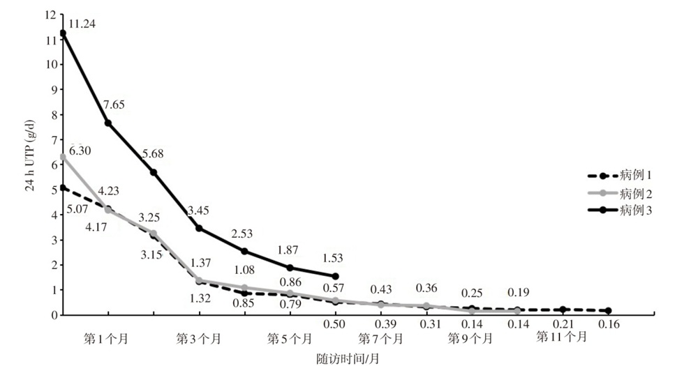
图4 3例PGNMID患者24 h UTP变化情况
Fig.4 Changes of 24 h UTP in three patients with PGNMID
| [1] | Li M, Xu G. An update of proliferative glomerulonephritis with monoclonal immunoglobulin deposits[J]. Clin Kidney J, 2022, 15(6):1041-1048. doi:10.1093/ckj/sfab269. |
| [2] | Gumber R, Cohen J B, Palmer M B, et al. A clone-directed approach may improve diagnosis and treatment of proliferative glomerulonephritis with monoclonal immunoglobulin deposits[J]. Kidney Int, 2018, 94(1):199-205. doi:10.1016/j.kint.2018.02.020. |
| [3] | Zhou H, Li M, Zeng C, et al. Efficacy of immunomodulatory drugs in combination with dexamethasone in proliferative glomerulonephritis with monoclonal immunoglobulin deposits[J]. Kidney Int Rep, 2022, 7(10):2166-2175. doi:10.1016/j.ekir.2022.07.009. |
| [4] | Bridoux F, Javaugue V, Nasr S H, et al. Proliferative glomerulonephritis with monoclonal immunoglobulin deposits:a nephrologist perspective[J]. Nephrol Dial Transplant, 2021, 36(2):208-215. doi:10.1093/ndt/gfz176. |
| [5] | Hou J, Haas M. Seeing the light(chains):a paradigm shift in PGNMID[J]. Kidney Int, 2025, 108(6):996-998. doi:10.1016/j.kint.2025.09.018. |
| [6] | Fourdinier O, Ulrich M, Karras A, et al. Glomerulonephritis with non-Randall-type,non-cryoglobulinaemic monoclonal immunoglobulin G deposits(PGNMID and ITG)[J]. Clin Kidney J, 2022, 15(9):1727-1736. doi:10.1093/ckj/sfac085. |
| [7] | Kousios A, Duncan N, Tam F W K, et al. Proliferative glomerulonephritis with monoclonal Ig deposits(PGNMID):diagnostic and treatment challenges for the nephrologist![J]. Kidney Int, 2019, 95(2):467-468. doi:10.1016/j.kint.2018.10.016. |
| [8] | Strati P, Nasr S H, Leung N, et al. Renal complications in chronic lymphocytic leukemia and monoclonal B-cell lymphocytosis:the Mayo Clinic experience[J]. Haematologica, 2015, 100(9):1180-1188. doi:10.3324/haematol.2015.128793. |
| [9] | Best Rocha A, Larsen C P. Membranous glomerulopathy with light chain-restricted deposits:A clinicopathological analysis of 28 cases[J]. Kidney Int Rep, 2017, 2(6):1141-1148. doi:10.1016/j.ekir.2017.07.008. |
| [10] | Liu M, Yu X, Wang S, et al. Proliferative glomerulonephritis with monoclonal immunoglobulin deposits:an entity associated with distinct diseases and comparison between IgG1 and IgG3 subtypes[J]. J Nephrol, 2022, 35(9):2363-2372. doi:10.1007/s40620-022-01317-w. |
| [11] | Aucouturier P, D'Agati V D, Ronco P. A fresh perspective on monoclonal gammopathies of renal significance[J]. Kidney Int Rep, 2021, 6(8):2059-2065. doi:10.1016/j.ekir.2021.04.026. |
| [12] | Bu L, Valeri A M, Said S M, et al. IgM variant of proliferative glomerulonephritis with monoclonal immunoglobulin deposits:A case series[J]. Am J Kidney Dis, 2025, 85(6):716-726.e1. doi:10.1053/j.ajkd.2025.01.009. |
| [13] | Javaugue V, Pascal V, Nasr S H, et al. Revisiting proliferative glomerulonephritis with monoclonal immunoglobulin deposits through immunoglobulin repertoire sequencing[J]. Kidney Int, 2025, 108(6):1146-1157. doi:10.1016/j.kint.2025.07.039. |
| [14] | Gowda K K, Nada R, Ramachandran R, et al. Proliferative glomerulonephritis with monoclonal immunoglobulin deposition disease:The utility of routine staining with immunoglobulin light chains[J]. Indian J Nephrol, 2015, 25(6):344-348. doi:10.4103/0971-4065.151354. |
| [15] | Xing G, Gillespie R, Bedri B, et al. Proliferative glomerulonephritis with monoclonal IgG deposits in children and young adults[J]. Pediatr Nephrol, 2018, 33(9):1531-1538. doi:10.1007/s00467-018-3949-8. |
| [16] | Gozzetti A, Guarnieri A, Zamagni E, et al. Monoclonal gammopathy of renal significance(MGRS):Real-world data on outcomes and prognostic factors[J]. Am J Hematol, 2022, 97(7):877-884. doi:10.1002/ajh.26566. |
| [17] | Santana De Roberts R, Batal I, Aljareh A, et al. Proliferative glomerulonephritis with monoclonal immunoglobulin deposits associated with parvovirus B19[J]. BMJ Case Rep, 2021, 14(6):e243061. doi:10.1136/bcr-2021-243061. |
| [18] | Nasr S H, Javaugue V. Insights into proliferative glomerulonephritis with monoclonal immunoglobulin deposits-is it really monoclonal or not?[J]. Curr Opin Nephrol Hypertens, 2025, 34(3):199-205. doi:10.1097/MNH.0000000000001061. |
| [19] | Amaador K, Peeters H, Minnema M C, et al. Monoclonal gammopathy of renal significance(MGRS)histopathologic classification, diagnostic workup, and therapeutic options[J]. Neth J Med, 2019, 77(7):243-254. |
| [20] | Zand L, Rajkumar S V, Leung N, et al. Safety and efficacy of daratumumab in patients with proliferative GN with monoclonal immunoglobulin deposits[J]. J Am Soc Nephrol, 2021, 32(5):1163-1173. doi:10.1681/ASN.2020101541. |
| [21] | Ji Q, Yu S, Zhou X, et al. Proliferative glomerulonephritis with monoclonal immunoglobulin deposits(PGNMID):three case reports and systematic review[J]. Front Med(Lausanne), 2025, 12:1637435. doi:10.3389/fmed.2025.1637435. |
| [22] | Bhutani G, Nasr S H, Said S M, et al. Hematologic characteristics of proliferative glomerulonephritides with nonorganized monoclonal immunoglobulin deposits[J]. Mayo Clin Proc, 2015, 90(5):587-596. doi:10.1016/j.mayocp.2015.01.024. |
| [23] | Fermand J P, Bridoux F, Kyle R A, et al. How I treat monoclonal gammopathy of renal significance(MGRS)[J]. Blood, 2013, 122(22):3583-3590. doi:10.1182/blood-2013-05-495929. |
| [24] | Lin L, Chen N. A Review on the diagnosis and treatment of proliferative glomerulonephritis with monoclonal immunoglobulin deposits[J]. Int J Gen Med, 2022, 15:8577-8582. doi:10.2147/IJGM.S386733. |
| [25] | Oki R, Unagami K, Taneda S, et al. Treatment with bortezomib for recurrent proliferative glomerulonephritis with monoclonal IgG deposits in kidney allograft. Case report and review of the literature[J]. J Nephrol, 2022, 35(4):1289-1293. doi:10.1007/s40620-022-01332-x. |
| [26] | Liu M Y, Yu X J, Wang S X, et al. Characteristics of complement protein deposition in proliferative glomerulonephritis with monoclonal immunoglobulin deposition[J]. Clin J Am Soc Nephrol, 2023, 18(12):1573-1582. doi:10.2215/CJN.0000000000000295. |
| [27] | Guiard E, Karras A, Plaisier E, et al. Patterns of noncryoglobulinemic glomerulonephritis with monoclonal Ig deposits:correlation with IgG subclass and response to rituximab[J]. Clin J Am Soc Nephrol, 2011, 6(7):1609-1616. doi:10.2215/CJN.10611110. |
| [1] | 苏雅, 张东伟, 宇明慧, 王春花, 吴月月. 复方苦参注射液联合靶向和免疫治疗对晚期肝癌的疗效观察[J]. 天津医药, 2026, 54(3): 324-328. |
| [2] | 李良, 张沁丽, 张亚倩, 庞瑾, 俞丽凝. 以卒中样症状为首发表现的抗谷氨酸脱羧酶抗体相关脑炎1例报告[J]. 天津医药, 2026, 54(10): 1104-1109. |
| [3] | 李荣琪, 李宇轩, 张燕, 李昕, 魏蔚. 血清干扰素调节因子1在类风湿关节炎中的临床意义[J]. 天津医药, 2026, 54(1): 84-87. |
| [4] | 李硕, 张云鹏, 黄艳, 曹美然, 贾兰芳, 胡桂才, 黄兰, 段书众. 系统性免疫炎症指数与IgA肾病患者临床病理特征的关系[J]. 天津医药, 2025, 53(9): 932-936. |
| [5] | 王婷, 王丹, 李瑛. 血清自身抗体变化对不明原因复发性流产患者妊娠结局的影响及预测模型构建[J]. 天津医药, 2025, 53(8): 831-835. |
| [6] | 王鑫, 赵晓静, 党琳, 门剑龙. 1例肝素诱导的血小板减少症患者多学科诊疗分析[J]. 天津医药, 2025, 53(2): 194-198. |
| [7] | 张超, 周天天, 李思雯, 汤为君, 胡伟军. 陕西省1~59岁人群白喉血清IgG抗体水平流行病学调查[J]. 天津医药, 2025, 53(10): 1081-1086. |
| [8] | 黄虹, 李恒, 范开元, 韦丽, 丁莉, 贾俊亚, 闫铁昆, 李栋. 体质量指数与特发性膜性肾病临床病理特征的关系[J]. 天津医药, 2024, 52(8): 815-819. |
| [9] | 白梦刻, 杨晓青, 李航, 王龙, 马继伟. 过敏性紫癜肾炎伴新月体形成患儿的实验室指标和病理特征分析[J]. 天津医药, 2024, 52(8): 820-824. |
| [10] | 尹韦玉, 廖红丽, 刘媛. SS相关抗体在特发性炎症性肌病及其相关间质性肺炎中的临床意义[J]. 天津医药, 2024, 52(7): 695-700. |
| [11] | 罗宴冉, 史晓飞, 韩磊, 张贝, 文路遥. 皮肌炎自身抗体在间质性肺病中的研究进展[J]. 天津医药, 2024, 52(7): 704-708. |
| [12] | 刘小艳, 卜锐, 陆健斐, 丁昱, 张幸. 术前Sonazoid超声造影评估肝细胞癌病理分化程度的价值[J]. 天津医药, 2024, 52(6): 658-662. |
| [13] | 王众, 赵静雯, 王天驰, 唐缨. 超声影像组学在肾炎组织学分型中的应用价值[J]. 天津医药, 2024, 52(10): 1100-1105. |
| [14] | 高歌, 张鑫越, 董奕君, 程根阳. 单纯膜性肾病与膜性肾病合并意义未明单克隆免疫球蛋白血症的差异性探讨[J]. 天津医药, 2023, 51(8): 878-882. |
| [15] | 曹越琦, 薛超. STAT4 rs7574865单核苷酸多态性与ANCA相关性血管炎的关系[J]. 天津医药, 2023, 51(1): 50-53. |
| 阅读次数 | ||||||
|
全文 |
|
|||||
|
摘要 |
|
|||||
