Tianjin Medical Journal ›› 2024, Vol. 52 ›› Issue (11): 1226-1232.doi: 10.11958/20240842
• Review • Previous Articles
LIU Song1( ), LI Wenbin1, SHAO Guo2, ZHANG Chunyang2, FENG Shijun2,△()
Received:2024-06-27
Revised:2024-08-10
Published:2024-11-15
Online:2024-11-12
Contact:
△E-mail:fsj18047211139@126.com
LIU Song, LI Wenbin, SHAO Guo, ZHANG Chunyang, FENG Shijun. Research progress on the mechanism of dura mater in the growth and development of skull/meninges/brain tissue system[J]. Tianjin Medical Journal, 2024, 52(11): 1226-1232.
CLC Number:
| 细胞因子 | 调控方式 | 影响方式 |
|---|---|---|
| FOXC1 | 激活/基因突变 | 硬脑膜发育的关键调控因子,FOXC1及其突变体的表达水平对顶部的硬脑膜具有高度的调控特异性[ |
| FOXC1ch/ch | 基因突变 | FOXC1的突变体,硬脑膜中FOXC1ch/ch导致脑膜间充质干细胞(MSCs)在细胞排布上异常紧密的相互贴合,致使硬脑膜发育缺陷[ |
| FGF | 激活/分化 | 形成神经胶质限制蛋白的基底膜(BM)蛋白的主要来源,直接调节骨化中心的形成[ |
| FAK | 机械力负荷 | FAK主要参与颅骨的发育过程,与维持颅腔和大气压压力差值密切相关,在维持硬脑膜结构完整性上起关键作用[ |
| BMP | 激活/去磷酸化/ 基因突变 | 与软脑膜发育关系密切,核心核因子Smad4调控皮质层的形成,参与皮质神经元的迁移[ |
| Wnt/β-catenin | 激活/敲除 | 当直接激活或移除β-catenin时,Wnt/β-catenin通路会导致外侧的脑膜发育不良与颅骨缺损[ |
| Twist1 | 基因缺失 | 维持硬脑膜与颅骨缝线处细胞保持低分化活性[ |
| TGF-β | 基因缺失 | TGF-β/Smad2/3信号转导通路与脑膜形成与发育密切相关,当胚胎中缺失关键调节因子TGF-β2,前脑脑膜和硬脑膜外膜不能发育[ |
| Runx2 | 激活/异二聚化 | 诱导成骨细胞增殖、成骨前细胞向成骨后分化[ |
| RA | 激活 | 参与皮质神经和脑血管发育[ |
Tab.1 The influence of cytokines on dural development
| 细胞因子 | 调控方式 | 影响方式 |
|---|---|---|
| FOXC1 | 激活/基因突变 | 硬脑膜发育的关键调控因子,FOXC1及其突变体的表达水平对顶部的硬脑膜具有高度的调控特异性[ |
| FOXC1ch/ch | 基因突变 | FOXC1的突变体,硬脑膜中FOXC1ch/ch导致脑膜间充质干细胞(MSCs)在细胞排布上异常紧密的相互贴合,致使硬脑膜发育缺陷[ |
| FGF | 激活/分化 | 形成神经胶质限制蛋白的基底膜(BM)蛋白的主要来源,直接调节骨化中心的形成[ |
| FAK | 机械力负荷 | FAK主要参与颅骨的发育过程,与维持颅腔和大气压压力差值密切相关,在维持硬脑膜结构完整性上起关键作用[ |
| BMP | 激活/去磷酸化/ 基因突变 | 与软脑膜发育关系密切,核心核因子Smad4调控皮质层的形成,参与皮质神经元的迁移[ |
| Wnt/β-catenin | 激活/敲除 | 当直接激活或移除β-catenin时,Wnt/β-catenin通路会导致外侧的脑膜发育不良与颅骨缺损[ |
| Twist1 | 基因缺失 | 维持硬脑膜与颅骨缝线处细胞保持低分化活性[ |
| TGF-β | 基因缺失 | TGF-β/Smad2/3信号转导通路与脑膜形成与发育密切相关,当胚胎中缺失关键调节因子TGF-β2,前脑脑膜和硬脑膜外膜不能发育[ |
| Runx2 | 激活/异二聚化 | 诱导成骨细胞增殖、成骨前细胞向成骨后分化[ |
| RA | 激活 | 参与皮质神经和脑血管发育[ |
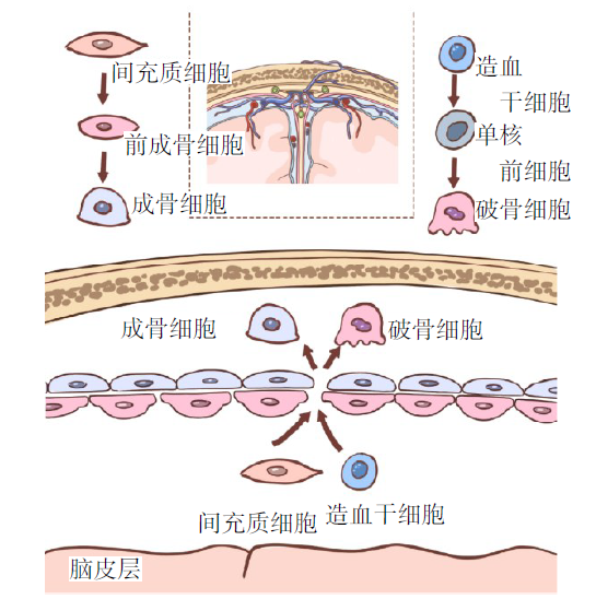
Fig.1 Schematic diagram of cell migration at cranial suture
| 组织/细胞 | 作用 时期 | 调控方式与成骨特点 |
|---|---|---|
| 硬脑膜 | 3个时期 | 颅骨发育起始位,调节颅骨发育的转录因子,骨基质蛋白和成骨细胞的生态位[ |
| SOM | 胚胎期 | 颅骨形成位点,增殖分化OPC[ |
| OPC | 胚胎期 | 成骨细胞增殖分化的祖细胞[ |
| 中胚层细胞 | 胚胎期 | 以膜内骨化的方式促进颅骨穹窿的发育和顶端扩张,分化顶骨和枕骨[ |
| 神经嵴细胞 | 胚胎期 | 以软骨内成骨的方式促进颅底及附近颅面骨的发育,以膜内骨化促进额骨发育[ |
Tab.2 The regulation of dural cells involved in cranial development
| 组织/细胞 | 作用 时期 | 调控方式与成骨特点 |
|---|---|---|
| 硬脑膜 | 3个时期 | 颅骨发育起始位,调节颅骨发育的转录因子,骨基质蛋白和成骨细胞的生态位[ |
| SOM | 胚胎期 | 颅骨形成位点,增殖分化OPC[ |
| OPC | 胚胎期 | 成骨细胞增殖分化的祖细胞[ |
| 中胚层细胞 | 胚胎期 | 以膜内骨化的方式促进颅骨穹窿的发育和顶端扩张,分化顶骨和枕骨[ |
| 神经嵴细胞 | 胚胎期 | 以软骨内成骨的方式促进颅底及附近颅面骨的发育,以膜内骨化促进额骨发育[ |
| 基因 | 作用时期 | 调控方式与成骨特点 |
|---|---|---|
| FOXC1 | 胚胎期 | FOXC1与Runx2增强子及其两个启动子结合直接促进 Runx2 表达,促进成骨分化[ |
| RA | 胚胎期 | 高水平的全反式维甲酸RA拮抗Wnt信号,抑制OPC形成前成骨细胞[ |
| GATA4 | 婴儿期与 青春期前 | 与FOXC1相互作用调节Runx2、OCN等成骨基因表达,FOXC1增强子可上调表达量;维持成年骨表型,敲除成骨细胞中的GATA4会导致骨小梁丢失[ |
| RAB23 | 胚胎期 | FGF和经典刺狸蛋白(Hh)/GLI1信号转导通路的上游负调节因子[ |
| BMP | 3个时期 | 亚型BMP-2促进矿化结节的形成和成骨分化;亚型BMP-4促进骨骼发育、重建和骨折愈合;亚型BMP-5促进骨折和软组织愈合;亚型BMP-7促进软骨形成;亚型BMP-9诱导成骨分化能力最强[ |
| FGF | 3个时期 | FGF-2通过丝裂原活化蛋白激酶(MAPK)通路激活和磷酸化股骨核心结合因子-a1(Cbfa1)/ Runx2,降低Wnt通路轴抑制蛋白2(axis inhibition protein 2,AXIN2)的活性;FGF9驱动软骨内骨化,基因突变后骨小梁形成增加和骨吸收减少,导致后额叶颅缝早闭[ |
| Wnt通路 | 3个时期 | 经典的Wnt通路主要控制细胞增殖,而非经典的Wnt通路调节细胞极性和介导神经嵴细胞与近轴中胚层细胞迁移;促进OPC增殖分化成骨基因[ |
| Prx1 | 婴儿期 | 在MSCs中表达,参与颅缝处骨生成与颅面骨生成[ |
| Runx2 | 3个时期 | 诱导脑膜缝线MSCs的增殖、成骨细胞分化和骨形成的主要因子[ |
| Twist1 | 3个时期 | Twist1-E-蛋白异二聚体激活中胚层和神经嵴细胞的分化,Twist1同型二聚体将干细胞维持在祖细胞状态,并阻断进入内胚层谱系[ |
| Gli1 | 婴儿期与青春 期前和损伤后 | 参与产生成人颅面骨,骨损伤后被激活,表达量减少引起颅缝早闭与颅骨生长停滞[ |
| FAK | 婴儿期与 青春期前 | 通过PI3K通路、MAPK通路和ERK通路调节成骨基因表达;增强Wnt / β-catenin信号通路,促进成骨细胞、祖细胞增殖和分化[ |
| Sp7 | 婴儿期与 青春期前 | 调节颅面骨增殖、分化和活性;促进骨矿化[ |
| COL1A1 | 青春期前 | 骨基质蛋白基因,参与骨量和骨脆性的调节,COL1A1通过FGFR2上调破骨细胞活性,导致小梁骨质丢失[ |
| OCN | 婴儿期与 青春期前 | 调节骨基质中的磷灰石晶体平行胶原纤维排列,增加新生骨强度[ |
| PSC | 正常发育和 损伤后 | 颅骨正常发育和颅骨损伤后分化成骨细胞;表达量下降会导致颅骨发育不良和矿化不足,以及颅缝线扩大[ |
Tab.3 The regulation of dural genes involved in cranial development
| 基因 | 作用时期 | 调控方式与成骨特点 |
|---|---|---|
| FOXC1 | 胚胎期 | FOXC1与Runx2增强子及其两个启动子结合直接促进 Runx2 表达,促进成骨分化[ |
| RA | 胚胎期 | 高水平的全反式维甲酸RA拮抗Wnt信号,抑制OPC形成前成骨细胞[ |
| GATA4 | 婴儿期与 青春期前 | 与FOXC1相互作用调节Runx2、OCN等成骨基因表达,FOXC1增强子可上调表达量;维持成年骨表型,敲除成骨细胞中的GATA4会导致骨小梁丢失[ |
| RAB23 | 胚胎期 | FGF和经典刺狸蛋白(Hh)/GLI1信号转导通路的上游负调节因子[ |
| BMP | 3个时期 | 亚型BMP-2促进矿化结节的形成和成骨分化;亚型BMP-4促进骨骼发育、重建和骨折愈合;亚型BMP-5促进骨折和软组织愈合;亚型BMP-7促进软骨形成;亚型BMP-9诱导成骨分化能力最强[ |
| FGF | 3个时期 | FGF-2通过丝裂原活化蛋白激酶(MAPK)通路激活和磷酸化股骨核心结合因子-a1(Cbfa1)/ Runx2,降低Wnt通路轴抑制蛋白2(axis inhibition protein 2,AXIN2)的活性;FGF9驱动软骨内骨化,基因突变后骨小梁形成增加和骨吸收减少,导致后额叶颅缝早闭[ |
| Wnt通路 | 3个时期 | 经典的Wnt通路主要控制细胞增殖,而非经典的Wnt通路调节细胞极性和介导神经嵴细胞与近轴中胚层细胞迁移;促进OPC增殖分化成骨基因[ |
| Prx1 | 婴儿期 | 在MSCs中表达,参与颅缝处骨生成与颅面骨生成[ |
| Runx2 | 3个时期 | 诱导脑膜缝线MSCs的增殖、成骨细胞分化和骨形成的主要因子[ |
| Twist1 | 3个时期 | Twist1-E-蛋白异二聚体激活中胚层和神经嵴细胞的分化,Twist1同型二聚体将干细胞维持在祖细胞状态,并阻断进入内胚层谱系[ |
| Gli1 | 婴儿期与青春 期前和损伤后 | 参与产生成人颅面骨,骨损伤后被激活,表达量减少引起颅缝早闭与颅骨生长停滞[ |
| FAK | 婴儿期与 青春期前 | 通过PI3K通路、MAPK通路和ERK通路调节成骨基因表达;增强Wnt / β-catenin信号通路,促进成骨细胞、祖细胞增殖和分化[ |
| Sp7 | 婴儿期与 青春期前 | 调节颅面骨增殖、分化和活性;促进骨矿化[ |
| COL1A1 | 青春期前 | 骨基质蛋白基因,参与骨量和骨脆性的调节,COL1A1通过FGFR2上调破骨细胞活性,导致小梁骨质丢失[ |
| OCN | 婴儿期与 青春期前 | 调节骨基质中的磷灰石晶体平行胶原纤维排列,增加新生骨强度[ |
| PSC | 正常发育和 损伤后 | 颅骨正常发育和颅骨损伤后分化成骨细胞;表达量下降会导致颅骨发育不良和矿化不足,以及颅缝线扩大[ |
| [1] | ZHAO F N, ZHU J L, DONG X H, et al. The influence of extracellular vesicles secreted by dural cells on osteoblasts[J]. Mol Biotechnol, 2023. doi:10.1007/s12033-023-00974-x.[Onlineaheadofprint]. |
| [2] | KO F C, SUMNER D R. How faithfully does intramembranous bone regeneration recapitulate embryonic skeletal development?[J]. Dev Dyn, 2021, 250(3):377-392. doi:10.1002/dvdy.240. |
| [3] | YAPIJAKIS C, PACHIS N, SOTIRIADOU T, et al. Molecular mechanisms involved in craniosynostosis[J]. In Vivo, 2023, 37(1):36-46. doi:10.21873/invivo.13052. |
| [4] | DESISTO J, O'ROURKE R, JONES H E, et al. Single-cell transcriptomic analyses of the developing meninges reveal meningeal fibroblast diversity and function[J]. Dev Cell, 2020, 54(1):43-59.e4. doi:10.1016/j.devcel.2020.06.009. |
| [5] | SMYTH L C D, XU D, OKAR S V, et al. Identification of direct connections between the dura and the brain[J]. Nature, 2024, 627(8002):165-173. doi:10.1038/s41586-023-06993-7. |
| [6] | AHN J H, CHO H, KIM J H, et al. Meningeal lymphatic vessels at the skull base brain cerebrospinal fluid[J]. Nature, 2019, 572(7767):62-66. doi:10.1038/s41586-019-1419-5. |
| [7] | LIAO J G, HUANG Y P, WANG Q, et al. Gene regulatory network from cranial neural crest cells to osteoblast differentiation and calvarial bone development[J]. Cell Mol Life Sci, 2022, 79(3):158. doi:10.1007/s00018-022-04208-2. |
| [8] | LI B, WANG Y G, FAN Y, et al. Cranial suture mesenchymal stem cells:insights and advances[J]. Biomolecules, 2021, 11(8):1129. doi:10.3390/biom11081129. |
| [9] | PATRICK E E, FLEETING C R, PATEL D R, et al. Corrigendum:modeling the volume of tissue activated in deep brain stimulation and its clinical influence:a review[J]. Front Hum Neurosci, 2024,18:1434402. doi:10.3389/fnhum.2024.1434402. |
| [10] | LOUVEAU A, SMIRNOV I, KEYES T J, et al. Structural and functional features of central nervous system lymphatic vessels[J]. Nature, 2015, 523(7560):337-341. doi:10.1038/nature14432. |
| [11] | AGARWAL N, LEWIS L D, HIRSCHLER L, et al. Current understanding of the anatomy,physiology,and magnetic resonance imaging of neurofluids:update from the 2022 "ISMRM imaging neurofluids study group" workshop in rome[J]. J Magn Reson Imaging, 2024, 59(2):431-449. doi:10.1002/jmri.28759. |
| [12] | AKKAYA B, SHEVACH E M. Regulatory T cells:master thieves of the immune system[J]. Cell Immunol, 2020,355:104160. doi:10.1016/j.cellimm.2020.104160. |
| [13] | LIU X H, GAO C, YUAN J Y, et al. Subdural haematomas drain into the extracranial lymphatic system through the meningeal lymphatic vessels[J]. Acta Neuropathol Commun, 2020, 8(1):16. doi:10.1186/s40478-020-0888-y. |
| [14] | DA MESQUITA S, PAPADOPOULOS Z, DYKSTRA T, et al. Meningeal lymphatics affect microglia responses and anti-Aβ immunotherapy[J]. Nature, 2021, 593(7858):255-260. doi:10.1038/s41586-021-03489-0. |
| [15] | GILLNäS S, GALLINI R, HE L, et al. Severe cerebellar malformations in mutant mice demonstrate a role for PDGF-C/PDGFRα signalling in cerebellar development[J]. Biol Open, 2022, 11(8):bio059431. doi:10.1242/bio.059431. |
| [16] | ZHANG P G M, FENG B, DAI G, et al. FOXC1 promotes osteoblastic differentiation of bone marrow mesenchymal stem cells via the Dnmt3b/CXCL12 Axis[J]. Biochem Genet, 2024, 62(1):176-192. doi:10.1007/s10528-023-10403-y. |
| [17] | ANG P S, MATRONGOLO M J, ZIETOWSKI M L, et al. Cranium growth,patterning and homeostasis[J]. Development, 2022, 149(22):dev201017. doi:10.1242/dev.201017. |
| [18] | SHROFF N P, XU P, KIM S, et al. Proliferation-driven mechanical compression induces signalling centre formation during mammalian organ development[J]. Nat Cell Biol, 2024, 26(4):519-529. doi:10.1038/s41556-024-01380-4. |
| [19] | HE W G, DENG Y X, KE K X, et al. Matricellular protein SMOC2 potentiates BMP9-Ⅰnduced osteogenic differentiation in mesenchymal stem cells through the enhancement of FAK/PI3K/AKT signaling[J]. Stem Cells Int, 2023,2023:5915988. doi:10.1155/2023/5915988. |
| [20] | ROWE C J, NWAOLU U, SALINAS D, et al. Inhibition of focal adhesion kinase 2 results in a macrophage polarization shift to M2 which attenuates local and systemic inflammation and reduces heterotopic ossification after polysystem extremity trauma[J]. Front Immunol, 2023,14:1280884. doi:10.3389/fimmu.2023.1280884. |
| [21] | ZHU L W, LIU Y Z, WANG A, et al. Application of BMP in bone tissue engineering[J]. Front Bioeng Biotechnol, 2022,10:810880. doi:10.3389/fbioe.2022.810880. |
| [22] | TIAN X, VATER C, RAINA D B, et al. Co-delivery of rhBMP-2 and zoledronic acid using calcium sulfate/hydroxyapatite carrier as a bioactive bone substitute to enhance and accelerate spinal fusion[J]. Bioact Mater, 2024, 36:256-271. doi:10.1016/j.bioactmat.2024.02.034. |
| [23] | BALL J R, SHELBY T, HERNANDEZ F, et al. Delivery of growth factors to enhance bone repair[J]. Bioengineering (Basel), 2023, 10(11):1252. doi:10.3390/bioengineering10111252. |
| [24] | CUNHA F B, POMINI K T, PLEPIS A, et al. In vivo biological behavior of polymer scaffolds of natural origin in the bone repair process[J]. Molecules, 2021, 26(6):1598. doi:10.3390/molecules26061598. |
| [25] | MOFFATT P, BORASCHI-DIAZ I, MARULANDA J, et al. Calvaria bone transcriptome in mouse models of osteogenesis imperfecta[J]. Int J Mol Sci, 2021, 22(10):5290. doi:10.3390/ijms22105290. |
| [26] | XIE B C, ZHOU H, LIU H Y, et al. Salidroside alleviates dexamethasone-induced inhibition of bone formation via transforming growth factor-beta/Smad2/3 signaling pathway[J]. Phytother Res, 2023, 37(5):1938-1950. doi:10.1002/ptr.7711. |
| [27] | CHEN H R, CUI Y J, ZHANG D M, et al. The role of fibroblast growth factor 8 in cartilage development and disease[J]. J Cell Mol Med, 2022, 26(4):990-999. doi:10.1111/jcmm.17174. |
| [28] | KOMORI T. Molecular mechanism of Runx2-dependent bone development[J]. Mol Cells, 2020, 43(2):168-175. doi:10.14348/molcells.2019.0244. |
| [29] | LI Y, JIE W, QI Y L, et al. Inhibition of RIPK1 alleviating vascular smooth muscle cells osteogenic transdifferentiation via Runx2[J]. iScience, 2024, 27(2):108766. doi:10.1016/j.isci.2023.108766. |
| [30] | COMO C N, KIM S, SIEGENTHALER J. Stuck on you:meninges cellular crosstalk in development[J]. Curr Opin Neurobiol, 2023,79:102676. doi:10.1016/j.conb.2023.102676. |
| [31] | CABRERA PEREIRA A, DASGUPTA K, HO T V, et al. Lineage-specific mutation of lmx1b provides new insights into distinct regulation of suture development in different areas of the calvaria[J]. Front Physiol, 2023,14:1225118. doi:10.3389/fphys.2023.1225118. |
| [32] | KOMORI T. Whole aspect of Runx2 functions in skeletal development[J]. Int J Mol Sci, 2022, 23(10):5776. doi:10.3390/ijms23105776. |
| [33] | SCHAEFFER S, IADECOLA C. Revisiting the neurovascular unit[J]. Nat Neurosci, 2021, 24(9):1198-1209. doi:10.1038/s41593-021-00904-7. |
| [34] | FURTADO L, FILHO J, FREITAS L S, et al. Anterior fontanelle closure and diagnosis of non-syndromic craniosynostosis:a comparative study using computed tomography[J]. J Pediatr (Rio J), 2022, 98(4):413-418. doi:10.1016/j.jped.2021.10.004. |
| [35] | FARMER D T, MLCOCHOVA H, ZHOU Y, et al. The developing mouse coronal suture at single-cell resolution[J]. Nat Commun, 2021, 12(1):4797. doi:10.1038/s41467-021-24917-9. |
| [36] | 寇正雄, 张海燕, 邵国, 等. fak/twist1信号通路在颅缝闭合过程中的作用机制研究[J]. 安徽医科大学学报, 2023, 58(1):60-66. |
| KOU Z X, ZHANG H Y, SHAO G, et al. The mechanism of FAK/Twist1 signal pathway in the closure of cranial suture[J]. Acta Universitatis Medicinalis Anhui, 2023, 58(1):60-66. doi:10.19405/j.cnki.issn1000-1492.2023.01.011. | |
| [37] | GUO J Q, YU S T, ZHANG H S, et al. Klf4 haploinsufficiency in Sp7+ lineage leads to underdeveloped mandibles and insufficient elongation of mandibular incisor[J]. Biochim Biophys Acta Mol Basis Dis, 2023, 1869(3):166636. doi:10.1016/j.bbadis.2022.166636. |
| [38] | EA C, HENNOCQ Q, PICARD A, et al. Growth charts in FGFR2- and FGFR3-related faciocraniosynostoses[J]. Bone Rep, 2022,16:101524. doi:10.1016/j.bonr.2022.101524. |
| [39] | MA L, CHANG Q, PEI F, et al. Skull progenitor cell-driven meningeal lymphatic restoration improves neurocognitive functions in craniosynostosis[J]. Cell Stem Cell, 2023, 30(11):1472-1485. doi:10.1016/j.stem.2023.09.012. |
| [40] | SONG C, LI T, ZHANG C, et al. RA-induced prominence-specific response resulted in distinctive regulation of wnt and osteogenesis[J]. Life Sci Alliance, 2023, 6(10):e202302013. doi:10.26508/lsa.202302013. |
| [41] | KHALID A B, PENCE J, SUTHON S, et al. GATA4 regulates mesenchymal stem cells via direct transcriptional regulation of the WNT signalosome[J]. Bone, 2021,144:115819. doi:10.1016/j.bone.2020.115819. |
| [42] | KOMORI T. What is the function of osteocalcin?[J]. J Oral Biosci, 2020, 62(3):223-227. doi:10.1016/j.job.2020.05.004. |
| [43] | HASAN M R, TAKATALO M, MA H, et al. RAB23 coordinates early osteogenesis by repressing FGF10-pERK1/2 and GLI1[J]. Elife, 2020,9:e55829. doi:10.7554/eLife.55829. |
| [44] | LEE S B, LEE H J, PARK J B. Bone morphogenetic protein-9 promotes osteogenic differentiation and mineralization in human stem-cell-derived spheroids[J]. Medicina (Kaunas), 2023, 59(7):1315. doi:10.3390/medicina59071315. |
| [45] | WANG H, QI L L, SHEMA C, et al. Advances in the role and mechanism of fibroblasts in fracture healing[J]. Front Endocrinol (Lausanne), 2024,15:1350958. doi:10.3389/fendo.2024.1350958. |
| [46] | LIU J Q, XIAO Q, XIAO J N, et al. Wnt/β-catenin signalling:function,biological mechanisms,and therapeutic opportunities[J]. Signal Transduct Target Ther, 2022, 7(1):3. doi:10.1038/s41392-021-00762-6. |
| [47] | ALDAWOOD Z A, MANCINELLI L, GENG X, et al. Expansion of the sagittal suture induces proliferation of skeletal stem cells and sustains endogenous calvarial bone regeneration[J]. Proc Natl Acad Sci U S A, 2023, 120(16):e2120826120. doi:10.1073/pnas.2120826120. |
| [48] | FAN X, WAARDENBERG A J, DEMUTH M, et al. TWIST1 homodimers and heterodimers orchestrate lineage-specific differentiation[J]. Mol Cell Biol, 2020, 40(11):e00663-00619. doi:10.1128/MCB.00663-19. |
| [49] | DI PIETRO L, BARBA M, PRAMPOLINI C, et al. GLI1 and AXIN2 are distinctive markers of human calvarial mesenchymal stromal cells in nonsyndromic craniosynostosis[J]. Int J Mol Sci, 2020, 21(12):4356. doi:10.3390/ijms21124356. |
| [50] | WU L D, LIU Z X, XIAO L, et al. The role of Gli1(+) mesenchymal stem cells in osteogenesis of craniofacial bone[J]. Biomolecules, 2023, 13(9):1351. doi:10.3390/biom13091351. |
| [51] | SHIN H R, KIM B S, KIM H J, et al. Excessive osteoclast activation by osteoblast paracrine factor RANKL is a major cause of the abnormal long bone phenotype in apert syndrome model mice[J]. J Cell Physiol, 2022, 237(4):2155-2168. doi:10.1002/jcp.30682. |
| [52] | DEBNATH S, YALLOWITZ A R, MCCORMICK J, et al. Discovery of a periosteal stem cell mediating intramembranous bone formation[J]. Nature, 2018, 562(7725):133-139. doi:10.1038/s41586-018-0554-8. |
| [53] | 张瑞欣, 董语迪, 肖建辉. lncRNA调控间充质干细胞向成骨细胞分化的研究进展[J]. 天津医药, 2021, 49(6):662-667. |
| ZHANG R X, DONG Y D, XIAO J H. Research progress on lncRNA regulation of mesenchymal stem cell differentiation into osteoblasts[J]. Tianjin Med J, 2021, 49(6):662-667. doi:10.11958/20203229. | |
| [54] | NESPOLI E, HAKANI M, HEIN T M, et al. Glial cells react to closed head injury in a distinct and spatiotemporally orchestrated manner[J]. Sci Rep, 2024, 14(1):2441. doi:10.1038/s41598-024-52337-4. |
| [55] | HU H X, ZHANG H, BU Z H, et al. Small extracellular vesicles released from bioglass/hydrogel scaffold promote vascularized bone regeneration by transferring miR-23a-3p[J]. Int J Nanomedicine, 2022, 17:6201-6220. doi:10.2147/ⅠJN.S389471. |
| [56] | SUN Y T, LI Y X, ZHANG Y, et al. A Polydopamine-assisted strontium-substituted apatite coating for titanium promotes osteogenesis and angiogenesis via FAK/MAPK and PI3K/AKT signaling pathways[J]. Mater Sci Eng C Mater Biol Appl, 2021,131:112482. doi:10.1016/j.msec.2021.112482. |
| [57] | 曹志威, 邵国, 张春阳. 硬脑膜对颅骨生长发育影响的研究现状[J]. 中华神经外科杂志, 2022, 38(5):537-540. |
| CAO Z W, SHAO G, ZHANG C Y. Research status of the influence of dura mater on skull growth and development[J]. Chinese Journal of Neurosurgery, 2022, 38(5):537-540. doi:10.3760/cma.j.cn112050-20210617-00290. | |
| [58] | PIBOUIN-FRAGNER L, EICHMANN A, PARDANAUD L. Environmental and intrinsic modulations of venous differentiation[J]. Cell Mol Life Sci, 2022, 79(9):491. doi:10.1007/s00018-022-04470-4. |
| [59] | DECIMO I, DOLCI S, PANUCCIO G, et al. Meninges:A widespread niche of neural progenitors for the brain[J]. Neuroscientist, 2021, 27(5):506-528. doi:10.1177/1073858420954826. |
| No related articles found! |
| Viewed | ||||||
|
Full text |
|
|||||
|
Abstract |
|
|||||