Tianjin Medical Journal ›› 2023, Vol. 51 ›› Issue (8): 869-872.doi: 10.11958/20221421
• Clinical Research • Previous Articles Next Articles
GUO Yufeng1( ), LIU Yumei1, BI Xiuzeng1, GUO Junlin1, SUN Li1, WANG Weixiu2, YANG Dingwei2
Received:2022-09-14
Revised:2023-02-06
Published:2023-08-15
Online:2023-08-10
GUO Yufeng, LIU Yumei, BI Xiuzeng, GUO Junlin, SUN Li, WANG Weixiu, YANG Dingwei. Correlation between macular retinal thickness and urinary protein in adult patients with nephrotic syndrome[J]. Tianjin Medical Journal, 2023, 51(8): 869-872.
CLC Number:
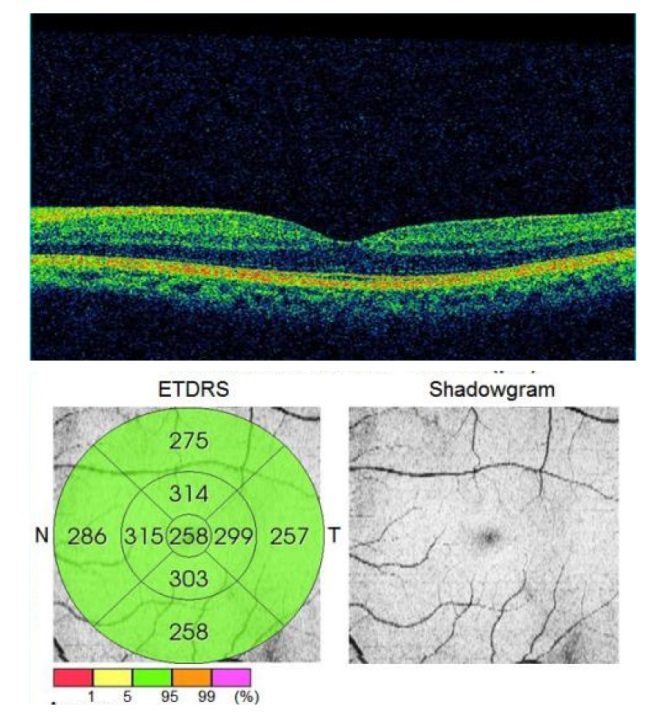
Fig.1 Retinal thickness in each division of macula regions
| 组别 | n | 男性/例(%) | 眼别(右/左)/例 | 年龄/岁 |
|---|---|---|---|---|
| A组 | 28 | 22(78.6) | 28/28 | 67.36±6.00 |
| B组 | 30 | 23(76.7) | 30/28 | 67.90±8.30 |
| C组 | 27 | 22(81.5) | 27/24 | 68.15±8.07 |
| D组 | 27 | 20(74.1) | 27/26 | 65.93±7.09 |
| χ2或F | 0.458 | 0.099 | 0.489 |
Tab.1 Comparison of clinical data between the four groups of patients
| 组别 | n | 男性/例(%) | 眼别(右/左)/例 | 年龄/岁 |
|---|---|---|---|---|
| A组 | 28 | 22(78.6) | 28/28 | 67.36±6.00 |
| B组 | 30 | 23(76.7) | 30/28 | 67.90±8.30 |
| C组 | 27 | 22(81.5) | 27/24 | 68.15±8.07 |
| D组 | 27 | 20(74.1) | 27/26 | 65.93±7.09 |
| χ2或F | 0.458 | 0.099 | 0.489 |
| 组别 | n | Center | InT | InS | |||||
|---|---|---|---|---|---|---|---|---|---|
| A组 | 56 | 223.02±17.05 | 280.09±17.09 | 292.93±16.34 | |||||
| B组 | 58 | 240.03±23.56a | 291.02±21.98a | 301.29±19.49 | |||||
| C组 | 51 | 227.00±30.21b | 275.53±31.49b | 285.04±23.33b | |||||
| D组 | 53 | 235.38±40.30a | 291.66±35.43ac | 298.75±30.71c | |||||
| F | 4.047* | 4.664* | 5.276* | ||||||
| 组别 | InN | InI | OutT | ||||||
| A组 | 292.79±17.85 | 285.95±19.77 | 250.41±14.27 | ||||||
| B组 | 304.24±18.73a | 298.57±22.20a | 252.47±19.71 | ||||||
| C组 | 286.94±28.18b | 280.53±30.86b | 245.41±25.32 | ||||||
| D组 | 300.09±28.12c | 293.91±33.98c | 250.11±33.81 | ||||||
| F | 5.775* | 4.792* | 0.811 | ||||||
| 组别 | OutS | OutI | OutN | ||||||
| A组 | 261.95±15.65 | 276.95±16.16 | 255.84±13.46 | ||||||
| B组 | 263.95±17.93 | 280.31±20.72 | 258.33±18.30 | ||||||
| C组 | 256.90±19.82 | 271.51±22.52 | 245.29±19.36ab | ||||||
| D组 | 263.19±29.54 | 279.79±28.52 | 251.15±33.58 | ||||||
| F | 1.169 | 1.722 | 3.555* | ||||||
Tab.2 Comparison of the mean retinal thickness in each division of the macular region between the four groups of patients
| 组别 | n | Center | InT | InS | |||||
|---|---|---|---|---|---|---|---|---|---|
| A组 | 56 | 223.02±17.05 | 280.09±17.09 | 292.93±16.34 | |||||
| B组 | 58 | 240.03±23.56a | 291.02±21.98a | 301.29±19.49 | |||||
| C组 | 51 | 227.00±30.21b | 275.53±31.49b | 285.04±23.33b | |||||
| D组 | 53 | 235.38±40.30a | 291.66±35.43ac | 298.75±30.71c | |||||
| F | 4.047* | 4.664* | 5.276* | ||||||
| 组别 | InN | InI | OutT | ||||||
| A组 | 292.79±17.85 | 285.95±19.77 | 250.41±14.27 | ||||||
| B组 | 304.24±18.73a | 298.57±22.20a | 252.47±19.71 | ||||||
| C组 | 286.94±28.18b | 280.53±30.86b | 245.41±25.32 | ||||||
| D组 | 300.09±28.12c | 293.91±33.98c | 250.11±33.81 | ||||||
| F | 5.775* | 4.792* | 0.811 | ||||||
| 组别 | OutS | OutI | OutN | ||||||
| A组 | 261.95±15.65 | 276.95±16.16 | 255.84±13.46 | ||||||
| B组 | 263.95±17.93 | 280.31±20.72 | 258.33±18.30 | ||||||
| C组 | 256.90±19.82 | 271.51±22.52 | 245.29±19.36ab | ||||||
| D组 | 263.19±29.54 | 279.79±28.52 | 251.15±33.58 | ||||||
| F | 1.169 | 1.722 | 3.555* | ||||||
| 指标 | Center× 分组 | InT× 分组 | InS× 分组 | InN× 分组 | InI× 分组 | OutN× 分组 |
|---|---|---|---|---|---|---|
| Eta | 0.232 | 0.248 | 0.262 | 0.274 | 0.251 | 0.218 |
| E2 | 0.054* | 0.061** | 0.069** | 0.075** | 0.063** | 0.047* |
Tab.3 Correlation between mean retinal thickness and urinary protein severity in macular area with differential zoning
| 指标 | Center× 分组 | InT× 分组 | InS× 分组 | InN× 分组 | InI× 分组 | OutN× 分组 |
|---|---|---|---|---|---|---|
| Eta | 0.232 | 0.248 | 0.262 | 0.274 | 0.251 | 0.218 |
| E2 | 0.054* | 0.061** | 0.069** | 0.075** | 0.063** | 0.047* |
| [1] | WONG C W, WONG T Y, CHENG C Y, et al. Kidney and eye diseases:common risk factors,etiological mechanisms,and pathways[J]. Kidney Int, 2014, 85(6):1290-1302. doi:10.1038/ki.2013.491. |
| [2] | FURSOVA A Z, DERBENEVA A S, VASILYEVA M A, et al. Development,clinical manifestations and diagnosis of retinal changes in chronic kidney disease[J]. Vestn Oftalmol, 2021, 137(1):107-114. doi:10.17116/oftalma2021137011107. |
| [3] | BILGE A D, YAYLALI S A, YAVUZ S, et al. Bilateral serous macular detachment in a patient with nephrotic syndrome[J]. Retin Cases Brief Rep, 2018, 12(3):260-262. doi:10.1097/ICB.0000000000000487. |
| [4] | HAGER A, WIEGAND W. Bilateral serous detachment of the neurosensory retina and retinal pigment epithelium with rip of the peripheral pigment epithelium[J]. Ophthalmologe, 2006, 103(11):966-970. doi:10.1007/s00347-006-1437-1. |
| [5] | IZZEDINE H, FARDEAU C, GAUTHIER M, et al. Bilateral serous retinal detachment as a presenting sign of nephrotic syndrome[J]. Intern Med, 2014, 53(22):2609-2613. doi:10.2169/internalmedicine.53.2720. |
| [6] | DE BENEDETTO U, PASTORE M R, BATTAGLIA PARODI M, et al. Retinal involvement in nephrotic syndrome secondary to minimal change disease[J]. Eur J Ophthalmol, 2012, 22(5):843-845. doi:10.5301/ejo.5000153. |
| [7] | GAMBATO T, FRANCESCUTTI L, LANZETTA P. Choroidal neovascularization in primary membranous nephropathy[J]. Am J Case Rep, 2020, 21:e923454. doi:10.12659/AJCR.923454. |
| [8] | FARRAH T E, DHILLON B, KEANE P A, et al. The eye,the kidney,and cardiovascular disease:old concepts,better tools,and new horizons[J]. Kidney Int, 2020, 98(2):323-342. doi:10.1016/j.kint.2020.01.039. |
| [9] | 梅长林. 肾病综合征[M]. 北京: 科学出版社, 2012:1-2. |
| MEI C L. Nephrotic Syndrome[M]. Beijing: Science Press, 2012:1-2. | |
| [10] | YAO T, HE Y, HUANG L, et al. Quantitative vessel density analysis of macular and peripapillary areas by optical coherence tomography angiography in adults with primary nephrotic syndrome[J]. Microvasc Res, 2022, 144:104407. doi:10.1016/j.mvr.2022.104407. |
| [11] | ZHANG W, ZHANG Y, KANG L, et al. Retinal and choroidal thickness in paediatric patients with hypoalbuminaemia caused by nephrotic syndrome[J]. BMC Ophthalmol, 2019, 19(1):44. doi:10.1186/s12886-019-1050-0. |
| [12] | 陶琳, 刘改灵, 邵凤民, 等. 老年肾病综合征患者临床特征及心血管事件发病率[J]. 中国老年学杂志, 2022, 42(11):2713-2715. |
| TAO L, LIU G L, SHAO F M, et al. Clinical characteristics and incidence of cardiovascular events in elderly patients with nephrotic syndrome[J]. Chinese Journal of Gerontology, 2022, 42(11):2713-2715. | |
| [13] | 刘玲玲. 老年肾病综合征的临床病理分析及不同治疗方法的疗效比较[D]. 承德: 承德医学院, 2021. |
| LIU L L. Clinicopathological analysis of elderly nephrotic syndrome and comparison of therapeutic effects under different treatment methods[D]. Chengde: Chengde Medical College, 2021. doi:10.27691/d.cnki.gcdyx.2021.000157. | |
| [14] | 杨光, 赵佳慧, 程庆砾. 老年肾病综合征的特点与诊治[J]. 中国临床保健杂志, 2020, 23(1):31-35. |
| YANG G, ZHAO J H, CHENG Q L. Characteristics and diagnosis of nephrotic syndrome in the elderly[J]. Chin J Clin Healthc, 2020, 23(1):31-35. | |
| [15] | UNGVARI Z, TARANTINI S, SOROND F, et al. Mechanisms of vascular aging,a geroscience perspective:JACC focus seminar[J]. J Am Coll Cardiol, 2020, 75(8):931-941. doi:10.1016/j.jacc.2019.11.061. |
| [16] | 滕娟, 余海跃, 陈志萍, 等. 2型糖尿病患者尿微量白蛋白含量与黄斑区视网膜微循环相关性分析[J]. 眼科新进展, 2020, 40(10):952-956. |
| TENG J, YU H Y, CHE Z P, et al. Correlation analysis between microalbumin changes and macular retinal microcirculation in type 2 diabetic patients[J]. Rec Adv Ophthal, 2020, 40(10):952-956. | |
| [17] | CANKURTARAN V, INANC M, TEKIN K, et al. Retinal microcirculation in predicting diabetic nephropathy in type 2 diabetic patients without retinopathy[J]. Ophthalmologica, 2020, 243(4):271-279. doi:10.1159/000504943. |
| [18] | OCHODNICKY P, HENNING R H, VAN DOKKUM R P, et al. Microalbuminuria and endothelial dysfunction:emerging targets for primary prevention of end-organ damage[J]. J Cardiovasc Pharmacol, 2006, 47(Suppl 2):S151-S176. doi:10.1097/00005344-200606001-00009. |
| [19] | DARUICH A, MATET A, MOULIN A, et al. Mechanisms of macular edema: beyond the surface[J]. Prog Retin Eye Res, 2018, 63:20-68. doi:10.1016/j.preteyeres.2017.10.006. |
| [20] | KHATRI A, PANDEY A, JOSHI K, et al. Redefining response in wet AMD to anti VEGF therapy based on non-OCTA versus OCTA evaluation[J]. Eur J Ophthalmol, 2021, 32(5):2719-2725. doi:10.1177/11206721211059349. |
| [21] | SCHOLL S, AUGUSTIN A, LOEWENSTEIN A, et al. General pathophysiology of macular edema[J]. Eur J Ophthalmol, 2011, 21(Suppl 6):S10-9. doi:10.5301/EJO.2010.6050. |
| Viewed | ||||||
|
Full text |
|
|||||
|
Abstract |
|
|||||