天津医药 ›› 2024, Vol. 52 ›› Issue (2): 142-147.doi: 10.11958/20230541
汪爱华( ), 张飞忠, 王红英
收稿日期:2023-04-18
修回日期:2023-06-25
出版日期:2024-02-15
发布日期:2024-01-26
作者简介:汪爱华(1978),女,主治医师,主要从事妇科疾病方面研究。E-mail:基金资助:
WANG Aihua(), ZHANG Feizhong, WANG Hongying
Received:2023-04-18
Revised:2023-06-25
Published:2024-02-15
Online:2024-01-26
汪爱华, 张飞忠, 王红英. 麝香酮调节SHH介导的自噬对卵巢癌细胞恶性进展的影响[J]. 天津医药, 2024, 52(2): 142-147.
WANG Aihua, ZHANG Feizhong, WANG Hongying. Impacts of muscone on malignant progression of ovarian cancer cells by regulating SHH mediated autophagy[J]. Tianjin Medical Journal, 2024, 52(2): 142-147.
摘要:
目的 探究麝香酮调节超音刺猬蛋白(SHH)介导的自噬对卵巢癌细胞恶性进展的影响。方法 检测0、2、4、8、16、24 μmol/L的麝香酮处理后的人卵巢癌细胞SKOV3存活率,筛选出麝香酮最佳细胞作用浓度。体外培养SKOV3细胞并构建其移植瘤小鼠模型,随机均分为对照组,麝香酮组,麝香酮+自噬抑制剂氯喹(CQ)组,麝香酮+空载组,麝香酮+SHH过表达组,按照分组分别以麝香酮、CQ、空载质粒及SHH过表达质粒处理后,检测各组移植瘤小鼠肿瘤体积和质量;分别以EdU染色、TUNEL染色、细胞划痕、Transwell侵袭实验、免疫印迹法、实时荧光定量PCR检测SKOV3细胞增殖、凋亡、迁移、侵袭、SKOV3细胞及其移植瘤小鼠肿瘤组织自噬相关蛋白(LC3Ⅱ/LC3Ⅰ、Beclin-1)与SHH表达。结果 与对照组相比,麝香酮组细胞增殖率、迁移率、侵袭数目、肿瘤体积和质量、肿瘤组织SHH mRNA及蛋白表达水平均降低(P<0.05),细胞凋亡率、细胞及肿瘤组织LC3Ⅱ/LC3Ⅰ和Beclin-1蛋白表达水平均升高(P<0.05)。与麝香酮组相比,麝香酮+CQ组和麝香酮+SHH过表达组细胞增殖率、迁移率、侵袭数目、肿瘤体积和质量升高(P<0.05),细胞凋亡率、细胞及肿瘤组织LC3Ⅱ/LC3Ⅰ和Beclin-1蛋白表达水平均降低(P<0.05);麝香酮+SHH过表达组细胞及肿瘤组织SHH mRNA及蛋白表达水平升高(P<0.05);麝香酮+空载组细胞各指标变化差异无统计学意义(P>0.05)。结论 麝香酮可通过下调SHH而促进卵巢癌细胞自噬,进而抑制其增殖、体内生长、迁移及侵袭,促进其凋亡,最终抑制其恶性进展。
中图分类号:
| 基因名称 | 引物序列(5′→3′) | 产物大小/bp |
|---|---|---|
| SHH | 上游:TGTCTGCTGCTAGTCCTCGTCTC 下游:GTGCCTCCTCTTCGAACCC | 265 |
| GAPDH | 上游:CAGGAGGCATTGCTGATGAT 下游:GAAGGCTGGGGCTCATTT | 178 |
表1 基因引物序列
Tab.1 Gene primer sequences
| 基因名称 | 引物序列(5′→3′) | 产物大小/bp |
|---|---|---|
| SHH | 上游:TGTCTGCTGCTAGTCCTCGTCTC 下游:GTGCCTCCTCTTCGAACCC | 265 |
| GAPDH | 上游:CAGGAGGCATTGCTGATGAT 下游:GAAGGCTGGGGCTCATTT | 178 |
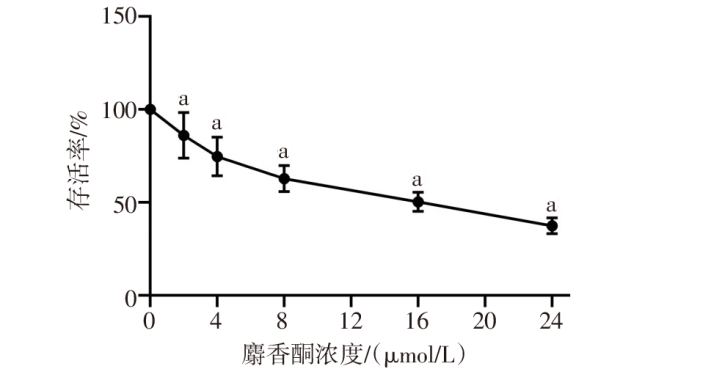
图1 不同浓度麝香酮对SKOV3细胞存活率的影响 F=83.820,n=6,P<0.05;a与0 μmol/L麝香酮比较,P<0.05。
Fig.1 Effects of different concentrations of muskone on survival rates of SKOV3 cells
| 组别 | 肿瘤体积/mm3 | 肿瘤质量/g |
|---|---|---|
| 对照组 | 914.36±78.10 | 0.81±0.08 |
| 麝香酮组 | 218.75±43.56a | 0.22±0.05a |
| 麝香酮+CQ组 | 780.84±92.81b | 0.65±0.09b |
| 麝香酮+空载组 | 240.13±51.73 | 0.29±0.06 |
| 麝香酮+SHH过表达组 | 847.82±85.64b | 0.74±0.07b |
| F | 219.700* | 142.100* |
表2 各组SKOV3移植瘤小鼠肿瘤体积和质量比较(n=10,$\bar{x}±s$)
Tab.2 Comparison of tumor volume and mass of SKOV3 transplanted tumor mice between five groups
| 组别 | 肿瘤体积/mm3 | 肿瘤质量/g |
|---|---|---|
| 对照组 | 914.36±78.10 | 0.81±0.08 |
| 麝香酮组 | 218.75±43.56a | 0.22±0.05a |
| 麝香酮+CQ组 | 780.84±92.81b | 0.65±0.09b |
| 麝香酮+空载组 | 240.13±51.73 | 0.29±0.06 |
| 麝香酮+SHH过表达组 | 847.82±85.64b | 0.74±0.07b |
| F | 219.700* | 142.100* |
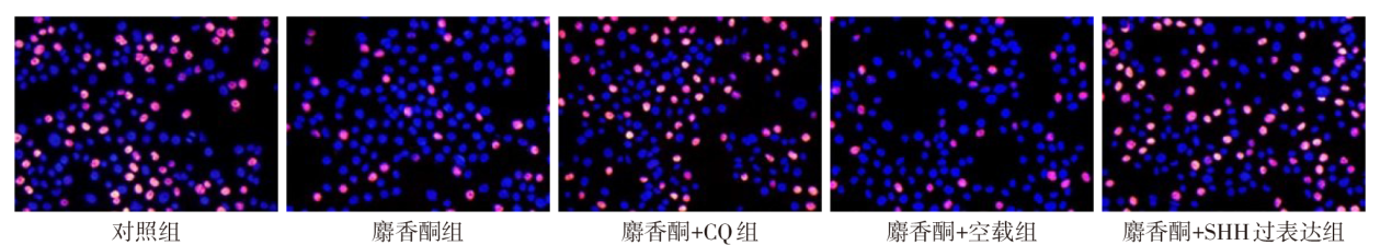
图2 EdU染色检测各组SKOV3细胞增殖(×200)
Fig.2 EdU staining for detecting SKOV3 cell proliferation in each group (×200)
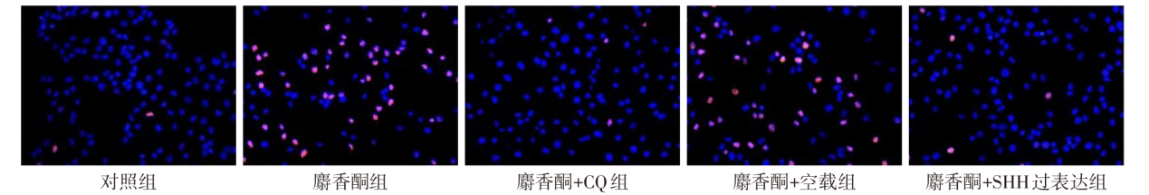
图3 TUNEL染色检测各组SKOV3细胞凋亡(×200)
Fig.3 TUNEL staining for detecting apoptosis of SKOV3 cells in each group (×200)
| 组别 | 增殖率 | 凋亡率 |
|---|---|---|
| 对照组 | 42.50±5.14 | 1.63±0.51 |
| 麝香酮组 | 11.76±3.42a | 62.45±10.26a |
| 麝香酮+CQ组 | 32.36±4.80b | 2.14±0.63b |
| 麝香酮+空载组 | 12.13±2.94 | 54.35±11.24 |
| 麝香酮+SHH过表达组 | 38.85±4.87b | 2.48±0.73b |
| F | 69.090* | 123.700* |
表3 各组SKOV3细胞增殖率和凋亡率比较(n=6,%,$\bar{x}±s$)
Tab.3 Comparison of the proliferation rate and apoptosis rate of SKOV3 cells between five groups
| 组别 | 增殖率 | 凋亡率 |
|---|---|---|
| 对照组 | 42.50±5.14 | 1.63±0.51 |
| 麝香酮组 | 11.76±3.42a | 62.45±10.26a |
| 麝香酮+CQ组 | 32.36±4.80b | 2.14±0.63b |
| 麝香酮+空载组 | 12.13±2.94 | 54.35±11.24 |
| 麝香酮+SHH过表达组 | 38.85±4.87b | 2.48±0.73b |
| F | 69.090* | 123.700* |
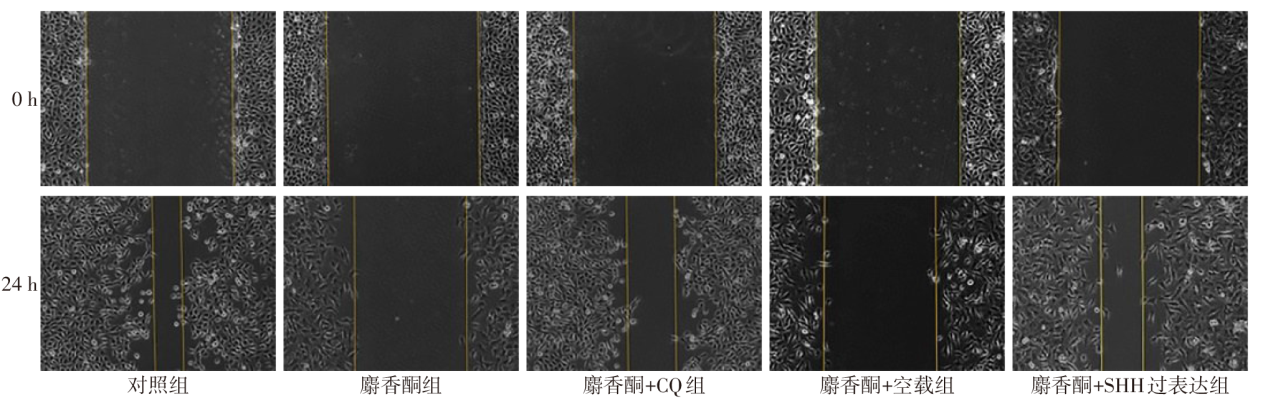
图4 细胞划痕实验检测各组SKOV3细胞迁移(×200)
Fig.4 Wounding healing assay for detecting SKOV3 cell migration in each group (×200)
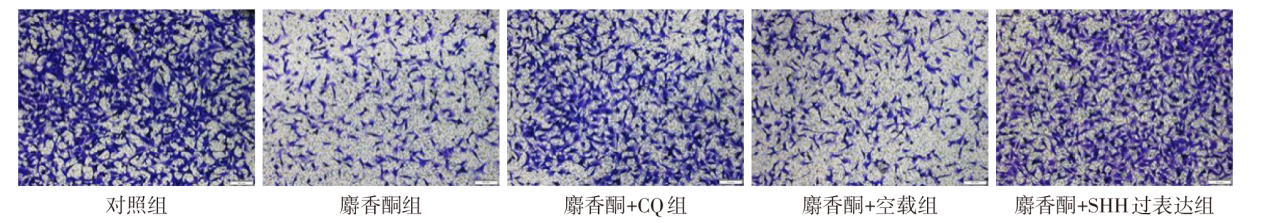
图5 Transwell侵袭实验检测各组SKOV3细胞侵袭(结晶紫染色,×200)
Fig.5 Transwell invasion assay for detecting invasion of SKOV3 cells in each group (crystal violet stain, ×200)
| 组别 | 迁移率/% | 侵袭数目/(个/视野) |
|---|---|---|
| 对照组 | 84.13±13.12 | 251.64±36.58 |
| 麝香酮组 | 19.79±3.51a | 85.42±12.23a |
| 麝香酮+CQ组 | 70.85±14.83b | 215.14±30.64b |
| 麝香酮+空载组 | 20.14±3.74 | 90.38±15.20 |
| 麝香酮+SHH过表达组 | 76.87±15.63b | 239.87±35.72b |
| F | 45.620* | 51.230* |
表4 各组SKOV3细胞迁移率与侵袭数目比较 (n=6,$\bar{x}±s$)
Tab.4 Comparison of SKOV3 cell migration rate and invasion number between five groups
| 组别 | 迁移率/% | 侵袭数目/(个/视野) |
|---|---|---|
| 对照组 | 84.13±13.12 | 251.64±36.58 |
| 麝香酮组 | 19.79±3.51a | 85.42±12.23a |
| 麝香酮+CQ组 | 70.85±14.83b | 215.14±30.64b |
| 麝香酮+空载组 | 20.14±3.74 | 90.38±15.20 |
| 麝香酮+SHH过表达组 | 76.87±15.63b | 239.87±35.72b |
| F | 45.620* | 51.230* |
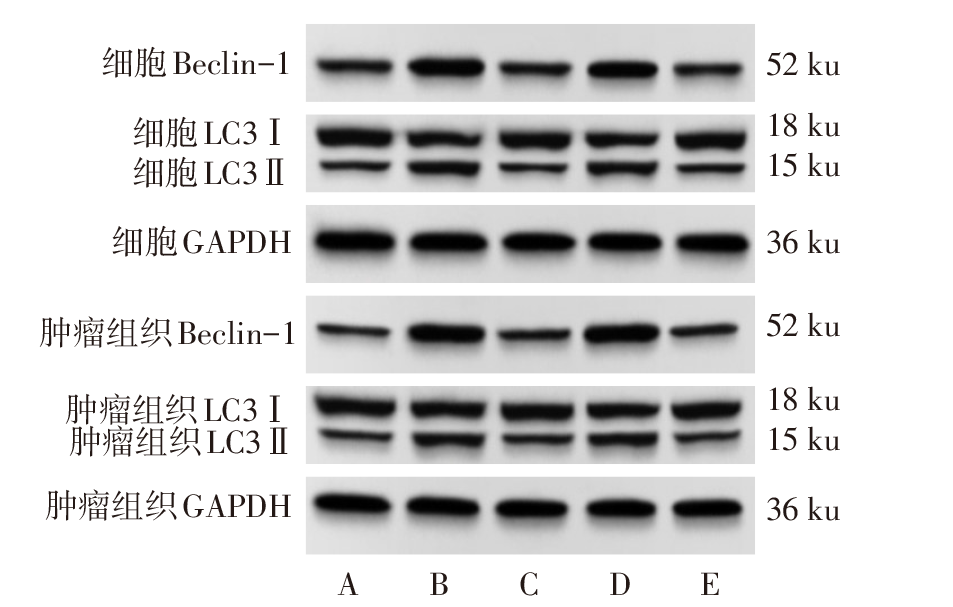
图6 免疫印迹检测各组SKOV3细胞及其移植瘤小鼠肿瘤组织自噬相关蛋白表达 A:对照组;B:麝香酮组;C:麝香酮+CQ组;D:麝香酮+空载组;E:麝香酮+SHH过表达组。
Fig.6 Immunoblotting detection of autophagy related protein expression in tumor tissue of SKOV3 cells and transplanted tumor mice in each group
| 组别 | 细胞(n=6) | 肿瘤组织(n=10) | ||
|---|---|---|---|---|
| Beclin-1 | LC3Ⅱ/ LC3Ⅰ | Beclin-1 | LC3Ⅱ/ LC3Ⅰ | |
| 对照组 | 0.51±0.08 | 0.33±0.04 | 0.37±0.04 | 0.41±0.08 |
| 麝香酮组 | 1.08±0.16a | 0.94±0.12a | 0.95±0.17a | 1.01±0.15a |
| 麝香酮+CQ组 | 0.58±0.07b | 0.36±0.06b | 0.43±0.06b | 0.46±0.07b |
| 麝香酮+空载组 | 1.10±0.13 | 0.92±0.15 | 0.96±0.13 | 1.02±0.14 |
| 麝香酮+SHH 过表达组 | 0.56±0.09b | 0.35±0.04b | 0.41±0.10b | 0.43±0.08b |
| F | 42.740* | 70.190* | 75.230* | 85.140* |
表5 各组SKOV3细胞及其移植瘤小鼠肿瘤组织自噬相关蛋白相对表达水平比较($\bar{x}±s$)
Tab.5 Comparison of relative expression levels of autophagy related proteins in tumor tissue of SKOV3 cells and transplanted tumor mice between five groups
| 组别 | 细胞(n=6) | 肿瘤组织(n=10) | ||
|---|---|---|---|---|
| Beclin-1 | LC3Ⅱ/ LC3Ⅰ | Beclin-1 | LC3Ⅱ/ LC3Ⅰ | |
| 对照组 | 0.51±0.08 | 0.33±0.04 | 0.37±0.04 | 0.41±0.08 |
| 麝香酮组 | 1.08±0.16a | 0.94±0.12a | 0.95±0.17a | 1.01±0.15a |
| 麝香酮+CQ组 | 0.58±0.07b | 0.36±0.06b | 0.43±0.06b | 0.46±0.07b |
| 麝香酮+空载组 | 1.10±0.13 | 0.92±0.15 | 0.96±0.13 | 1.02±0.14 |
| 麝香酮+SHH 过表达组 | 0.56±0.09b | 0.35±0.04b | 0.41±0.10b | 0.43±0.08b |
| F | 42.740* | 70.190* | 75.230* | 85.140* |
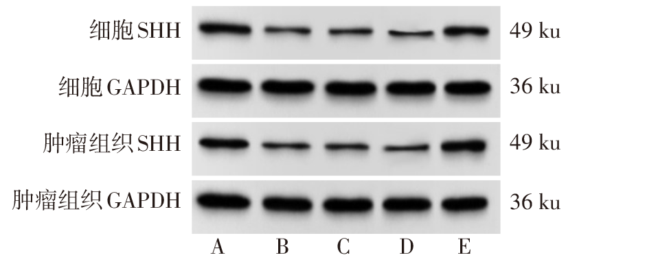
图7 免疫印迹检测各组SKOV3细胞及其移植瘤小鼠肿瘤组织SHH蛋白表达 A:对照组;B:麝香酮组;C:麝香酮+CQ组;D:麝香酮+空载组;E:麝香酮+SHH过表达组。
Fig.7 Immunoblotting detection of SHH protein expression in tumor tissue of SKOV3 cells and transplanted tumor mice in each group
| 组别 | 细胞(n=6) | 肿瘤组织(n=10) | ||
|---|---|---|---|---|
| SHH mRNA | SHH蛋白 | SHH mRNA | SHH蛋白 | |
| 对照组 | 0.99±0.17 | 0.67±0.05 | 0.97±0.14 | 0.71±0.08 |
| 麝香酮组 | 0.38±0.06a | 0.14±0.03a | 0.32±0.07a | 0.20±0.05a |
| 麝香酮+CQ组 | 0.37±0.04 | 0.13±0.02 | 0.33±0.06 | 0.19±0.04 |
| 麝香酮+空载组 | 0.39±0.05 | 0.15±0.04 | 0.34±0.08 | 0.21±0.06 |
| 麝香酮+SHH 过表达组 | 0.94±0.13b | 0.62±0.08b | 0.91±0.19b | 0.65±0.11b |
| F | 45.090* | 123.800* | 71.100* | 83.400* |
表6 各组SKOV3细胞及其移植瘤小鼠肿瘤组织SHH mRNA、蛋白相对表达水平比较($\bar{x}±s$)
Tab.6 Comparison of relative expression levels of SHH mRNA and protein in tumor tissue of SKOV3 cells and transplanted tumor mice between five groups
| 组别 | 细胞(n=6) | 肿瘤组织(n=10) | ||
|---|---|---|---|---|
| SHH mRNA | SHH蛋白 | SHH mRNA | SHH蛋白 | |
| 对照组 | 0.99±0.17 | 0.67±0.05 | 0.97±0.14 | 0.71±0.08 |
| 麝香酮组 | 0.38±0.06a | 0.14±0.03a | 0.32±0.07a | 0.20±0.05a |
| 麝香酮+CQ组 | 0.37±0.04 | 0.13±0.02 | 0.33±0.06 | 0.19±0.04 |
| 麝香酮+空载组 | 0.39±0.05 | 0.15±0.04 | 0.34±0.08 | 0.21±0.06 |
| 麝香酮+SHH 过表达组 | 0.94±0.13b | 0.62±0.08b | 0.91±0.19b | 0.65±0.11b |
| F | 45.090* | 123.800* | 71.100* | 83.400* |
| [1] | PORTER R, MATULONIS U A. Immunotherapy for ovarian cancer[J]. Clin Adv Hematol Oncol, 2022, 20(4):240-253. |
| [2] | KHANLARKHANI N, AZIZI E, AMIDI F, et al. Metabolic risk factors of ovarian cancer:a review[J]. JBRA Assist Reprod, 2022, 26(2):335-347. doi:10.5935/1518-0557.20210067. |
| [3] | FENG C, YUAN X X. Role of autophagy and its regulation by noncoding RNAs in ovarian cancer[J]. Exp Biol Med, 2023:15353702231151958. doi:10.1177/15353702231151958. |
| [4] | VIDONI C, FERRARESI A, VALLINO L, et al. Glycolysis inhibition of autophagy drives malignancy in ovarian cancer:exacerbation by IL-6 and attenuation by resveratrol[J]. Int J Mol Sci, 2023, 24(2):1723. doi:10.3390/ijms24021723. |
| [5] | 齐娜, 段文娟, 李雅婧, 等. 麝香酮药理作用的研究进展[J]. 世界科学技术-中医药现代化, 2020, 22(8):3042-3047. |
| QI N, DUAN W J, LI Y J, et al. Research Progress on the Pharmacological Action of Muscone[J]. Modernization of Traditional Chinese Medicine and Materia Medica-World Science and Technology, 2020, 22(8):3042-3047. doi:10.11842/wst.20181223004. | |
| [6] | ZHAO Y R, TAO S X, WANG Q, et al. A network-based pharmacological study on the mechanism of action of muscone in breast cancer[J]. Transl Cancer Res, 2022, 11(5):1195-1206. doi:10.21037/tcr-22-667. |
| [7] | 卢鹏, 樊晶晶, 罗旭, 等. 麝香酮对肺癌细胞的顺铂耐药和小鼠体内的肿瘤生长的作用[J]. 广西医科大学学报, 2020, 37(11):1948-1953. |
| LU P, FAN J J, LUO X, et al. The effect of Muscone on lung cancer cells resistance to cisplatin and tumor growth in mice[J]. J Guangxi Med Univ, 2020, 37(11):1948-1953. doi:10.16190/j.cnki.45-1211/r.2020.11.003. | |
| [8] | ZHANG M, TAO Z Y, GAO L J, et al. Toosendanin inhibits colorectal cancer cell growth through the Hedgehog pathway by targeting Shh[J]. Drug Dev Res, 2022, 83(5):1201-1211. doi:10.1002/ddr.21951. |
| [9] | LONDERO A P, ORSARIA M, VIOLA L, et al. Survivin,sonic hedgehog,krüppel-like factors,and p53 pathway in serous ovarian cancer:an immunohistochemical study[J]. Hum Pathol, 2022, 127:92-101. doi:10.1016/j.humpath.2022.06.023. |
| [10] | 赵飞, 于新平, 赵涵, 等. GLI1及Shh在卵巢型子宫内膜异位症恶变过程中的表达及其意义[J]. 中华妇产科杂志, 2022, 57(2):125-132. |
| ZHAO F, YU X P, ZHAO H, et al. Expression and significance of GLI1 and Shh in the malignant transformation of ovarian endometriosis[J]. Chin J Obstet Gynecol, 2022, 57(2):125-132. doi:10.3760/cma.j.cn112141-20211219-00736. | |
| [11] | ZHANG X Y, LIU Q B, ZHANG T T, et al. Bone-targeted nanoplatform enables efficient modulation of bone tumor microenvironment for prostate cancer bone metastasis treatment[J]. Drug Deliv, 2022, 29(1):889-905. doi:10.1080/10717544.2022.2050845. |
| [12] | PAN Y B, ZHOU J N, ZHANG W D, et al. The Sonic Hedgehog signaling pathway regulates autophagy and migration in ovarian cancer[J]. Cancer Med, 2021, 10(13):4510-4521. doi:10.1002/cam4.4018. |
| [13] | NAKAI H, MATSUMURA N. The roles and limitations of bevacizumab in the treatment of ovarian cancer[J]. Int J Clin Oncol, 2022, 27(7):1120-1126. doi:10.1007/s10147-022-02169-x. |
| [14] | BOSE S, SAHA P, CHATTERJEE B, et al. Chemokines driven ovarian cancer progression,metastasis and chemoresistance:potential pharmacological targets for cancer therapy[J]. Semin Cancer Biol, 2022, 86(Pt 2):568-579. doi:10.1016/j.semcancer.2022.03.028. |
| [15] | 王志, 谢建絮, 李婉斯, 等. 麝香及西黄方中麝香酮在正常、乳腺癌癌前病变大鼠体内药动学的比较[J]. 中成药, 2020, 42(10):2545-2550. |
| WANG Z, XIE J X, LI W S, et al. Comparison of in vivo pharmacokinetics of muscone from Moschus and Xihuang Decoction in normal rats and rats with precancerous lesions of breast cancer[J]. Chin Tradit Pat Med, 2020, 42(10):2545-2550. doi:10.3969/j.issn.1001-1528.2020.10.001. | |
| [16] | 任瑶, 江一鸣, 项蓉蓉, 等. 西黄丸组分中药调节肿瘤微环境中Treg细胞PI3K/AKT通路的抗肿瘤作用机制研究[J]. 药物评价研究, 2019, 42(3):437-443. |
| REN Y, JIANG Y M, XIANG R R, et al. Antitumor mechanism of Xihuang Pills component-based Chinese medicine by regulating Treg cells PI3K/AKT pathway in tumor microenvironment[J]. Drug Eval Res, 2019, 42(3):437-443. doi:CNKI:SUN:YWPJ.0.2019-03-010. | |
| [17] | CHEN S, GAO Y, ZHU P, et al. Anti-cancer drug anlotinib promotes autophagy and apoptosis in breast cancer[J]. Front Biosci (Landmark Ed), 2022, 27(4):125. doi:10.31083/j.fbl2704125. |
| [18] | ZHANG M J, YUE H D, HUANG X, et al. Novel platinum nanoclusters activate PI3K/AKT/mTOR signaling pathway-mediated autophagy for cisplatin-resistant ovarian cancer therapy[J]. ACS Appl Mater Interfaces, 2022, 14(43):48502-48514. doi:10.1021/acsami.2c15143. |
| [19] | SNEHA S, NAGARE R P, SIDHANTH C, et al. The hedgehog pathway regulates cancer stem cells in serous adenocarcinoma of the ovary[J]. Cell Oncol, 2020, 43(4):601-616. doi:10.1007/s13402-020-00504-w. |
| [20] | CHEN L P, LI Y, SONG Z H, et al. O-GlcNAcylation promotes cerebellum development and medulloblastoma oncogenesis via SHH signaling[J]. Proc Natl Acad Sci USA, 2022, 119(34):e2202821119. doi:10.1073/pnas.2202821119. |
| [1] | 杨怡, 朵鸿, 杨亚男, 刘云, 梁凤仪, 杨雪琴. 基于肿瘤标志物的实体瘤疗效评价标准在晚期卵巢癌疗效评估中的价值[J]. 天津医药, 2026, 54(1): 46-51. |
| [2] | 黄伟, 王健键, 黄英, 杨俊. 复方苦参注射液联合化疗及贝伐珠单抗对卵巢癌患者近期疗效的影响[J]. 天津医药, 2026, 54(1): 88-92. |
| [3] | 黄慧琦, 伍秋苑, 张昆, 李佩贤, 熊亚明, 叶国麟, 周丹. 川楝素联合奥拉帕尼在三阴性乳腺癌中的抗肿瘤机制研究[J]. 天津医药, 2025, 53(9): 897-902. |
| [4] | 林义伟, 魏谭军, 陈飞, 肖成, 袁烈, 王毅. 圣草酚调控UBA52表达对代谢相关脂肪性肝病的体内和体外作用[J]. 天津医药, 2025, 53(9): 916-922. |
| [5] | 马春梅, 于鹏, 张其程, 杨磊, 李棣华, 谭建, 孟召伟. 异硫氰酸苄酯联合索拉非尼治疗未分化甲状腺癌机制探讨[J]. 天津医药, 2025, 53(5): 449-455. |
| [6] | 吕丽凤, 高亚克, 王丽. 上皮性卵巢癌患者外周血T淋巴细胞水平和病理参数对预后的预测价值[J]. 天津医药, 2025, 53(5): 488-491. |
| [7] | 李冰心, 许军英, 张雅茹, 周小兵. 冬虫夏草通过调控AMPK/mTOR通路保护高糖诱导的足细胞损伤[J]. 天津医药, 2025, 53(3): 225-229. |
| [8] | 方杰, 黄芮, 郑红慧, 贾倩倩, 鲍静. miR-9-5p靶向TIMP2诱导多发性骨髓瘤细胞自噬和凋亡的机制[J]. 天津医药, 2024, 52(8): 785-790. |
| [9] | 李大强, 李坚, 陆哲明, 曹阳. 毛蕊异黄酮对脊髓损伤大鼠神经元自噬及凋亡的影响[J]. 天津医药, 2024, 52(8): 798-803. |
| [10] | 钟敏, 施震, 周劲松, 李晋杰. GABA信号通路对脓毒症大鼠急性肺损伤内质网应激和线粒体自噬的影响[J]. 天津医药, 2024, 52(7): 733-737. |
| [11] | 王柯, 叶寒露. 隐丹参酮调节HIF-1α/BNIP3信号通路对兔膝骨关节炎模型软骨细胞自噬和凋亡的影响[J]. 天津医药, 2024, 52(4): 372-378. |
| [12] | 何颖, 张广华, 田立东, 于泳浩. 富氢液通过增加自噬治疗大鼠神经病理性疼痛[J]. 天津医药, 2024, 52(3): 261-265. |
| [13] | 赵元元, 吴小华. 基于PI3K/Akt/mTOR信号通路探讨LINC00173对多囊卵巢综合征颗粒细胞自噬的影响[J]. 天津医药, 2024, 52(11): 1121-1126. |
| [14] | 喻萍, 周敏, 苏丹. 卵巢癌化疗耐药预测模型的建立及效果评价[J]. 天津医药, 2024, 52(11): 1177-1182. |
| [15] | 张睿, 陈思思, 王彤丹, 于珮. KLF4通过促进自噬减轻高糖浓度下巨噬细胞胆固醇沉积[J]. 天津医药, 2024, 52(10): 1014-1019. |
| 阅读次数 | ||||||
|
全文 |
|
|||||
|
摘要 |
|
|||||