
天津医药 ›› 2026, Vol. 54 ›› Issue (10): 1009-1017.doi: 10.11958/20261146
• 实验研究 • 下一篇
冯丽娜1( ), 阎亮2,△(), 薛水娜3, 王上增3
收稿日期:2026-04-30
修回日期:2026-05-29
出版日期:2026-10-15
发布日期:2026-10-10
通讯作者:
△E-mail:33946314@qq.com
作者简介:冯丽娜(1984),女,主治医师,主要从事关节运动损伤方面研究。E-mail:flnlina@126.com
基金资助:
FENG Lina1(), YAN Liang2,△(), XUE Shuina3, WANG Shangzeng3
Received:2026-04-30
Revised:2026-05-29
Published:2026-10-15
Online:2026-10-10
Contact:
△E-mail:33946314@qq.com
冯丽娜, 阎亮, 薛水娜, 王上增. 基于STING/ACSL4通路探讨复元活血汤抑制膝骨关节炎大鼠软骨细胞铁死亡的作用机制[J]. 天津医药, 2026, 54(10): 1009-1017.
FENG Lina, YAN Liang, XUE Shuina, WANG Shangzeng. Mechanism of Fuyuan Huoxue decoction in inhibiting chondrocyte ferroptosis of rats with knee osteoarthritis via the STING/ACSL4 pathway[J]. Tianjin Medical Journal, 2026, 54(10): 1009-1017.
摘要:
目的 基于干扰素基因刺激因子(STING)/长链酰基辅酶A合成酶家族成员4(ACSL4)通路探讨复元活血汤(FHD)抑制膝骨关节炎(KOA)大鼠软骨细胞铁死亡的作用机制。方法 将SD大鼠随机分为假手术(Sham)组、模型(KOA)组、FHD低剂量(FHD-L)组、FHD高剂量(FHD-H)组、塞来昔布阳性对照(PC)组及FHD-H+STING激活剂(FHD-H+DMXAA)组,每组12只。采用改良Hulth法建立KOA模型。造模成功后各组分别给予相应药物灌胃或尾静脉注射干预,连续8周。采用Lequesne MG评分评估膝关节功能;HE染色观察软骨组织形态;番红O-固绿染色结合OARSI评分评估软骨退变程度;酶联免疫吸附试验检测软骨组织中白细胞介素(IL)-1β、肿瘤坏死因子(TNF)-α、基质金属蛋白酶(MMP)-13及血小板反应蛋白解整合素金属肽酶-5(ADAMTS-5)水平;生化试剂盒测定软骨组织Fe2+、丙二醛(MDA)、谷胱甘肽(GSH)及超氧化物歧化酶(SOD)水平;DCFH-DA探针法检测软骨组织活性氧(ROS)水平;普鲁士蓝染色观察铁沉积;透射电镜观察软骨细胞超微结构;免疫组化法检测软骨组织谷胱甘肽过氧化物酶4(GPX4)、溶质载体家族7成员11(SLC7A11)、环氧化酶-2(COX-2)蛋白表达;Western blot法检测软骨组织STING及ACSL4蛋白表达。结果 与Sham组相比,KOA组Lequesne MG评分,OARSI评分,软骨组织炎症及基质降解因子(IL-1β、TNF-α、MMP-13、ADAMTS-5),铁死亡相关指标(Fe2+、MDA、ROS),普鲁士蓝染色阳性面积比,COX-2阳性表达面积及STING、ACSL4蛋白表达均显著升高(P<0.05),而GSH、SOD水平及GPX4、SLC7A11阳性表达面积均显著降低(P<0.05);HE染色、番红O-固绿染色及透射电镜显示KOA组软骨结构严重破坏,软骨基质丢失,线粒体萎缩、嵴断裂。与KOA组相比,FHD-L组、FHD-H组及PC组上述指标均有不同程度改善(P<0.05),其中FHD-H组效果优于FHD-L组(P<0.05)。与FHD-H组相比,FHD-H+DMXAA组上述指标的改善效应被显著逆转(P<0.05)。结论 FHD可能通过抑制STING/ACSL4通路减轻软骨细胞铁死亡,从而改善KOA大鼠的关节功能与软骨损伤。
中图分类号:
| 组别 | Lequesne MG评分(n=12) | OARSI评分(n=6) | |
|---|---|---|---|
| 给药前 | 给药后 | ||
| Sham组 | 0.08±0.02 | 0.22±0.08 | 1.03±0.52 |
| KOA组 | 8.81±1.34a | 8.67±1.23a | 14.67±2.16a |
| FHD-L组 | 8.69±1.22a | 5.92±0.71b | 10.83±1.94b |
| FHD-H组 | 8.75±1.18a | 2.67±0.59bc | 5.17±1.47bc |
| PC组 | 8.78±1.25a | 2.83±0.44bc | 5.83±1.60bc |
| FHD-H+DMXAA组 | 8.72±1.16a | 7.08±0.86d | 12.50±2.07d |
| F | 118.941** | 220.018** | 54.232** |
表1 各组大鼠给药前后Lequesne MG、OARSI评分比较 (分,$\bar{x}±s$)
Tab.1 Comparison of Lequesne MG score and OARSI score before and after drug administration between six groups of rats
| 组别 | Lequesne MG评分(n=12) | OARSI评分(n=6) | |
|---|---|---|---|
| 给药前 | 给药后 | ||
| Sham组 | 0.08±0.02 | 0.22±0.08 | 1.03±0.52 |
| KOA组 | 8.81±1.34a | 8.67±1.23a | 14.67±2.16a |
| FHD-L组 | 8.69±1.22a | 5.92±0.71b | 10.83±1.94b |
| FHD-H组 | 8.75±1.18a | 2.67±0.59bc | 5.17±1.47bc |
| PC组 | 8.78±1.25a | 2.83±0.44bc | 5.83±1.60bc |
| FHD-H+DMXAA组 | 8.72±1.16a | 7.08±0.86d | 12.50±2.07d |
| F | 118.941** | 220.018** | 54.232** |
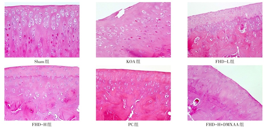
图1 各组大鼠膝关节软骨HE染色(×200)
Fig.1 HE staining of knee joint cartilage of each group of rats (×200)
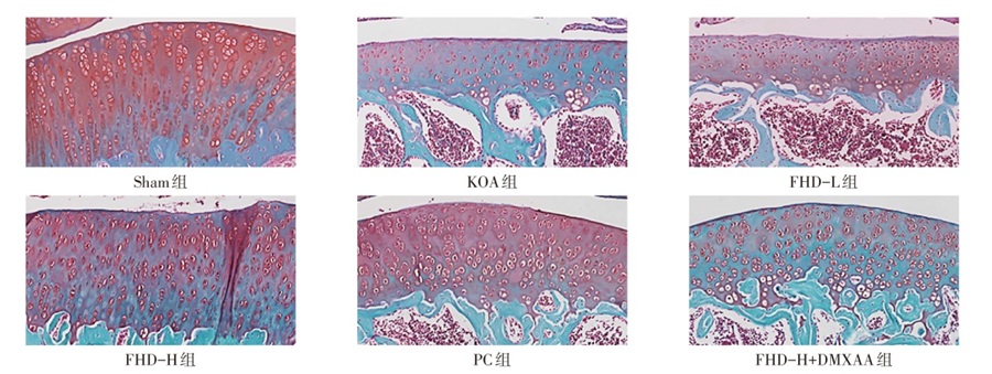
图2 各组大鼠膝关节软骨番红O-固绿染色(×100)
Fig.2 Safranin O-Grimmaux staining of knee joint cartilage in each group of rats (×100)
| 组别 | IL-1β/ (pg/mg) | TNF-α/ (pg/mg) | MMP-13/ (ng/mg) | ADAMTS-5/(ng/mg) |
|---|---|---|---|---|
| Sham组 | 12.47±2.15 | 8.23±1.56 | 3.42±0.58 | 2.15±0.42 |
| KOA组 | 86.34±7.82a | 72.56±6.93a | 28.67±3.14a | 18.93±2.08a |
| FHD-L组 | 58.26±6.14b | 48.37±5.25b | 18.42±2.56b | 12.67±1.85b |
| FHD-H组 | 28.53±3.67bc | 22.18±3.42bc | 8.35±1.27bc | 5.28±0.96bc |
| PC组 | 31.42±4.05bc | 24.56±3.78bc | 9.64±1.48bc | 6.12±1.03bc |
| FHD-H+DMXAA组 | 67.38±6.52d | 56.29±5.84d | 22.53±2.87d | 15.47±1.92d |
| F | 158.243** | 153.825** | 115.575** | 114.235** |
表2 各组大鼠软骨组织IL-1β、TNF-α、MMP-13、ADAMTS-5表达比较 (n=6,$\bar{x}±s$)
Tab.2 Comparison of IL-1β, TNF-α, MMP-13 and ADAMTS-5 expression in cartilage tissue between six groups of rats
| 组别 | IL-1β/ (pg/mg) | TNF-α/ (pg/mg) | MMP-13/ (ng/mg) | ADAMTS-5/(ng/mg) |
|---|---|---|---|---|
| Sham组 | 12.47±2.15 | 8.23±1.56 | 3.42±0.58 | 2.15±0.42 |
| KOA组 | 86.34±7.82a | 72.56±6.93a | 28.67±3.14a | 18.93±2.08a |
| FHD-L组 | 58.26±6.14b | 48.37±5.25b | 18.42±2.56b | 12.67±1.85b |
| FHD-H组 | 28.53±3.67bc | 22.18±3.42bc | 8.35±1.27bc | 5.28±0.96bc |
| PC组 | 31.42±4.05bc | 24.56±3.78bc | 9.64±1.48bc | 6.12±1.03bc |
| FHD-H+DMXAA组 | 67.38±6.52d | 56.29±5.84d | 22.53±2.87d | 15.47±1.92d |
| F | 158.243** | 153.825** | 115.575** | 114.235** |
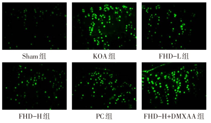
图3 各组大鼠关节软骨DCFH-DA染色(×200)
Fig.3 DCFH-DA staining of articular cartilage in each group of rats (×200)
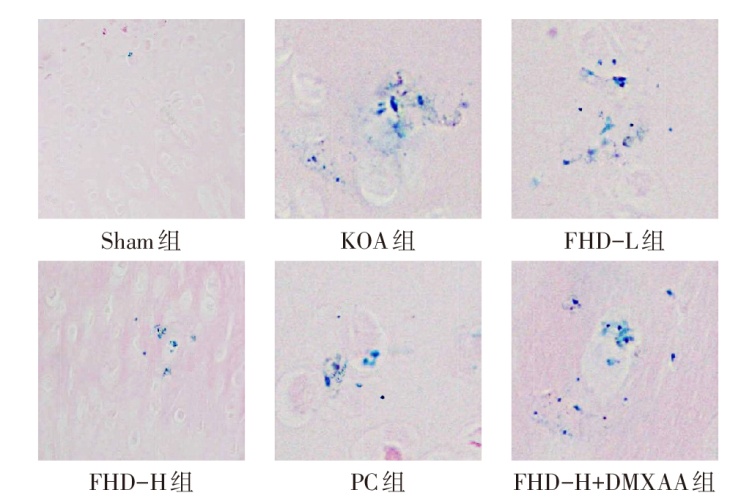
图4 各组大鼠关节软骨组织普鲁士蓝染色(×200)
Fig.4 Prussian blue staining of articular cartilage tissue of each group of rats (×200)
| 组别 | Fe2+/(μmol/g) | MDA/(nmol/g) | ROS(荧光强度) | ||
|---|---|---|---|---|---|
| Sham组 | 0.28±0.05 | 1.32±0.21 | 100.00±8.56 | ||
| KOA组 | 1.86±0.17a | 5.78±0.52a | 286.54±18.63a | ||
| FHD-L组 | 1.28±0.14b | 4.12±0.43b | 218.36±15.42b | ||
| FHD-H组 | 0.58±0.08bc | 2.18±0.26bc | 138.27±11.35bc | ||
| PC组 | 0.65±0.09bc | 2.35±0.28bc | 145.38±12.17bc | ||
| FHD-H+DMXAA组 | 1.52±0.15d | 4.86±0.47d | 248.65±16.28d | ||
| F | 155.527** | 126.533** | 157.966** | ||
| 组别 | GSH/ (μmol/g) | SOD/ (U/mg) | 普鲁士蓝染色 阳性面积比/% | ||
| Sham组 | 8.64±0.87 | 42.38±3.86 | 0.42±0.11 | ||
| KOA组 | 2.35±0.31a | 15.28±1.94a | 8.76±0.94a | ||
| FHD-L组 | 3.86±0.42b | 24.63±2.35b | 5.83±0.72b | ||
| FHD-H组 | 6.72±0.58bc | 36.47±2.86bc | 1.85±0.34bc | ||
| PC组 | 6.48±0.62bc | 35.26±2.74bc | 2.12±0.41bc | ||
| FHD-H+DMXAA组 | 3.12±0.38d | 20.18±2.13d | 6.94±0.85d | ||
| F | 114.110** | 90.650** | 163.643** | ||
表3 各组大鼠软骨组织铁死亡相关指标比较 (n=6,$\bar{x}±s$)
Tab.3 Comparison of iron death-related indicators in cartilage tissue between six groups of rats
| 组别 | Fe2+/(μmol/g) | MDA/(nmol/g) | ROS(荧光强度) | ||
|---|---|---|---|---|---|
| Sham组 | 0.28±0.05 | 1.32±0.21 | 100.00±8.56 | ||
| KOA组 | 1.86±0.17a | 5.78±0.52a | 286.54±18.63a | ||
| FHD-L组 | 1.28±0.14b | 4.12±0.43b | 218.36±15.42b | ||
| FHD-H组 | 0.58±0.08bc | 2.18±0.26bc | 138.27±11.35bc | ||
| PC组 | 0.65±0.09bc | 2.35±0.28bc | 145.38±12.17bc | ||
| FHD-H+DMXAA组 | 1.52±0.15d | 4.86±0.47d | 248.65±16.28d | ||
| F | 155.527** | 126.533** | 157.966** | ||
| 组别 | GSH/ (μmol/g) | SOD/ (U/mg) | 普鲁士蓝染色 阳性面积比/% | ||
| Sham组 | 8.64±0.87 | 42.38±3.86 | 0.42±0.11 | ||
| KOA组 | 2.35±0.31a | 15.28±1.94a | 8.76±0.94a | ||
| FHD-L组 | 3.86±0.42b | 24.63±2.35b | 5.83±0.72b | ||
| FHD-H组 | 6.72±0.58bc | 36.47±2.86bc | 1.85±0.34bc | ||
| PC组 | 6.48±0.62bc | 35.26±2.74bc | 2.12±0.41bc | ||
| FHD-H+DMXAA组 | 3.12±0.38d | 20.18±2.13d | 6.94±0.85d | ||
| F | 114.110** | 90.650** | 163.643** | ||
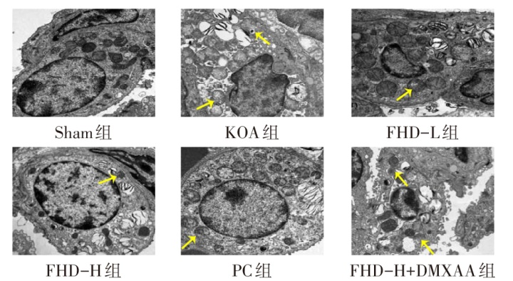
图5 各组大鼠关节软骨细胞线粒体超微结构(透射电镜,×7 000) 黄色箭头所示为线粒体萎缩及嵴断裂。
Fig.5 Ultrastructure of mitochondria in articular cartilage cells of each group of rats (transmission electron microscope, ×7 000)
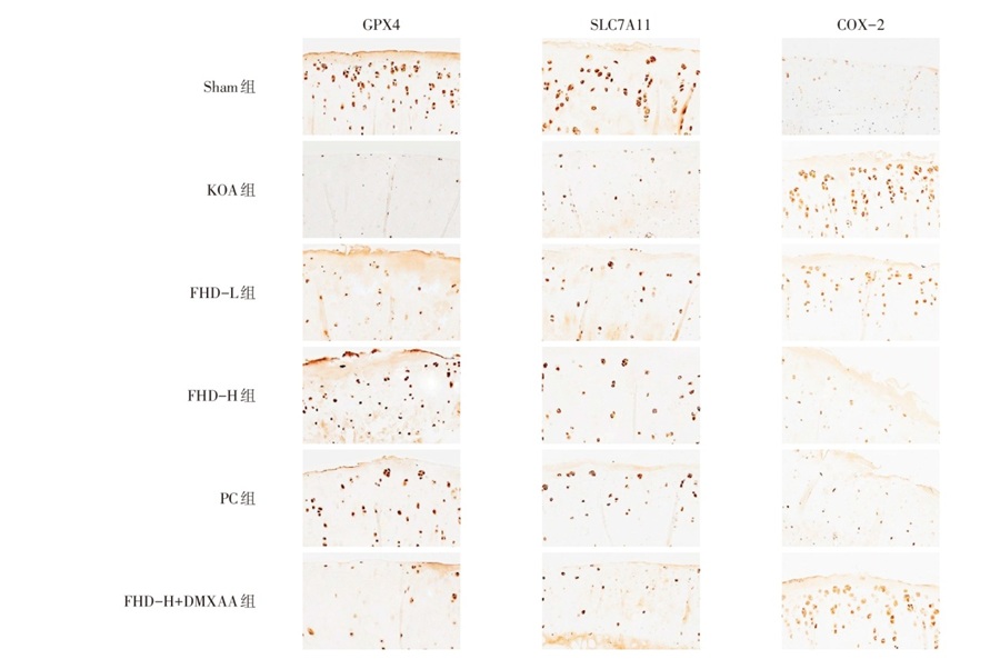
图6 各组大鼠软骨组织GPX4、SLC7A11、COX-2免疫组化染色(×200)
Fig.6 Immunohistochemical staining of GPX4, SLC7A11 and COX-2 in cartilage tissue of each group of rats (×200)
| 组别 | GPX4 阳性面积 | SLC7A11 阳性面积 | COX-2 阳性面积 |
|---|---|---|---|
| Sham组 | 18.64±2.15 | 15.32±1.86 | 2.86±0.52 |
| KOA组 | 3.28±0.64a | 4.15±0.73a | 21.48±2.35a |
| FHD-L组 | 7.56±1.08b | 7.83±1.12b | 14.36±1.87b |
| FHD-H组 | 15.38±1.64bc | 13.26±1.45bc | 5.27±0.94bc |
| PC组 | 14.52±1.58bc | 12.48±1.52bc | 5.86±1.03bc |
| FHD-H+DMXAA组 | 4.86±0.92d | 5.68±0.98d | 17.52±2.08d |
| F | 116.887** | 69.423** | 133.647** |
表4 各组大鼠软骨组织GPX4、SLC7A11、COX-2阳性表达面积比较 (n=6,%,$\bar{x}±s$)
Tab.4 Comparison of the positive expression areas of GPX4, SLC7A11 and COX-2 in cartilage tissue between six groups of rats
| 组别 | GPX4 阳性面积 | SLC7A11 阳性面积 | COX-2 阳性面积 |
|---|---|---|---|
| Sham组 | 18.64±2.15 | 15.32±1.86 | 2.86±0.52 |
| KOA组 | 3.28±0.64a | 4.15±0.73a | 21.48±2.35a |
| FHD-L组 | 7.56±1.08b | 7.83±1.12b | 14.36±1.87b |
| FHD-H组 | 15.38±1.64bc | 13.26±1.45bc | 5.27±0.94bc |
| PC组 | 14.52±1.58bc | 12.48±1.52bc | 5.86±1.03bc |
| FHD-H+DMXAA组 | 4.86±0.92d | 5.68±0.98d | 17.52±2.08d |
| F | 116.887** | 69.423** | 133.647** |
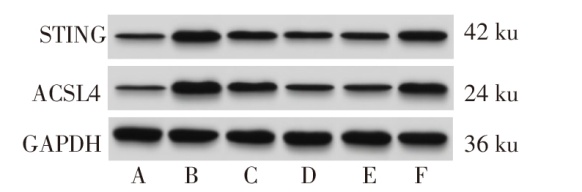
图7 Western blot检测各组大鼠软骨组织STING、ACSL4蛋白表达 A:Sham组;B:KOA组;C:FHD-L组;D:FHD-H组;E:PC组;F:FHD-H+DMXAA组。
Fig.7 Expression of STING and ACSL4 proteins in cartilage tissue of each group of rats detected by Western blot assay
| 组别 | STING | ACSL4 |
|---|---|---|
| Sham组 | 0.21±0.04 | 0.18±0.03 |
| KOA组 | 0.86±0.09a | 0.92±0.10a |
| FHD-L组 | 0.62±0.07b | 0.67±0.08b |
| FHD-H组 | 0.32±0.05bc | 0.28±0.04bc |
| PC组 | 0.35±0.06bc | 0.31±0.05bc |
| FHD-H+DMXAA组 | 0.73±0.08d | 0.78±0.09d |
| F | 88.884** | 114.061** |
表5 各组大鼠软骨组织STING、ACSL4蛋白相对表达量比较 (n=6,$\bar{x}±s$)
Tab.5 Comparison of relative expression levels of STING and ACSL4 proteins in cartilage tissue between six groups of rats
| 组别 | STING | ACSL4 |
|---|---|---|
| Sham组 | 0.21±0.04 | 0.18±0.03 |
| KOA组 | 0.86±0.09a | 0.92±0.10a |
| FHD-L组 | 0.62±0.07b | 0.67±0.08b |
| FHD-H组 | 0.32±0.05bc | 0.28±0.04bc |
| PC组 | 0.35±0.06bc | 0.31±0.05bc |
| FHD-H+DMXAA组 | 0.73±0.08d | 0.78±0.09d |
| F | 88.884** | 114.061** |
| [1] | Chen L, Huang F L, Tang Q, et al. Targeted therapy for knee osteoarthritis:from basic to clinics[J]. Medicine (Baltimore), 2025, 104(33):e43686. doi:10.1097/MD.0000000000043686. |
| [2] | Hu Z, Chen L, Zhao J, et al. Lipoxin A4 ameliorates knee osteoarthritis progression in rats by antagonizing ferroptosis through activation of the ESR2/LPAR3/Nrf2 axis in synovial fibroblast-like synoviocytes[J]. Redox Biol, 2024, 73:103143. doi:10.1016/j.redox.2024.103143. |
| [3] | Sun K, Shi Y, Yan C, et al. Glycolysis-derived lactate induces ACSL4 expression and lactylation to activate ferroptosis during intervertebral disc degeneration[J]. Adv Sci (Weinh), 2025, 12(21):e2416149. doi:10.1002/advs.202416149. |
| [4] | Lan J, Deng Z, Wang Q, et al. Neuropeptide substance P attenuates colitis by suppressing inflammation and ferroptosis via the cGAS-STING signaling pathway[J]. Int J Biol Sci, 2024, 20(7):2507-2531. doi:10.7150/ijbs.94548. |
| [5] | Gao L, Zhang J, Yang T, et al. STING/ACSL4 axis-dependent ferroptosis and inflammation promote hypertension-associated chronic kidney disease[J]. Mol Ther, 2023, 31(10):3084-3103. doi:10.1016/j.ymthe.2023.07.026. |
| [6] | 哈思远, 张巍. 超声引导下股神经阻滞联合复元活血汤对全膝关节置换术后疼痛及膝关节功能的影响[J]. 河南中医, 2024, 44(3):402-406. |
| Ha S Y, Zhang W. The influence of ultrasound-guided femoral nerve block combined with original qi-restoring and blood-moving decoction on pain and knee function after total knee arthroplasty[J]. Henan Tradit Chin Med, 2024, 44(3):402-406. doi:10.16367/j.issn.1003-5028.2024.03.0077. | |
| [7] | 陈世琳, 陈文恒, 王光辉, 等. 异甘草素对膝关节骨关节炎大鼠软骨细胞铁死亡的影响及其作用机制[J]. 中华骨科杂志, 2025, 45(4):230-240. |
| Chen S L, Chen W H, Wang G H, et al. Effect and mechanism of isoliquiritigenin on ferroptosis of chondrocytes in rats with knee osteoarthritis[J]. Chin J Orthop, 2025, 45(4):230-240. doi:10.3760/cma.j.cn121113-20240910-00496. | |
| [8] | 周欣华, 杨琪琪, 李蔚然, 等. 电针调控PI3K/Akt/mTOR通路对膝关节骨关节炎大鼠膝关节滑膜组织自噬的影响[J]. 针刺研究, 2026, 51(1):59-67. |
| Zhou X H, Yang Q Q, Li W R, et al. Effects of electroacupuncture on autophagy of the synovial tissue in the knee joint of rats with knee osteoarthritis by regulating PI3K/Akt/mTOR pathway[J]. Acupunct Res, 2026, 51(1):59-67. doi:10.13702/j.1000-0607.20241225. | |
| [9] | 张永波, 林伟, 苏冬冬, 等. 复元活血汤对大鼠骨质疏松性椎体骨折愈合和血管化的作用[J]. 中国骨质疏松杂志, 2025, 31(8):1178-1184. |
| Zhang Y B, Lin W, Su D D, et al. Effect of Fuyuan Huoxue Decoction on healing and vascularization of osteoporotic vertebral fracture in rats[J]. Chin J Osteoporosis, 2025, 31(8):1178-1184. doi:10.3969/j.issn.1006-7108.2025.08.013. | |
| [10] | 方红育, 黄涛, 周少怀, 等. 川陈皮素通过抑制STING/NF-κB信号通路促进骨质疏松大鼠的骨折愈合[J]. 免疫学杂志, 2023, 39(10):857-864. |
| Fang H Y, Huang T, Zhou S H, et al. Nobiletin promotes fracture healing in osteoporosis rats by inhibiting STING/NF-κB signal pathway[J]. Immunol J, 2023, 39(10):857-864. doi:10.13431/j.cnki.immunol.j.20230111. | |
| [11] | Yu L, Su Z, Tian D, et al. Piezo1 induces mitochondrial autophagy dysfunction leading to cartilage injury in knee osteoarthritis[J]. Mol Med, 2025, 31(1):272. doi:10.1186/s10020-025-01335-x. |
| [12] | 张健哲, 杨永菊, 张宇, 等. 膝骨关节炎中医辨证存在的问题与对策[J]. 中华中医药学刊, 2023, 41(12):137-141. |
| Zhang J Z, Yang Y J, Zhang Y, et al. Problems and countermeasures in syndrome differentiation of knee-osteoarthritis in Traditional Chinese Medicine[J]. Chin Arch Tradit Chin Med, 2023, 41(12):137-141. doi:10.13193/j.issn.1673-7717.2023.12.026. | |
| [13] | 蒲晓东, 杨艳梅. 膝骨关节炎中医病名再辨识[J]. 时珍国医国药, 2017, 28(12):2972-2973. |
| Pu X D, Yang Y M. Re-identification of the TCM sickness name of knee osteoarthritis[J]. Lishizhen Med Mater Med Res, 2017, 28(12):2972-2973. doi:10.3969/j.issn.1008-0805.2017.12.055. | |
| [14] | 唐海波, 叶贵生, 王旭, 等. 周长征教授基于久病入络理论治疗膝骨关节炎临证经验[J]. 中国民族民间医药, 2024, 33(7):62-65. |
| Tang H B, Ye G S, Wang X, et al. Professor Zhou Changzheng's clinical experience in treating knee osteoarthritis based on the theory of chronic disease entering collaterals[J]. Chin J Ethnomed Ethnopharm, 2024, 33(7):62-65. doi:10.3969/j.issn.1007-8517.2024.07.zgmzmjyyzz202407014. | |
| [15] | 王阶, 董艳, 熊兴江, 等. 活血化瘀治疗冠脉微循环障碍的研究进展[J]. 中国科学基金, 2024, 38(4):677-686. |
| Wang J, Dong Y, Xiong X J, et al. Research progress on promoting blood circulation and removing blood stasis in the treatment of coronary microvascular dysfunction[J]. Bull Natl Nat Sci Found China, 2024, 38(4):677-686. doi:10.16262/j.cnki.1000-8217.2024.04.008. | |
| [16] | 司元龙, 陈志伟, 申建军. 基于OPG/RANKL/RANK信号通路探讨中医“血瘀”与膝骨关节炎[J]. 中医学报, 2026, 41(5):1031-1036. |
| Si Y L, Chen Z W, Shen J J. "Blood Stasis" and knee osteoarthritis Traditional Chinese Medicine based on OPG/RANKL/RANK signaling pathway[J]. Acta Chin Med, 2026, 41(5):1031-1036. doi:10.16368/j.issn.1674-8999.2026.05.153. | |
| [17] | Jin M, Mi L, Li H W, et al. The value of super microvascular imaging and shear wave elastography in evaluating synovial lesions in knee osteoarthritis[J]. J Back Musculoskelet Rehabil, 2025, 38(6):1342-1351. doi:10.1177/10538127251328901. |
| [18] | Zhuang S, Chen Q, Guo X, et al. Mechanism of action of total saponin Achyranthes in treating knee osteoarthritis explored using network pharmacology and animal experimentation[J]. Iran J Basic Med Sci, 2025, 28(6):762-771. doi:10.22038/ijbms.2025.83153.17974. |
| [19] | Duan Z, Gao H, Xiao X, et al. Comparison of passive and active motion:effect of myokine irisin on cartilage in knee osteoarthritis rats[J]. Front Physiol, 2025, 16:1639174. doi:10.3389/fphys.2025.1639174. |
| [20] | Zhang Y, Li J, Liu J, et al. Ferroptosis in osteoarthritis:towards novel therapeutic strategy[J]. Cell Prolif, 2025, 58(3):e13779. doi:10.1111/cpr.13779. |
| [21] | Wang R, Shi D, Pan X, et al. Epigenetic mechanisms of Nsd1-mediated histone methylation modifications in chondrocyte ferroptosis in knee osteoarthritis[J]. Biomol Biomed, 2025, 25(4):894-904. doi:10.17305/bb.2024.10879. |
| [22] | Jin X, He R, Lin Y, et al. Shenshuaifu granule attenuates acute kidney injury by inhibiting ferroptosis mediated by p53/SLC7A11/GPX4 pathway[J]. Drug Des Devel Ther, 2023, 17:3363-3383. doi:10.2147/DDDT.S433994. |
| [23] | Teng X, Sheng S, Pan M, et al. Homoplantaginin protects pancreatic tissue in severe acute pancreatitis mice via inhibiting ferroptosis by modulating the circDNMT3B/miR-20b-5p/SLC7A11 axis[J]. BMC Gastroenterol, 2026, 26(1):120. doi:10.1186/s12876-025-04600-9. |
| [24] | Marques E, Burr S P, Casey A M, et al. An inherited mitochondrial DNA mutation remodels inflammatory cytokine responses in macrophages and in vivo in mice[J]. Nat Commun, 2025, 16(1):10222. doi:10.1038/s41467-025-65023-4. |
| [25] | Zeng L, Yang K, Yu G, et al. Advances in research on immunocyte iron metabolism,ferroptosis,and their regulatory roles in autoimmune and autoinflammatory diseases[J]. Cell Death Dis, 2024, 15(7):481. doi:10.1038/s41419-024-06807-2. |
| [26] | Chen J, Chen P, Song Y, et al. STING upregulation mediates ferroptosis and inflammatory response in lupus nephritis by upregulating TBK1 and activating NF-κB signal pathway[J]. J Biosci, 2024, 49:9. |
| [27] | Cao S, Wei Y, Xiong A, et al. Paeonol inhibits ACSL4 to protect chondrocytes from ferroptosis and ameliorates osteoarthritis progression[J]. J Orthop Translat, 2024, 50:1-13. doi:10.1016/j.jot.2024.10.005. |
| [28] | Xu T, Xie Z, Zhao Y, et al. Melatonin impedes NCOA4/ACSL4-dependent ferroptosis by targeting STING to ameliorate diabetic cognitive dysfunction[J]. Bioorg Chem, 2026, 169:109357. doi:10.1016/j.bioorg.2025.109357. |
| [29] | 闵洁煜, 应流念, 刘美言, 等. STING/ACSL4信号通路在环泊酚减轻OGD/R诱导肾小管上皮细胞铁死亡中的作用[J]. 中华麻醉学杂志, 2025, 45(5):603-610. |
| Min J Y, Ying L N, Liu M Y, et al. Role of STING/ACSL4 signaling pathway in alleviation of oxygen-glucose deprivation and restoration-induced ferroptosis in renal tubular epithelial cells[J]. Chin J Anesthesiol, 2025, 45(5):603-610. doi:10.3760/cma.j.cn131073-20240709-00515. | |
| [30] | Kuang B, Yang K, Zhong X, et al. Celecoxib in oncology:targeting the COX-2/PGE2 axis to reprogram the tumor immune microenvironment and enhance multimodal therapy[J]. Front Pharmacol, 2025, 16:1691392. doi:10.3389/fphar.2025.1691392. |
| [31] | Waseem M, Imtiaz A, Alexander A, et al. Crosstalk between oxidative stress,mitochondrial dysfunction,chromosome instability,and the activation of the cGAS-STING/IFN pathway in systemic sclerosis[J]. Ageing Res Rev, 2025, 110:102812. doi:10.1016/j.arr.2025.102812. |
| [32] | Algieri C, Nesci S, Oppedisano F. Mitochondrial dysfunction acts as a modulator of the immunometabolic route for activating the cytosolic DNA sensor pathway in triggering innate immunosurveillance[J]. J Transl Med, 2025, 23(1):1321. doi:10.1186/s12967-025-07392-4. |
| [1] | 刘琳琳, 梁冬雪, 刘丽华, 田颖, 孙丽丽. 槐杞黄调控Jun/SLC7A11介导的气道上皮细胞铁死亡抑制哮喘大鼠气道重塑及胶原沉积[J]. 天津医药, 2026, 54(9): 902-907. |
| [2] | 刘振陶, 孙国栋, 徐伟, 马帅, 康林林. 毫米波辅助治疗膝骨关节炎的疗效观察[J]. 天津医药, 2026, 54(9): 987-991. |
| [3] | 吕大鹏, 周红荣, 李丹, 周娜. 瑞芬太尼调节HIF-1α/BNIP3通路对骨关节炎大鼠软骨损伤的影响[J]. 天津医药, 2026, 54(7): 699-704. |
| [4] | 邓新宜, 杨晨, 曹建林, 杨磊, 谭建, 贾强, 孟召伟. 金诺芬抑制葡萄糖摄取并诱导未分化甲状腺癌铁死亡[J]. 天津医药, 2026, 54(6): 570-576. |
| [5] | 艾希睿, 赵亚男, 刘鹏, 闫景明, 朱藜, 苑成浩. 膝关节腔内注射PRP联合间歇性低氧训练治疗早期膝骨关节炎的疗效及安全性[J]. 天津医药, 2026, 54(4): 384-389. |
| [6] | 高峰, 陈达, 惠文文. 钩吻素子调节AMPK/NLRP3信号通路对膝骨关节炎大鼠软骨损伤的影响[J]. 天津医药, 2026, 54(2): 158-163. |
| [7] | 赵兰君, 李良惠, 马馨, 巩娇娇, 郑臣辉, 石琳. 穿心莲内酯调控STAT3/GPX4通路对骨髓瘤细胞增殖和凋亡的影响[J]. 天津医药, 2026, 54(1): 8-13. |
| [8] | 何仁豪, 张聘, 尹识渊, 张杰. 关节镜探查联合单髁置换术对膝关节骨性关节炎患者膝关节功能的影响[J]. 天津医药, 2025, 53(7): 725-729. |
| [9] | 苏红见, 张春艳, 张卫东, 韩利, 乔亚红. 鸢尾素调控EBF3/ALOX15通路影响肺腺癌细胞增殖和迁移[J]. 天津医药, 2025, 53(4): 337-342. |
| [10] | 贾方, 李平, 李静, 尹路. 全膝关节置换术后神经认知功能障碍的影响因素及列线图模型的构建[J]. 天津医药, 2025, 53(3): 287-292. |
| [11] | 崔琳, 李海晴, 陈雨彤, 朱佳璇, 王璐怡. 经皮神经电刺激联合动态关节松动术对膝骨关节炎疗效观察[J]. 天津医药, 2025, 53(12): 1285-1289. |
| [12] | 高辰, 乔云阳, 季嘉玲, 王娥, 霍影, 张爱青. 蟾蜍灵对高糖诱导的肾小管上皮细胞细胞外基质合成的影响[J]. 天津医药, 2025, 53(10): 1009-1015. |
| [13] | 李波, 林红, 黄兰英, 李玲钰. HMOX1和MAPK14与脓毒症急性肺损伤患儿发病和预后的关系[J]. 天津医药, 2025, 53(10): 1052-1056. |
| [14] | 吴斌, 刘兆祥, 张月红, 王昌耀. 过度机械应力调控Piezo1介导成软骨细胞铁死亡的机制探讨[J]. 天津医药, 2025, 53(1): 14-18. |
| [15] | 张训功, 杨光辉, 杜增利, 薛培, 马梓昆. 铁死亡与老年骨折患者术后认知功能障碍的相关性[J]. 天津医药, 2025, 53(1): 47-51. |
| 阅读次数 | ||||||
|
全文 |
|
|||||
|
摘要 |
|
|||||
