
天津医药 ›› 2026, Vol. 54 ›› Issue (8): 802-807.doi: 10.11958/20260964
刘冰雁1,2( ), 黄冬雪1,2, 张越1, 刘海迎1, 谢克亮1, 庄淑梅2, 沈悦好1,△()
收稿日期:2026-04-14
修回日期:2026-04-27
出版日期:2026-08-15
发布日期:2026-08-07
通讯作者:
△E-mail:作者简介:刘冰雁(2002),女,硕士在读,主要从事重症护理方面研究。E-mail:基金资助:
LIU Bingyan1,2(), HUANG Dongxue1,2, ZHANG Yue1, LIU Haiying1, XIE Keliang1, ZHUANG Shumei2, SHEN Yuehao1,△()
Received:2026-04-14
Revised:2026-04-27
Published:2026-08-15
Online:2026-08-07
Contact:
△E-mail: 刘冰雁, 黄冬雪, 张越, 刘海迎, 谢克亮, 庄淑梅, 沈悦好. 富氢生理盐水对3期压力性损伤大鼠创面修复的影响[J]. 天津医药, 2026, 54(8): 802-807.
LIU Bingyan, HUANG Dongxue, ZHANG Yue, LIU Haiying, XIE Keliang, ZHUANG Shumei, SHEN Yuehao. Effect of hydrogen-rich normal saline on wound repair in rats with stage 3 pressure injury[J]. Tianjin Medical Journal, 2026, 54(8): 802-807.
摘要:
目的 观察富氢生理盐水局部应用对3期压力性损伤大鼠创面修复的影响。方法 选取36只SPF级SD雄性大鼠,建立3期压力性损伤模型后(其中4只用于评判建模是否成功),随机分为对照组与干预组,每组16只。对照组使用生理盐水清创,干预组使用富氢生理盐水(氢含量≥2 mg/L)清创,均2次/d。于干预第0、3、7、10、14天观察创面愈合率;第3天检测血清肿瘤坏死因子-α(TNF-α)、白细胞介素-6(IL-6)及血管内皮生长因子(VEGF)水平;第7、14天取创面组织行HE、Masson染色及Ki-67免疫组化检测,评估组织病理、胶原沉积与细胞增殖情况。结果 与对照组相比,干预组在第3、7、10、14天的创面愈合率均升高(P<0.05);干预第3天,干预组血清TNF-α、IL-6水平降低,VEGF水平升高(P<0.05)。HE和Masson染色显示,干预组创面愈合质量更优,第7、14天胶原体积分数均高于对照组(P<0.05)。免疫组化显示,干预组第7天Ki-67阳性细胞比率高于对照组(P<0.05)。结论 富氢生理盐水局部应用可促进3期压力性损伤创面愈合,其机制可能与减轻氧化应激与炎症反应、促进胶原合成与细胞增殖有关。
中图分类号:
| 组别 | 第3天 | 第7天 | 第10天 | 第14天 |
|---|---|---|---|---|
| 对照组 | 23.85±3.87 | 40.13±6.17 | 65.07±5.31 | 81.54±1.70 |
| 干预组 | 36.98±3.29 | 67.28±6.97 | 78.15±3.67 | 90.69±3.73 |
| t | 5.205** | 5.893** | 3.992** | 4.654** |
表1 2组大鼠干预不同时间点创面愈合率比较
Tab.1 Comparison of wound healing rates at different time points between two groups of rats (n=4,%,$\bar{x}±s$)
| 组别 | 第3天 | 第7天 | 第10天 | 第14天 |
|---|---|---|---|---|
| 对照组 | 23.85±3.87 | 40.13±6.17 | 65.07±5.31 | 81.54±1.70 |
| 干预组 | 36.98±3.29 | 67.28±6.97 | 78.15±3.67 | 90.69±3.73 |
| t | 5.205** | 5.893** | 3.992** | 4.654** |
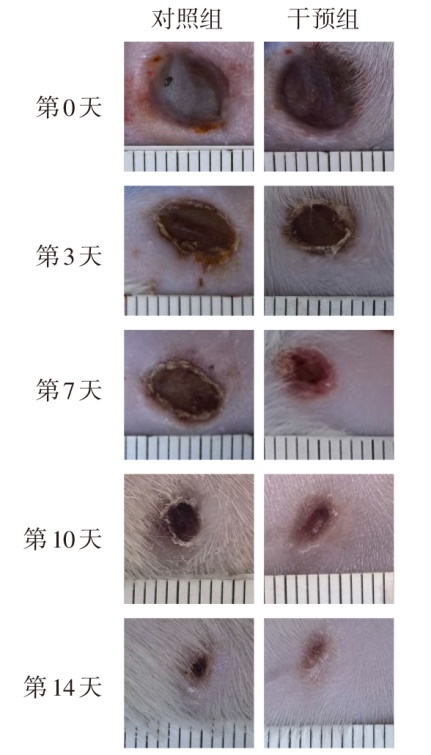
图1 2组大鼠不同时间点创面愈合情况 图中标尺长度均为1 cm。
Fig.1 Wound healing status at different time points in two groups of rats
| 组别 | TNF-α | IL-6 | VEGF |
|---|---|---|---|
| 对照组 | 20.33±2.23 | 48.70±8.74 | 41.43±2.06 |
| 干预组 | 13.76±0.69 | 23.94±5.50 | 61.60±3.87 |
| t | 5.405** | 4.925** | 8.960** |
表2 2组大鼠TNF-α、IL-6、VEGF水平比较
Tab.2 Comparison of systemic inflammatory factors TNF-α,IL-6 and VEGF between two groups of rats (n=4,ng/L,$\bar{x}±s$)
| 组别 | TNF-α | IL-6 | VEGF |
|---|---|---|---|
| 对照组 | 20.33±2.23 | 48.70±8.74 | 41.43±2.06 |
| 干预组 | 13.76±0.69 | 23.94±5.50 | 61.60±3.87 |
| t | 5.405** | 4.925** | 8.960** |
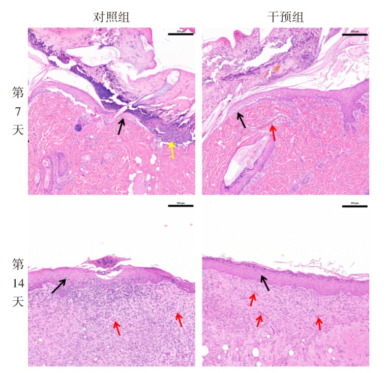
图2 2组大鼠不同时间创面组织HE染色情况(×100) 黑色箭头示再上皮化边缘,红色箭头示新生血管,黄色箭头示坏死组织。
Fig.2 HE staining of wound tissue at different time points in two groups of rats (×100)
| 组别 | 第7天 | 第14天 |
|---|---|---|
| 对照组 | 33.92±2.65 | 54.92±9.24 |
| 干预组 | 48.84±6.66 | 75.31±8.19 |
| t | 4.009* | 3.206* |
表3 2组大鼠不同时间创面组织CVF比较
Tab.3 Comparison of CVF in wound tissue at different time points between two groups of rats (n=4,%,$\bar{x}±s$)
| 组别 | 第7天 | 第14天 |
|---|---|---|
| 对照组 | 33.92±2.65 | 54.92±9.24 |
| 干预组 | 48.84±6.66 | 75.31±8.19 |
| t | 4.009* | 3.206* |
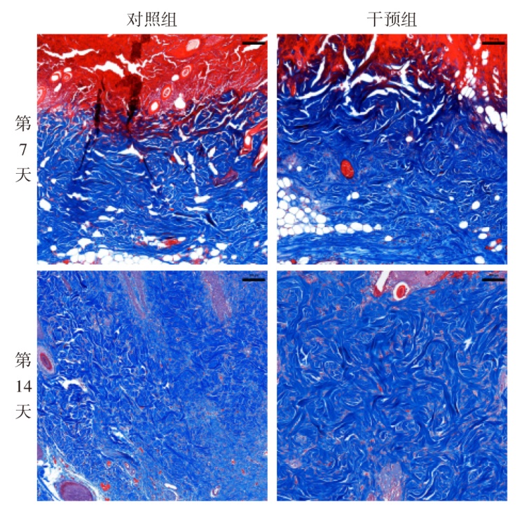
图3 2组大鼠不同时间创面组织Masson染色情况(×100)
Fig.3 Masson staining of wound tissue at different time points in two groups of rats (×100)
| 组别 | 第7天 | 第14天 |
|---|---|---|
| 对照组 | 28.16±1.70 | 22.72±2.84 |
| 干预组 | 34.57±3.88 | 25.87±4.02 |
| t | 2.966* | 1.241 |
表4 2组大鼠不同时间创面组织Ki-67阳性细胞比率比较
Tab.4 Comparison of Ki-67 positive cell ratio in wound tissue at different time points between two groups of rats (n=4,%,$\bar{x}±s$)
| 组别 | 第7天 | 第14天 |
|---|---|---|
| 对照组 | 28.16±1.70 | 22.72±2.84 |
| 干预组 | 34.57±3.88 | 25.87±4.02 |
| t | 2.966* | 1.241 |
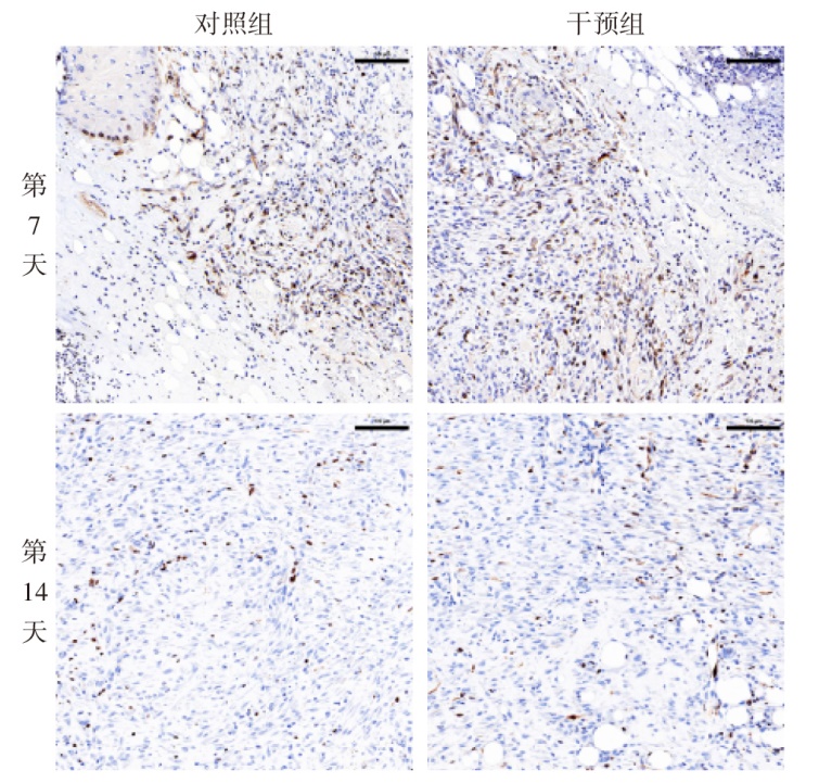
图4 2组大鼠不同时间创面组织免疫组化Ki-67染色情况(×200)
Fig.4 Immunohistochemical Ki-67 staining of wound tissue at different time points in two groups of rats (×200)
| [1] | Li Z, Lin F, Thalib L, et al. Global prevalence and incidence of pressure injuries in hospitalised adult patients:A systematic review and meta-analysis[J]. Int J Nurs Stud, 2020, 105:103546. doi:10.1016/j.ijnurstu.2020.103546. |
| [2] | Padula W V, Black J M, Davidson P M, et al. Adverse effects of the medicare PSI-90 hospital penalty system on revenue-neutral hospital-acquired conditions[J]. J Patient Saf, 2020, 16(2):e97-e102. doi:10.1097/PTS.0000000000000517. |
| [3] | Labeau S O, Afonso E, Benbenishty J, et al. Prevalence,associated factors and outcomes of pressure injuries in adult intensive care unit patients:the DecubICUs study[J]. Intensive Care Medicine, 2021, 47(2):160-169. doi:10.1007/s00134-020-06234-9. |
| [4] | Wang Z, Fan J, Chen L, et al. Strategies to preventing pressure injuries among intensive care unit patients mechanically ventilated in prone position:a systematic review and a Delphi study[J]. Front Med(Lausanne), 2023, 10:1131270. doi:10.3389/fmed.2023.1131270. |
| [5] | Gould L J, Alderden J, Aslam R, et al. WHS guidelines for the treatment of pressure ulcers-2023 update[J]. Wound Repair Regen, 2024, 32(1):6-33. doi:10.1111/wrr.13130. |
| [6] | Niemiec S M, Louiselle A E, Liechty K W, et al. Role of microRNAs in pressure ulcer immune response,pathogenesis,and treatment[J]. Int J Mol Sci, 2020, 22(1):64. doi:10.3390/ijms22010064. |
| [7] | Kottner J, Cuddigan J, Carville K, et al. Prevention and treatment of pressure ulcers/injuries:The protocol for the second update of the international Clinical Practice Guideline 2019[J]. J Tissue Viability, 2019, 28(2):51-58. doi:10.1016/j.jtv.2019.01.001. |
| [8] | Ohsawa I, Ishikawa M, Takahashi K, et al. Hydrogen acts as a therapeutic antioxidant by selectively reducing cytotoxic oxygen radicals[J]. Nat Med, 2007, 13(6):688-694. doi:10.1038/nm1577. |
| [9] | Zhang A, Jiang X, Xiong B, et al. Sustained-release photothermal microneedles for postoperative incisional analgesia and wound healing via hydrogen therapy[J]. Adv Sci(Weinh), 2025, 12(35):e03698. doi:10.1002/advs.202503698. |
| [10] | Bu Y, Liu Y, Liu M, et al. TRIM55 aggravates cardiomyocyte apoptosis after myocardial infarction via modulation of the Nrf2/HO-1 pathway[J]. JACC Basic Transl Sci, 2024, 9(9):1104-1122. doi:10.1016/j.jacbts.2024.05.006. |
| [11] | 张亚楠, 李贤, 严德云, 等. 构建各期大鼠压疮模型方法的实验研究[J]. 护理研究, 2017, 31(31):3945-3948. |
| Zhang Y N, Li X, Yan D Y, et al. Experimental study on establishing method of pressure ulcer model of rats[J]. Chinese Nursing Research, 2017, 31(31):3945-3948. doi:10.3969/j.issn.1009-6493.2017.31.013. | |
| [12] | Van Damme N, Van Hecke A, Remue E, et al. Physiological processes of inflammation and edema initiated by sustained mechanical loading in subcutaneous tissues:A scoping review[J]. Wound Repair Regen, 2020, 28(2):242-265. doi:10.1111/wrr.12777. |
| [13] | Krzyszczyk P, Schloss R, Palmer A, et al. The role of macrophages in acute and chronic wound healing and interventions to promote pro-wound healing phenotypes[J]. Front Physiol, 2018, 9:419. doi:10.3389/fphys.2018.00419. |
| [14] | 蔡灯塔, 常静宜, 贾姗姗, 等. 罗沙司他通过抑制凋亡和炎症反应改善小鼠心肌缺血再灌注损伤[J]. 天津医药, 2024, 52(11):1146-1151. |
| Cai D T, Chang J Y, Jia S S, et al. Roxadustat ameliorates myocardial ischemia-reperfusion injury in mice by inhibiting apoptosis and inflammatory response[J]. Tianjin Med J, 2024, 52(11):1146-1151. doi:10.11958/20240445. | |
| [15] | Jin L, Tan S, Fan K, et al. Research progress of hydrogen on chronic nasal inflammation[J]. J Inflamm Res, 2023, 16:2149-2157. doi:10.2147/JIR.S413179. |
| [16] | Qiu X, Dong K, Guan J, et al. Hydrogen attenuates radiation-induced intestinal damage by reducing oxidative stress and inflammatory response[J]. Int Immunopharmacol, 2020, 84:106517. doi:10.1016/j.intimp.2020.106517. |
| [17] | Sin T K, Yung B Y, Yip S P, et al. SIRT1-dependent myoprotective effects of resveratrol on muscle injury induced by compression[J]. Front Physiol, 2015, 6:293. doi:10.3389/fphys.2015.00293. |
| [18] | 韦佳琪, 王甜, 穆昕, 等. 石斛碱激活Akt/mTOR信号通路缓解高糖诱导的HTR8细胞铁死亡[J]. 南开大学学报(自然科学版), 2025, 58(5):6-10. |
| Wei J Q, Wang T, Mu X, et al. Dendrobium activates Akt/mTOR signaling pathway to alleviate ferroptosis in HTR8 cells induced by high glucose[J]. Journal of Nankai University(Natural Science), 2025, 58(5):6-10. doi:10.3969/j.issn.0465-7942.2025.05.002. | |
| [19] | Li H, Chen O, Ye Z, et al. Inhalation of high concentrations of hydrogen ameliorates liver ischemia/reperfusion injury through A(2A)receptor mediated PI3K-Akt pathway[J]. Biochem Pharmacol, 2017, 130:83-92. doi:10.1016/j.bcp.2017.02.003. |
| [20] | Song Y, Cui X, Zhao R, et al. Emodin protects against lipopolysaccharide-induced inflammatory injury in HaCaT cells through upregulation of miR-21[J]. Artif Cells Nanomed Biotechnol, 2019, 47(1):2654-2661. doi:10.1080/21691401.2019.1629951. |
| [21] | Cui Y, Meng S, Zhang N, et al. High-concentration hydrogen inhalation mitigates sepsis-associated encephalopathy in mice by improving mitochondrial dynamics[J]. CNS Neurosci Ther, 2024, 30(9):e70021. doi:10.1111/cns.70021. |
| [22] | Wigner-Jeziorska P, Janik-Karpińska E, Niwald M, et al. Effect of SARS-CoV-2 infection and BNT162b2 vaccination on the mRNA expression of genes associated with angiogenesis[J]. Int J Mol Sci, 2023, 24(22):16094. doi:10.3390/ijms242216094. |
| [23] | Endo-Takahashi Y, Negishi Y, Nakamura A, et al. Systemic delivery of miR-126 by miRNA-loaded Bubble liposomes for the treatment of hindlimb ischemia[J]. Sci Rep, 2014, 4:3883. doi:10.1038/srep03883. |
| [24] | Luo Y L, Luo S F, Luo L, et al. Effect of hydrocolloid dressing on pressure ulcer in patients with non-invasive positive pressure ventilation:A meta-analysis[J]. Int Wound J, 2023, 21(2):e14442. doi:10.1111/iwj.14442. |
| [25] | Rodríguez D, Morrison C J, Overall C M. Matrix metalloproteinases:what do they not do? New substrates and biological roles identified by murine models and proteomics[J]. Biochim Biophys Acta, 2010, 1803(1):39-54. doi:10.1016/j.bbamcr.2009.09.015. |
| [26] | Cui Y, Liu J, Song Y, et al. High concentration hydrogen protects sepsis-associated encephalopathy by enhancing Pink1/Parkin-mediated mitophagy and inhibiting cGAS-STING-IRF3 pathway[J]. CNS Neurosci Ther, 2025, 31(2):e70305. doi:10.1111/cns.70305. |
| [27] | Peng Y, He D, Ge X, et al. Construction of heparin-based hydrogel incorporated with Cu5.4O ultrasmall nanozymes for wound healing and inflammation inhibition[J]. Bioact Mater, 2021, 6(10):3109-3124. doi:10.1016/j.bioactmat.2021.02.006. |
| [28] | Jiang X, Xia C, Zhao R, et al. Recent progress in hydrogen-mediated neuroprotection via modulation of mitochondrial quality control mechanisms in brain injury[J]. Curr Neurovasc Res, 2025, 22(5):347-364. doi:10.2174/0115672026430179251224095358. |
| [29] | Li J, Hong Z, Liu H, et al. Hydrogen-rich saline promotes the recovery of renal function after ischemia/reperfusion injury in rats via anti-apoptosis and anti-inflammation[J]. Front Pharmacol, 2016, 7:106. doi:10.3389/fphar.2016.00106. |
| [30] | Ge Y S, Zhang Q Z, Li H, et al. Hydrogen-rich saline protects against hepatic injury induced by ischemia-reperfusion and laparoscopic hepatectomy in swine[J]. Hepatobiliary Pancreat Dis Int, 2019, 18(1):48-61. doi:10.1016/j.hbpd.2018.12.001. |
| [31] | Yu P, Hong N, Wang G, et al. Hydrogen gas therapy:A promising approach for sepsis management post-burn injury by modulating inflammation, oxidative stress, and wound healing[J]. Cytojournal, 2025, 22:46. doi:10.25259/Cytojournal_253_2024. |
| [32] | Yu L, Wang Q, Liu N, et al. Circular RNA circ-Ttc3 protects HaCaT cells from hypoxic injury by downregulation of miR-449a[J]. IUBMB Life, 2020, 72(3):505-514. doi:10.1002/iub.2236. |
| [1] | 梁东阁, 曹欢, 张婷, 何倩倩, 黄晗, 仝瑞防. 地榆皂苷Ⅰ调节RhoA/ROCK信号通路对支原体肺炎幼鼠肺组织炎性损伤的影响[J]. 天津医药, 2026, 54(8): 808-814. |
| [2] | 付楠楠, 刘涛, 李娟, 赵煜. 雷公藤红素对肝癌细胞氧化应激和衰老的调控作用[J]. 天津医药, 2026, 54(7): 681-686. |
| [3] | 王雪岩, 尚葛础, 王冠, 王会乐, 张启杨. 小骨窗开颅术与传统大骨瓣开颅术治疗中老年自发性脑出血患者的临床疗效比较[J]. 天津医药, 2026, 54(5): 473-477. |
| [4] | 郑建发, 杨洋, 张晓红. 全身炎症反应指数联合心房心肌病标志物对AMI后新发心房颤动的预测价值[J]. 天津医药, 2026, 54(5): 498-503. |
| [5] | 张婧, 魏玉英, 宁海虹, 韦红梅, 王嘉玮, 曹薇, 吴宾. DUSP9在2型糖尿病心肌病小鼠心肌损伤中的保护作用及其机制[J]. 天津医药, 2026, 54(3): 238-244. |
| [6] | 王文文, 董垚, 赵璐, 任鲁宁, 杜红阳. 火把花根片对中度特应性皮炎患者的临床疗效研究[J]. 天津医药, 2026, 54(1): 92-96. |
| [7] | 孔翠文, 路延双, 孙丽萍, 于芬芬. LncRNA SNHG14靶向miR-30a-5p对高糖诱导的足细胞损伤的影响[J]. 天津医药, 2025, 53(9): 903-909. |
| [8] | 周玉科, 孙伟, 高庆, 贺军. 七氟醚调节ROS/TXNIP/NLRP3通路对脊髓损伤大鼠神经细胞炎症的影响[J]. 天津医药, 2025, 53(9): 910-915. |
| [9] | 林义伟, 魏谭军, 陈飞, 肖成, 袁烈, 王毅. 圣草酚调控UBA52表达对代谢相关脂肪性肝病的体内和体外作用[J]. 天津医药, 2025, 53(9): 916-922. |
| [10] | 李硕, 张云鹏, 黄艳, 曹美然, 贾兰芳, 胡桂才, 黄兰, 段书众. 系统性免疫炎症指数与IgA肾病患者临床病理特征的关系[J]. 天津医药, 2025, 53(9): 932-936. |
| [11] | 于晓猛, 索睿, 杜昕桃, 索莹, 阿亚拉·阿斯哈尔, 郝天旭, 赵晓赟. 人脐带来源的间充质干细胞对慢性间歇低氧小鼠的影响[J]. 天津医药, 2025, 53(8): 814-819. |
| [12] | 陈慧, 赵凯, 刘振国, 常瑛, 巨康璐. 金丝桃素通过调控NLRP3炎症小体改善小鼠急性胰腺炎的机制探讨[J]. 天津医药, 2025, 53(8): 820-825. |
| [13] | 李迎杰, 张航, 郝少婷, 许菲, 郭魏超, 宋惠, 王艳芬. 生脉散合加味升陷汤对气虚血瘀型射血分数降低的心力衰竭的疗效观察[J]. 天津医药, 2025, 53(8): 860-864. |
| [14] | 李诚诚, 李龙骧, 于立恒. 灯盏细辛注射液联合阿托伐他汀钙治疗急性缺血性脑卒中的疗效观察[J]. 天津医药, 2025, 53(8): 865-869. |
| [15] | 丁杨娟, 夏时海, 许威, 高庆红, 刘涛涛. 氧化应激及抗氧化剂在急性胰腺炎中的研究进展[J]. 天津医药, 2025, 53(4): 444-448. |
| 阅读次数 | ||||||
|
全文 |
|
|||||
|
摘要 |
|
|||||
