Tianjin Medical Journal ›› 2026, Vol. 54 ›› Issue (1): 8-13.doi: 10.11958/20252757
• Cell and Molecular Biology • Previous Articles Next Articles
ZHAO Lanjun1( ), LI Lianghui1, MA Xin1, GONG Jiaojiao1, ZHENG Chenhui1, SHI Lin2,△()
Received:2025-08-19
Revised:2025-09-25
Published:2026-01-15
Online:2026-01-19
Contact:
△ E-mail:ZHAO Lanjun, LI Lianghui, MA Xin, GONG Jiaojiao, ZHENG Chenhui, SHI Lin. The effect of andrographolide regulating STAT3/GPX4 pathway on proliferation and apoptosis of myeloma cells[J]. Tianjin Medical Journal, 2026, 54(1): 8-13.
CLC Number:
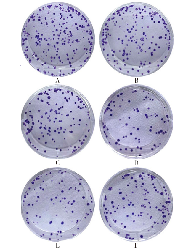
Fig.1 Results of the colony formation experiment on U266 cells
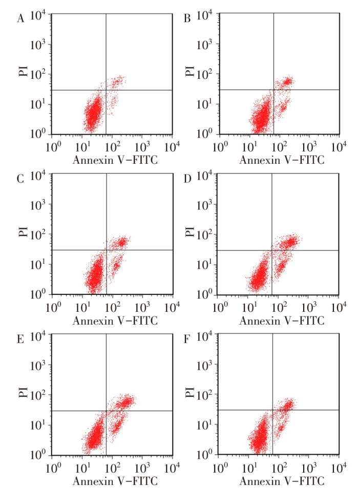
Fig.2 Flow cytometry observation of apoptosis in U266 cells
| 组别 | OD450值 | 克隆形成率/% | |||
|---|---|---|---|---|---|
| 骨髓瘤组 | 1.11±0.10 | 66.53±3.78 | |||
| Andro低浓度组 | 0.91±0.09a | 55.46±2.93a | |||
| Andro中浓度组 | 0.73±0.08ab | 41.77±2.56ab | |||
| Andro高浓度组 | 0.41±0.05abc | 28.65±1.58abc | |||
| Stattic组 | 0.36±0.04a | 25.44±1.39a | |||
| Andro高浓度+Colivelin组 | 0.69±0.07d | 36.95±2.01d | |||
| F | 88.740** | 239.300** | |||
| 组别 | 细胞凋亡率/% | 线粒体膜电位/% | |||
| 骨髓瘤组 | 4.58±0.26 | 56.57±3.19 | |||
| Andro低浓度组 | 8.99±0.43a | 47.78±2.75a | |||
| Andro中浓度组 | 13.78±0.71ab | 37.76±2.44ab | |||
| Andro高浓度组 | 23.44±1.38abc | 29.68±1.67abc | |||
| Stattic组 | 24.06±1.41a | 27.71±1.83a | |||
| Andro高浓度+Colivelin组 | 16.62±0.93d | 35.56±2.05d | |||
| F | 394.000** | 129.800** |
Tab.1 Comparison of OD450 values, colony formation rate, apoptosis rate and mitochondrial membrane potential of U266 cells between six groups (n=6,$\bar{x}±s$)
| 组别 | OD450值 | 克隆形成率/% | |||
|---|---|---|---|---|---|
| 骨髓瘤组 | 1.11±0.10 | 66.53±3.78 | |||
| Andro低浓度组 | 0.91±0.09a | 55.46±2.93a | |||
| Andro中浓度组 | 0.73±0.08ab | 41.77±2.56ab | |||
| Andro高浓度组 | 0.41±0.05abc | 28.65±1.58abc | |||
| Stattic组 | 0.36±0.04a | 25.44±1.39a | |||
| Andro高浓度+Colivelin组 | 0.69±0.07d | 36.95±2.01d | |||
| F | 88.740** | 239.300** | |||
| 组别 | 细胞凋亡率/% | 线粒体膜电位/% | |||
| 骨髓瘤组 | 4.58±0.26 | 56.57±3.19 | |||
| Andro低浓度组 | 8.99±0.43a | 47.78±2.75a | |||
| Andro中浓度组 | 13.78±0.71ab | 37.76±2.44ab | |||
| Andro高浓度组 | 23.44±1.38abc | 29.68±1.67abc | |||
| Stattic组 | 24.06±1.41a | 27.71±1.83a | |||
| Andro高浓度+Colivelin组 | 16.62±0.93d | 35.56±2.05d | |||
| F | 394.000** | 129.800** |
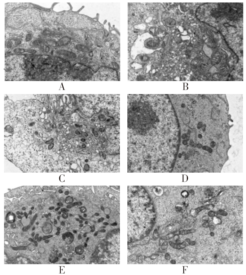
Fig.3 Observation of mitochondrial morphology in U266 cells detected by transmission electron microscopy (×8 000)
| 组别 | Fe2+/(nmol/106 cell) | 4-HNE/(mmol/g) | |||
|---|---|---|---|---|---|
| 骨髓瘤组 | 0.23±0.03 | 0.45±0.05 | |||
| Andro低浓度组 | 0.54±0.06a | 0.89±0.09a | |||
| Andro中浓度组 | 0.95±0.10ab | 1.63±0.15ab | |||
| Andro高浓度组 | 1.89±0.17abc | 2.44±0.19abc | |||
| Stattic组 | 2.04±0.16a | 2.51±0.21a | |||
| Andro高浓度+Colivelin组 | 1.08±0.12d | 1.53±0.14d | |||
| F | 224.300** | 182.200** | |||
| 组别 | MDA/(μmol/g) | GSH/(μmol/g) | ROS/% | ||
| 骨髓瘤组 | 20.33±1.58 | 9.86±0.51 | 4.38±0.25 | ||
| Andro低浓度组 | 28.77±1.69a | 7.73±0.39a | 9.65±0.51a | ||
| Andro中浓度组 | 37.73±1.98ab | 5.45±2.90ab | 16.69±0.97ab | ||
| Andro高浓度组 | 46.65±2.82abc | 2.63±0.15abc | 35.76±1.89abc | ||
| Stattic组 | 49.44±3.15a | 2.43±0.13a | 37.26±2.34a | ||
| Andro高浓度+Colivelin组 | 35.51±1.87d | 4.88±0.26d | 18.88±1.06d | ||
| F | 139.600** | 33.890** | 572.000** | ||
Tab.2 Comparison of Fe2+, 4-HNE, MDA, GSH and ROS levels in U266 cells between six groups (n=6,$\bar{x}±s$)
| 组别 | Fe2+/(nmol/106 cell) | 4-HNE/(mmol/g) | |||
|---|---|---|---|---|---|
| 骨髓瘤组 | 0.23±0.03 | 0.45±0.05 | |||
| Andro低浓度组 | 0.54±0.06a | 0.89±0.09a | |||
| Andro中浓度组 | 0.95±0.10ab | 1.63±0.15ab | |||
| Andro高浓度组 | 1.89±0.17abc | 2.44±0.19abc | |||
| Stattic组 | 2.04±0.16a | 2.51±0.21a | |||
| Andro高浓度+Colivelin组 | 1.08±0.12d | 1.53±0.14d | |||
| F | 224.300** | 182.200** | |||
| 组别 | MDA/(μmol/g) | GSH/(μmol/g) | ROS/% | ||
| 骨髓瘤组 | 20.33±1.58 | 9.86±0.51 | 4.38±0.25 | ||
| Andro低浓度组 | 28.77±1.69a | 7.73±0.39a | 9.65±0.51a | ||
| Andro中浓度组 | 37.73±1.98ab | 5.45±2.90ab | 16.69±0.97ab | ||
| Andro高浓度组 | 46.65±2.82abc | 2.63±0.15abc | 35.76±1.89abc | ||
| Stattic组 | 49.44±3.15a | 2.43±0.13a | 37.26±2.34a | ||
| Andro高浓度+Colivelin组 | 35.51±1.87d | 4.88±0.26d | 18.88±1.06d | ||
| F | 139.600** | 33.890** | 572.000** | ||
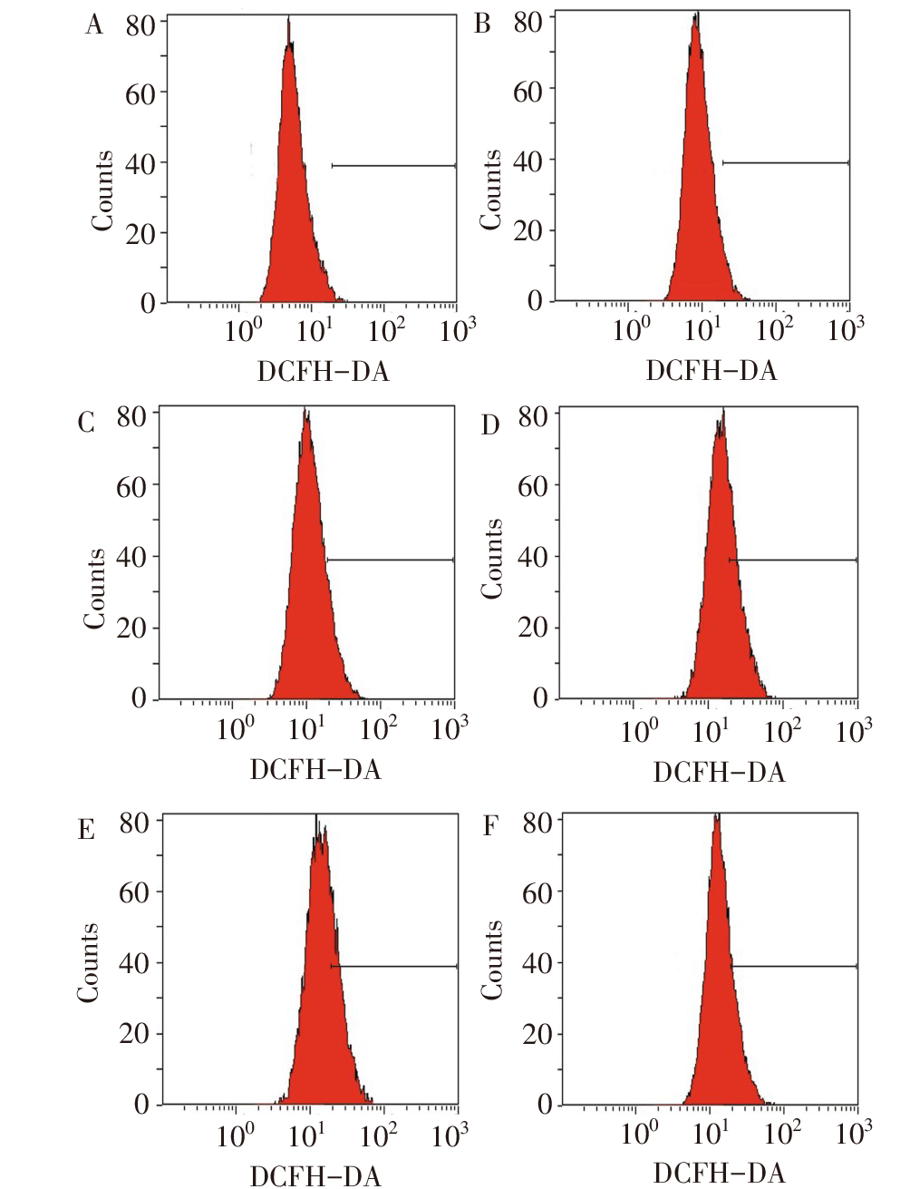
Fig.4 ROS levels in U266 cells observed by flow cytometry
| 组别 | PCNA/GAPDH | Bcl-2/GAPDH | Bax/GAPDH | Bcl-2/Bax | SLC7A11/GAPDH | p-STAT3/STAT3 | GPX4/GAPDH |
|---|---|---|---|---|---|---|---|
| 骨髓瘤组 | 1.89±0.21 | 0.98±0.10 | 0.16±0.02 | 6.13±0.34 | 1.34±0.15 | 0.89±0.10 | 1.21±0.13 |
| Andro低浓度组 | 1.61±0.18a | 0.75±0.08a | 0.34±0.04a | 2.21±0.12a | 1.02±0.11a | 0.71±0.08a | 0.95±0.10a |
| Andro中浓度组 | 1.03±0.11ab | 0.51±0.06ab | 0.53±0.06ab | 0.96±0.10ab | 0.83±0.09ab | 0.54±0.06ab | 0.74±0.08ab |
| Andro高浓度组 | 0.67±0.08abc | 0.23±0.03abc | 0.89±0.09abc | 0.26±0.03abc | 0.54±0.06abc | 0.26±0.03abc | 0.11±0.02abc |
| Stattic组 | 0.69±0.09a | 0.26±0.03a | 0.91±0.10a | 0.29±0.03a | 0.50±0.06a | 0.22±0.03a | 0.13±0.02a |
| Andro高浓度+Colivelin组 | 0.98±0.11d | 0.54±0.06d | 0.56±0.07d | 0.96±0.09d | 0.88±0.09d | 0.46±0.05d | 0.76±0.08d |
| F | 77.770** | 117.100** | 110.900** | 1 200.000** | 60.690** | 99.060** | 175.300** |
Tab.3 Comparison of PCNA, Bcl-2/Bax, SLC7A11, p-STAT3 and GPX4 proteins in U266 cells between six groups (n=6,$\bar{x}±s$)
| 组别 | PCNA/GAPDH | Bcl-2/GAPDH | Bax/GAPDH | Bcl-2/Bax | SLC7A11/GAPDH | p-STAT3/STAT3 | GPX4/GAPDH |
|---|---|---|---|---|---|---|---|
| 骨髓瘤组 | 1.89±0.21 | 0.98±0.10 | 0.16±0.02 | 6.13±0.34 | 1.34±0.15 | 0.89±0.10 | 1.21±0.13 |
| Andro低浓度组 | 1.61±0.18a | 0.75±0.08a | 0.34±0.04a | 2.21±0.12a | 1.02±0.11a | 0.71±0.08a | 0.95±0.10a |
| Andro中浓度组 | 1.03±0.11ab | 0.51±0.06ab | 0.53±0.06ab | 0.96±0.10ab | 0.83±0.09ab | 0.54±0.06ab | 0.74±0.08ab |
| Andro高浓度组 | 0.67±0.08abc | 0.23±0.03abc | 0.89±0.09abc | 0.26±0.03abc | 0.54±0.06abc | 0.26±0.03abc | 0.11±0.02abc |
| Stattic组 | 0.69±0.09a | 0.26±0.03a | 0.91±0.10a | 0.29±0.03a | 0.50±0.06a | 0.22±0.03a | 0.13±0.02a |
| Andro高浓度+Colivelin组 | 0.98±0.11d | 0.54±0.06d | 0.56±0.07d | 0.96±0.09d | 0.88±0.09d | 0.46±0.05d | 0.76±0.08d |
| F | 77.770** | 117.100** | 110.900** | 1 200.000** | 60.690** | 99.060** | 175.300** |
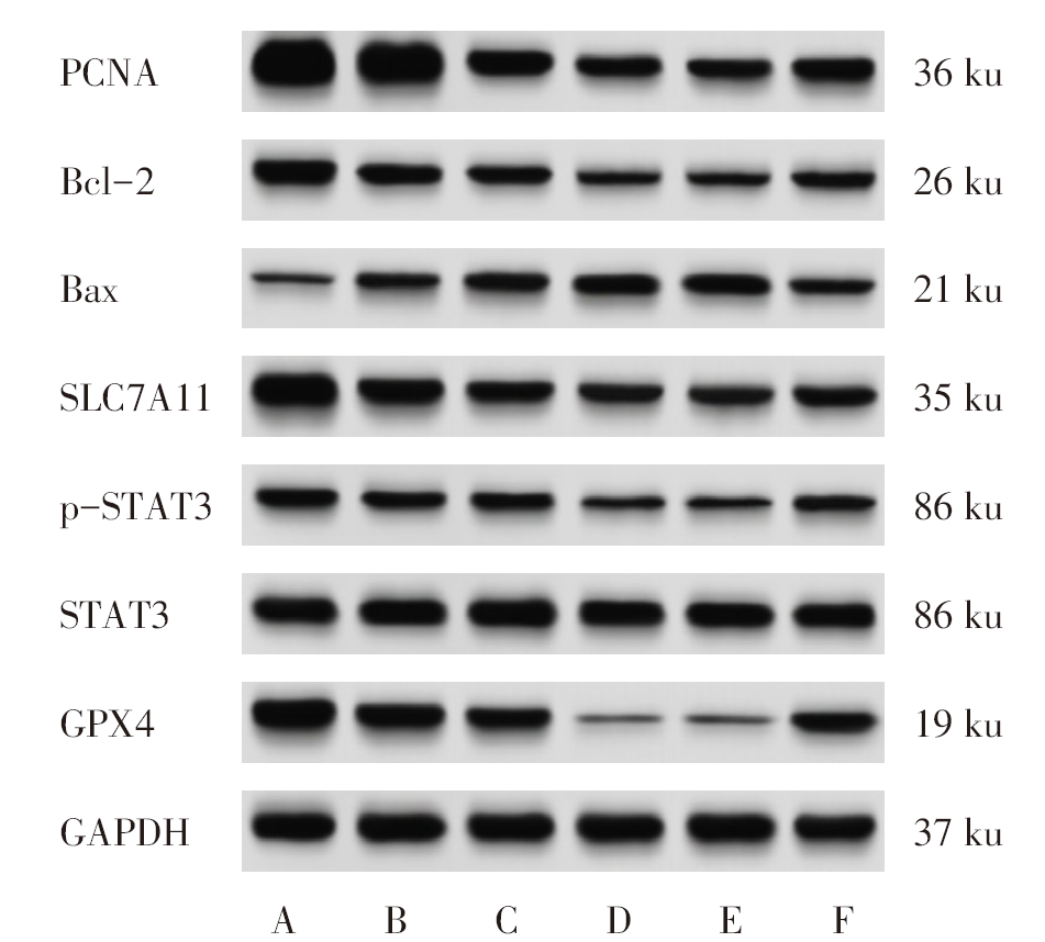
Fig.5 Western blot analysis of PCNA, Bcl-2, Bax, SLC7A11, p-STAT3 and GPX4 protein expressions in U266 cells
| [1] | XU Y, WANG T, WAN J, et al. Long non-coding RNA NEAT1 promotes multiple myeloma malignant transformation via targeting miR-485-5p/ABCB8[J]. Hematology, 2024, 29(1):2422153. doi:10.1080/16078454.2024.2422153. |
| [2] | GUI H, FAN X. Anti-tumor effect of dandelion flavone on multiple myeloma cells and its mechanism[J]. Discov Oncol, 2024, 15(1):215. doi:10.1007/s12672-024-01076-z. |
| [3] | JIANG W, ZHOU M. Analysis of the role of dihydromyricetin derived from vine tea (Ampelopsis grossedentata) on multiple myeloma by activating STAT1/RIG-I axis[J]. Oncol Res, 2024, 32(8):1359-1368. doi:10.32604/or.2024.043423. |
| [4] | HU J, LI Y, XIE X, et al. The therapeutic potential of andrographolide in cancer treatment[J]. Biomed Pharmacother, 2024,180:117438. doi:10.1016/j.biopha.2024.117438. |
| [5] | DOI H, MATSUI T, DIJKSTRA J M, et al. Andrographolide,isolated from Andrographis paniculata,induces apoptosis in monocytic leukemia and multiple myeloma cells via augmentation of reactive oxygen species production[J]. F1000Res, 2021,10:542. doi:10.12688/f1000research.53595.3. |
| [6] | YU H, KOU Q, YUAN H, et al. Alkannin triggered apoptosis and ferroptosis in gastric cancer by suppressing lipid metabolism mediated by the c-Fos/SREBF1 axis[J]. Phytomedicine, 2025,140:156604. doi:10.1016/j.phymed.2025.156604. |
| [7] | LI W, YIN X, FU H, et al. Ethanol extract of Eclipta prostrata induces multiple myeloma ferroptosis via Keap1/Nrf2/HO-1 axis[J]. Phytomedicine, 2024,128:155401. doi:10.1016/j.phymed.2024.155401. |
| [8] | LI G, FENG J, HUANG S, et al. LncRNA-PVT1 inhibits ferroptosis through activating STAT3/GPX4 axis to promote osteosarcoma progression[J]. Front Biosci (Landmark Ed), 2024, 29(6):207. doi:10.31083/j.fbl2906207. |
| [9] | 韦佳, 李蓉, 王卉妍, 等. 秦巴硒菇提取物FA-2-b-β通过STAT3/GPX4信号通路诱导伯基特淋巴瘤细胞铁死亡[J]. 中国病理生理杂志, 2025, 41(3):453-462. |
| WEI J, LI R, WANG H Y, et al. Agaricus blazei extract FA-2-b-β induces ferroptosis of Burkitt lymphoma cells through STAT3/GPX4 signaling pathway[J]. Chin J Pathophysiol, 2025, 41(3):453-462. doi:10.3969/j.issn.1000-4718.2025.03.005. | |
| [10] | SUN L, LI X, XIAO Y, et al. Mfsd2a suppresses colorectal cancer progression and liver metastasis via the S100A14/STAT3 axis[J]. J Transl Med, 2025, 23(1):59. doi:10.1186/s12967-024-05994-y. |
| [11] | WU H, QIAN J, ZHOU L, et al. FHND004 inhibits malignant proliferation of multiple myeloma by targeting PDZ-binding kinase in MAPK pathway[J]. Aging (Albany NY), 2024, 16(5):4811-4831. doi:10.18632/aging.205634. |
| [12] | WANG Z, CHEN H, CAI X, et al. Andrographolide induces protective autophagy and targeting DJ-1 triggers reactive oxygen species-induced cell death in pancreatic cancer[J]. PeerJ, 2024,12:e17619. doi:10.7717/peerj.17619. |
| [13] | LUO Y, HU J, JIAO Y, et al. Andrographolide anti-proliferation and metastasis of hepatocellular carcinoma through LncRNA MIR22HG regulation[J]. J Nat Med, 2024, 78(1):123-145. doi:10.1007/s11418-023-01752-4. |
| [14] | LI W, FU H, FANG L, et al. Andrographolide induced ferroptosis in multiple myeloma cells by regulating the P38/Nrf2/HO-1 pathway[J]. Arch Biochem Biophys, 2023,742:109622. doi:10.1016/j.abb.2023.109622. |
| [15] | ARBEL-GROISSMAN M, LIEFSHITZ B, KATZ N, et al. PCNA unloading is crucial for the bypass of DNA lesions using homologous recombination[J]. Int J Mol Sci, 2024, 25(6):3359. doi:10.3390/ijms25063359. |
| [16] | WAN C, LIU S, ZHAO L, et al. Curcumin protects rat endplate chondrocytes against IL-1β-induced apoptosis via Bcl-2/Bax regulation[J]. J Mol Histol, 2025, 56(2):111. doi:10.1007/s10735-025-10390-x. |
| [17] | GLOVER H L, SCHREINER A, DEWSON G, et al. Mitochondria and cell death[J]. Nat Cell Biol, 2024, 26(9):1434-1446. doi:10.1038/s41556-024-01429-4. |
| [18] | YANG L, LIU X, CHEN S, et al. Scutellarin ameliorates mitochondrial dysfunction and apoptosis in OGD/R-insulted HT22 cells through mitophagy induction[J]. Biomed Pharmacother, 2024,179:117340. doi:10.1016/j.biopha.2024.117340. |
| [19] | PENG X, SUN B, TANG C, et al. HMOX1-LDHB interaction promotes ferroptosis by inducing mitochondrial dysfunction in foamy macrophages during advanced atherosclerosis[J]. Dev Cell, 2025, 60(7):1070-1086.e8. doi:10.1016/j.devcel.2024.12.011. |
| [20] | DIXON S J, OLZMANN J A. The cell biology of ferroptosis[J]. Nat Rev Mol Cell Biol, 2024, 25(6):424-442. doi:10.1038/s41580-024-00703-5. |
| [21] | JIWA H, XIE Z, QU X, et al. Casticin induces ferroptosis in human osteosarcoma cells through Fe2+overload and ROS production mediated by HMOX1 and LC3-NCOA4[J]. Biochem Pharmacol, 2024,226:116346. doi:10.1016/j.bcp.2024.116346. |
| [22] | KIM N Y, SHIVANNE GOWDA S G, LEE S G, et al. Cannabidiol induces ERK activation and ROS production to promote autophagy and ferroptosis in glioblastoma cells[J]. Chem Biol Interact, 2024,394:110995. doi:10.1016/j.cbi.2024.110995. |
| [23] | ZHANG M, ZHENG H, ZHU X, et al. Synchronously evoking disulfidptosis and ferroptosis via systematical glucose deprivation targeting SLC7A11/GSH/GPX4 antioxidant axis[J]. ACS Nano, 2025, 19(14):14233-14248. doi:10.1021/acsnano.5c00730. |
| [24] | ZHONG X, ZHANG W, ZHANG W, et al. FASN contributes to ADM resistance of diffuse large B-cell lymphoma by inhibiting ferroptosis via NF-κB/STAT3/GPX4 axis[J]. Cancer Biol Ther, 2024, 25(1):2403197. doi:10.1080/15384047.2024.2403197. |
| [25] | SUN Q, LU H, YUAN F, et al. SLC10A3 regulates ferroptosis of glioblastoma through the STAT3/GPX4 pathway[J]. Sci Rep, 2025, 15(1):21259. doi:10.1038/s41598-025-05866-5. |
| [26] | ZHANG W, GONG M, ZHANG W, et al. Thiostrepton induces ferroptosis in pancreatic cancer cells through STAT3/GPX4 signalling[J]. Cell Death Dis, 2022, 13(7):630. doi:10.1038/s41419-022-05082-3. |
| Viewed | ||||||
|
Full text |
|
|||||
|
Abstract |
|
|||||