
Tianjin Medical Journal ›› 2026, Vol. 54 ›› Issue (7): 693-698.doi: 10.11958/20260440
• Experimental Research • Previous Articles Next Articles
HAN Wanting( ), FU Tong, LIU Lindong, NI Lingling, WU Xinying()
Received:2026-02-10
Revised:2026-03-22
Published:2026-07-15
Online:2026-07-13
Contact:
△E-mail:rebeccahxt@163.com
HAN Wanting, FU Tong, LIU Lindong, NI Lingling, WU Xinying. An experimental study of multimodal MRI technology for evaluating the impact of tanshinone ⅡA on glymphatic system function in rats with Alzheimer's disease[J]. Tianjin Medical Journal, 2026, 54(7): 693-698.
CLC Number:
| 组别 | ADC/(×10-3 mm2/s) | DDC/(×10-3 mm2/s) | 异质性指数α |
|---|---|---|---|
| Control组 | 1.11±0.03 | 0.83±0.10 | 0.84±0.02 |
| AD组 | 0.61±0.04a | 0.42±0.05a | 0.64±0.03a |
| AD+Tgn-073组 | 0.91±0.08b | 0.69±0.03b | 0.73±0.10b |
| AD+Tan-ⅡA组 | 0.76±0.20 | 0.55±0.13 | 0.67±0.05 |
| F | 11.030** | 14.120** | 28.250** |
Tab.1 Comparison of diffusion parameters(ADC,DDC and α) in brain of rats between the four groups (n=3,$\stackrel{-}{x}\pm s$)
| 组别 | ADC/(×10-3 mm2/s) | DDC/(×10-3 mm2/s) | 异质性指数α |
|---|---|---|---|
| Control组 | 1.11±0.03 | 0.83±0.10 | 0.84±0.02 |
| AD组 | 0.61±0.04a | 0.42±0.05a | 0.64±0.03a |
| AD+Tgn-073组 | 0.91±0.08b | 0.69±0.03b | 0.73±0.10b |
| AD+Tan-ⅡA组 | 0.76±0.20 | 0.55±0.13 | 0.67±0.05 |
| F | 11.030** | 14.120** | 28.250** |
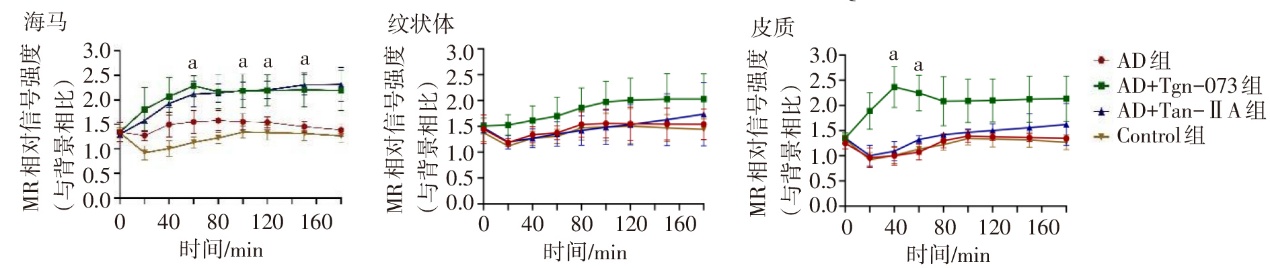
Fig.1 Temporal trends of signal intensity variations in hippocampus, striatum and cortex of each group
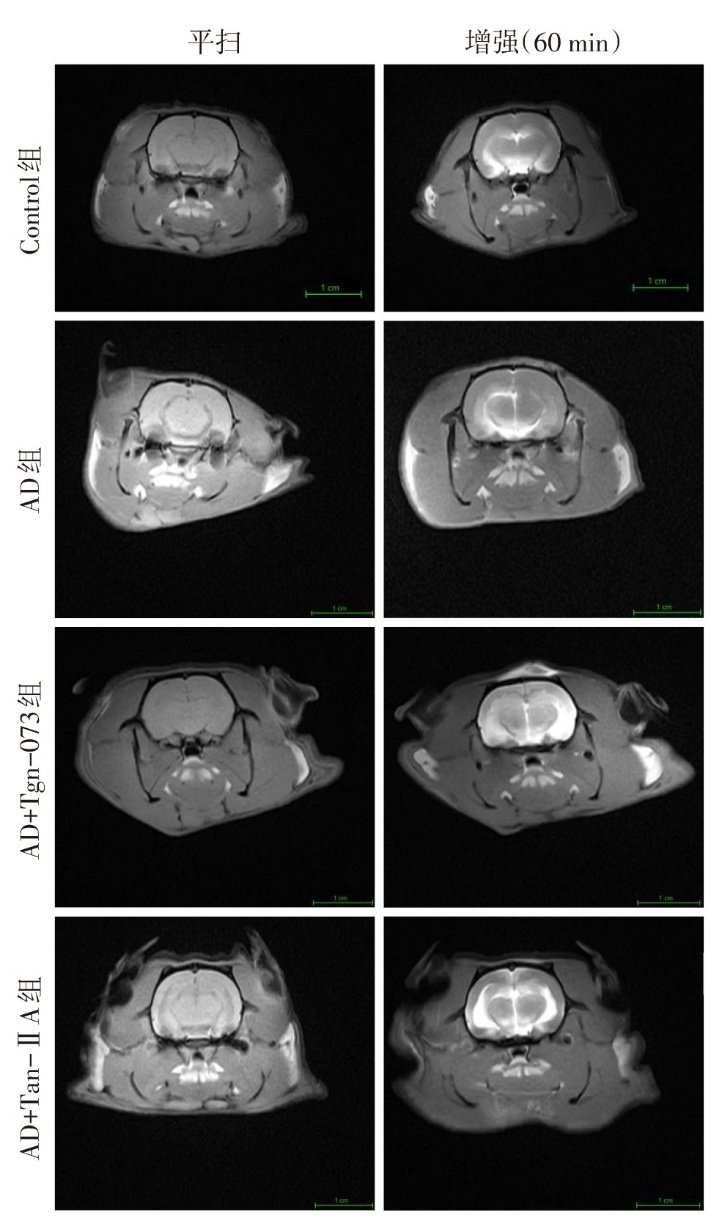
Fig.2 Representative T1-weighted MR plain scan combined with contrast-enhanced images between the four groups
| 组别 | 海马 | 纹状体 | 皮质 |
|---|---|---|---|
| Control组 | 0.98±0.08 | 0.99±0.05 | 1.00±0.04 |
| AD组 | 1.28±0.08a | 0.98±0.13 | 0.94±0.10 |
| AD+Tgn-073组 | 1.11±0.12b | 0.81±0.07b | 0.84±0.06b |
| AD+Tan-ⅡA组 | 1.09±0.14b | 0.88±0.04b | 0.91±0.05 |
| F | 14.790** | 13.210** | 10.670** |
Tab.2 Comparison of AQP4 in hippocampus,striatum and cortex of rats between the four groups (n=12,$\stackrel{-}{x}\pm s$)
| 组别 | 海马 | 纹状体 | 皮质 |
|---|---|---|---|
| Control组 | 0.98±0.08 | 0.99±0.05 | 1.00±0.04 |
| AD组 | 1.28±0.08a | 0.98±0.13 | 0.94±0.10 |
| AD+Tgn-073组 | 1.11±0.12b | 0.81±0.07b | 0.84±0.06b |
| AD+Tan-ⅡA组 | 1.09±0.14b | 0.88±0.04b | 0.91±0.05 |
| F | 14.790** | 13.210** | 10.670** |
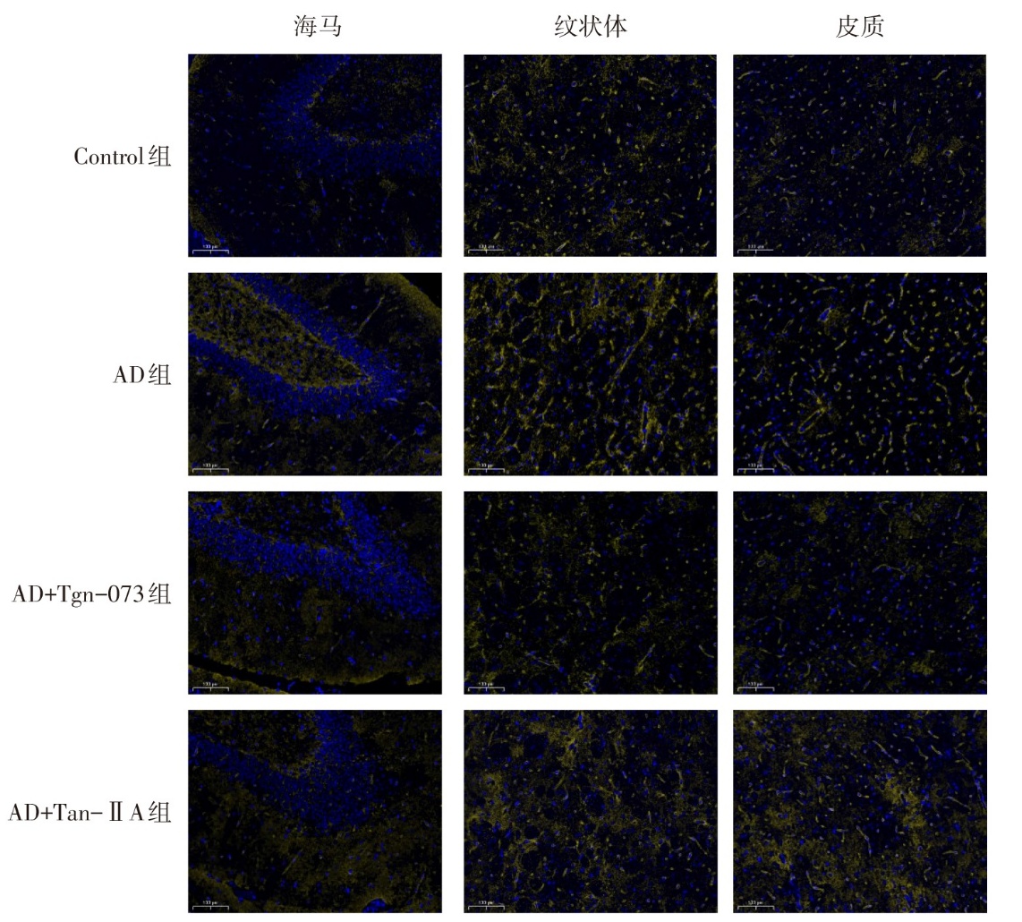
Fig.3 Expression of AQP4 in the hippocampus, striatum and caudal cortex of rats in four groups (immunofluorescence staining, ×200)
| [1] | Rostagno A A. Pathogenesis of Alzheimer's disease[J]. Int J Mol Sci, 2022, 24(1):1070. doi:10.3390/ijms24010107. |
| [2] | Buccellato F R, D'Anca M, Serpente M, et al. The role of glymphatic system in Alzheimer's and Parkinson's Disease pathogenesis[J]. Biomedicines, 2022, 10(9):2261. doi:10.3390/biomedicines10092261. |
| [3] | Chong P, Garic D, Shen M D, et al. Sleep,cerebrospinal fluid,and the glymphatic system:a systematic review[J]. Sleep Med Rev, 2022, 61:101572. doi:10.1016/j.smrv.2021.101572. |
| [4] | Hablitz L M, Nedergaard M. The glymphatic system:a novel component of fundamental neurobiology[J]. J Neurosci, 2021, 41(37):7698-7711. doi:10.1523/JNEUROSCI.0619-21.2021. |
| [5] | Harrison I F, Ismail O, Machhada A, et al. Impaired glymphatic function and clearance of tau in an Alzheimer's disease model[J]. Brain, 2020, 143(8):2576-2593. doi:10.1093/brain/awaa179. |
| [6] | Yamada K, Iwatsubo T. Involvement of the glymphatic/meningeal lymphatic system in Alzheimer's disease:insights into proteostasis and future directions[J]. Cell Mol Life Sci, 2024, 81(1):192. doi:10.1007/s00018-024-05225-z. |
| [7] | 翟书鹏, 贾航, 苗统, 等. 帕金森病患者血清Trx1、PDCD4、AQP4水平与认知功能的关系[J]. 天津医药, 2025, 53(2):146-150. |
| Zhai S P, Jia H, Miao T, et al. The relationship between serum Trx1,PDCD4,AQP4 expression and cognitive function in patients with Parkinson's disease[J]. Tianjin Med J, 2025, 53(2):146-150. doi:10.11958/20241632. | |
| [8] | Liu X Q, Hu T, Wu G L, et al. Tanshinone IIA,the key compound in Salvia miltiorrhiza,improves cognitive impairment by upregulating Aβ-degrading enzymes in APP/PS1 mice[J]. Int J Biol Macromol, 2024, 254(Pt 2):127923. doi:10.1016/j.ijbiomac.2023.127923. |
| [9] | Peng X, Chen L, Wang Z, et al. Tanshinone IIA regulates glycogen synthase kinase-3β-related signaling pathway and ameliorates memory impairment in APP/PS1 transgenic mice[J]. Eur J Pharmacol, 2022, 918:174772. doi:10.1016/j.ejphar.2022.174772. |
| [10] | Alghanimy A, Martin C, Gallagher L, et al. The effect of a novel AQP4 facilitator,TGN-073,on glymphatic transport captured by diffusion MRI and DCE-MRI[J]. PLoS One, 2023, 18(3):e0282955. doi:10.1371/journal.pone.0282955. |
| [11] | 朱青春, 崔国红, 邵水金, 等. 阿尔茨海默病大鼠自噬相关蛋白Beclin-1和凋亡相关蛋白p53表达变化及电针的调控作用[J]. 中国中医药信息杂志, 2014(9):68-71. |
| Zhu Q C, Cui G H, Shao S J, et al. Expression changes of autophagy associated protein Beclin-1 and apoptosis associated protein p53 in rats with Alzheimer's disease and regulatory effects of electroacupuncture[J]. Chinese Journal of Information on TCM, 2014(9):68-71. doi:10.3969/j.issn.1005-5304.2014.09.020. | |
| [12] | Yang P, Wang Z, Zhang Z, et al. The extended application of the rat brain in stereotaxic coordinates in rats of various body weight[J]. J Neurosci Methods, 2018, 307:60-69. doi:10.1016/j.jneumeth.2018.06.026. |
| [13] | Lopes D M, Wells J A, Ma D, et al. Glymphatic inhibition exacerbates tau propagation in an Alzheimer's disease model[J]. Alzheimers Res Ther, 2024, 16(1):71. doi:10.1186/s13195-024-01439-2. |
| [14] | Xavier A L R, Hauglund N L, Von Holstein-Rathlou S, et al. Cannula implantation into the cisterna magna of rodents[J]. J Vis Exp, 2018, 135:57378. doi:10.3791/57378. |
| [15] | Guo R, Lu F, Lin J, et al. Multi-b-value DWI to evaluate the synergistic antiproliferation and anti-heterogeneity effects of bufalin plus sorafenib in an orthotopic HCC model[J]. Eur Radiol Exp, 2024, 8(1):43. doi:10.1186/s41747-024-00448-y. |
| [16] | Zhang G, Yan R, Liu W, et al. Use of biexponential and stretched exponential models of intravoxel incoherent motion and dynamic contrast-enhanced magnetic resonance imaging to assess the proliferation of endometrial carcinoma[J]. Quant Imaging Med Surg, 2023, 13(4):2568-2581. doi:10.21037/qims-22-688. |
| [17] | Yushkevich P A, Piven J, Hazlett H C, et al. User-guided 3D active contour segmentation of anatomical structures: significantly improved efficiency and reliability[J]. Neuroimage, 2006, 31(3):1116-1128. doi:10.1016/j.neuroimage.2006.01.015. |
| [18] | 陈宗勤, 鲍奕仿, 李郁欣. 早期阿尔茨海默病Aβ单抗治疗中淀粉样蛋白相关影像异常的研究进展[J]. 国际医学放射学杂志, 2025, 48(1):59-63. |
| Chen Z Q, Bao Y F, Li Y X. Research progress on amyloid-related imaging abnormalities in early Alzheimer’s disease treated with Aβ monoclonal antibody[J]. Int J Med Radiol, 2025, 48(1):59-63. doi:10.19300/j.2025.Z21811. | |
| [19] | Zhang H, Wei W, Zhao M, et al. Interaction between Aβ and Tau in the pathogenesis of Alzheimer's disease[J]. Int J Biol Sci, 2021, 17(9):2181-2192. doi:10.7150/ijbs.57078. |
| [20] | Silva I, Silva J, Ferreira R, et al. Glymphatic system,AQP4,and their implications in Alzheimer's disease[J]. Neurol Res Pract, 2021, 3(1):5. doi:10.1186/s42466-021-00102-7. |
| [21] | Peng W, Achariyar T M, Li B, et al. Suppression of glymphatic fluid transport in a mouse model of Alzheimer's disease[J]. Neurobiol Dis, 2016, 93:215-225. doi:10.1016/j.nbd.2016.05.015. |
| [22] | Fang L, Cheng H, Chen W, et al. Therapeutic effects of tanshinone IIA and Tetramethylpyrazine nanoemulsions on cognitive impairment and neuronal damage in Alzheimer's disease rat models[J]. J Pharm Pharmacol, 2024, 76(9):1169-1177. doi:10.1093/jpp/rgae069. |
| [23] | Yang L X, Luo M, Li S Y. Tanshinone IIA improves Alzheimer's disease via RNA nuclear-enriched abundant transcript 1/microRNA-291a-3p/member RAS oncogene family Rab22a axis[J]. World J Psychiatry, 2024, 14(4):563-581. doi:10.5498/wjp.v14.i4.563. |
| [24] | Du L, Xu B, Zhao Z, et al. Identification and classification of Alzheimer's disease patients using novel fractional motion model[J]. Front Neurosci, 2020, 14:767. doi:10.3389/fnins.2020.00767. |
| [25] | Xue Y, Zhang Z, Wen C, et al. Characterization of Alzheimer's disease using ultra-high b-values apparent diffusion coefficient and diffusion kurtosis imaging[J]. Aging Dis, 2019, 10(5):1026-1036. doi:10.14336/AD.2018.1129. |
| [26] | Syková E, VoríSek I, Antonova T, et al. Changes in extracellular space size and geometry in APP23 transgenic mice:a model of Alzheimer's disease[J]. Proc Natl Acad Sci U S A, 2005, 102(2):479-484. doi:10.1073/pnas.0408235102. |
| [27] | Mueggler T, Meyer-Luehmann M, Rausch M, et al. Restricted diffusion in the brain of transgenic mice with cerebral amyloidosis[J]. Eur J Neurosci, 2004, 20(3):811-817. doi:10.1111/j.1460-9568.2004.03534.x. |
| [28] | Chahardehi A M, Arefnezhad R, Pourbafrani A, et al. MicroRNAs modulation by curcumin,catalpol,and other natural products in Alzheimer's disease: a review[J]. Mol Biol Rep, 2025, 52(1):445. doi:10.1007/s11033-025-10543-x. |
| [29] | Salman M M, Kitchen P, Halsey A, et al. Emerging roles for dynamic aquaporin-4 subcellular relocalization in CNS water homeostasis[J]. Brain, 2022, 145(1):64-75. doi:10.1093/brain/awab311. |
| [30] | Hablitz L M, Plá V, Giannetto M, et al. Circadian control of brain glymphatic and lymphatic fluid flow[J]. Nat Commun, 2020, 11(1):4411. doi:10.1038/s41467-020-18115-2. |
| [31] | Barichello De Quevedo J L, Leffa D T, Pascoal T A. Glymphatic system waste clearance and Alzheimer's disease[J]. Braz J Psychiatry, 2023, 45(5):385-386. doi:10.47626/1516-4446-2023-0049. |
| [1] | REN Baolong, JIAO Aiju, ZHU Ruolan, ZHAO Weijing. The predictive value of lipoprotein-associated phospholipase A2 combined with the total burden of MRI scores on cognitive impairment in patients with cerebral small vessel disease [J]. Tianjin Medical Journal, 2026, 54(3): 245-248. |
| [2] | GU Cheng, SHEN Xinyu, SUN Jinghua, YAN Saike, WANG Haiping. The evaluation value of dynamic MRI imaging technology for LARS after anorectal preservation surgery in low rectal cancer [J]. Tianjin Medical Journal, 2024, 52(6): 653-657. |
| [3] | ZHANG Xue, WANG Jiarui, CHEN Kangyin. Effects of glycyrrhizin on myocardial HMGB1/TLR4/NF-κB/HIF-1α signaling pathway in chronic kidney disease rats [J]. Tianjin Medical Journal, 2023, 51(2): 155-159. |
| [4] | YU Yongzhen, CHENG Tianhao, ZOU Xiulan, ZHANG Mengyi, YU Yangyang, ZOU Yuping, PANG Long. Experimental study of chronic retinal damage induced by blue light exposure in Brown Norway rats [J]. Tianjin Medical Journal, 2023, 51(11): 1193-1198. |
| [5] | SUN Haibei, BAO Yitegele, ZHANG Mohan. Inhibitory effect of Rotigaptide on autonomic contraction of gastric smooth muscle in diabetic rats [J]. Tianjin Medical Journal, 2022, 50(7): 693-697. |
| [6] | LIAO Yidong, MING Jiang, ZHANG Yu, LIAO Yifei, XU Kaya, △. Extraction and identification of primary cortical neurons of suckling rats by papain and DNA enzyme [J]. Tianjin Medical Journal, 2022, 50(6): 583-587. |
| [7] | ZHANG Jing, SUN Hui, ZHU Lijun, FENG Ziren, DU Lin, MENG Aiguo△. Relationship between miR-27a expression and ferroptosis in the early stage of ischemic stroke of rats [J]. Tianjin Medical Journal, 2022, 50(6): 595-600. |
| [8] | LI Yubai, WANG Jun△, YIN Jing. Mechanism of Wnt 5a in bronchopulmonary dysplasia model [J]. Tianjin Medical Journal, 2022, 50(6): 566-570. |
| [9] | ZHAI Yajun, YANG Han, CHEN Wanli, LIU Yue, WEI Liping, LIU Keqiang, QI Xin△. Study on the effects of allisartan isoproxil on stabilizing antihypertension and myocardial protection in spontaneous hypertensive rats [J]. Tianjin Medical Journal, 2022, 50(5): 481-486. |
| [10] | TANG Yinglin, XU Qiongfang, XU Liting . Effects of fasudil on oligodendrocyte maturation and myelination after white matter injury in preterm rats #br# [J]. Tianjin Medical Journal, 2022, 50(2): 160-166. |
| [11] | GAO Linlin, WANG Jun, GUO Yanyan, LIU Xiaomei△. Effects of folic acid on expression of VEGF and receptor 1 in placenta of IUGR rats [J]. Tianjin Medical Journal, 2022, 50(1): 73-77. |
| [12] | CHEN Hui-xia, YAN Yu-han, YU Hui, WANG Zhan-li, HU Hai. Analysis of serum metabonomics in renovascular hypertensive rats based on GC-TOF/MS [J]. Tianjin Medical Journal, 2021, 49(6): 583-587. |
| [13] | ZHANG Xiao-hong, WANG Hong-min, LI Yang, HU Chao-yang, LI Feng-zhi, CHANG Rui, HUANG Han, JIN Li . Effects of emodin on oxidative stress damage and miR-34a/SIRT1 axis in rat model of streptococcus pneumoniae pneumonia [J]. Tianjin Medical Journal, 2021, 49(6): 588-592. |
| [14] | HE Yong-hong, QIANG Li, WANG Song-ping. The effect of atorvastatin calcium on pulmonary vascular remodeling in chronic obstructive pulmonary disease rats #br# [J]. Tianjin Medical Journal, 2021, 49(6): 598-602. |
| [15] | HOU Li-ping, GENG Jian-lin, GU Wei, LIU Qing-qing. Effects of selenium yeast combined with vitamin D on thyroid related hormones and antibodies in experimental autoimmune thyroiditis rats #br# [J]. Tianjin Medical Journal, 2021, 49(2): 136-141. |
| Viewed | ||||||
|
Full text |
|
|||||
|
Abstract |
|
|||||
