
Tianjin Medical Journal ›› 2024, Vol. 52 ›› Issue (12): 1291-1295.doi: 10.11958/20241245
• Clinical Research • Previous Articles Next Articles
DENG Wenwen( ), MENG Xianghong, SUN Zhenye, YANG Qilong, WANG Zhi△()
Received:2024-08-30
Revised:2024-10-13
Published:2024-12-15
Online:2024-12-17
Contact:
△E-mail:DENG Wenwen, MENG Xianghong, SUN Zhenye, YANG Qilong, WANG Zhi. QCT analysis of the effect of knee varus on bone mineral density of medial and lateral femoral tibial compartments in knee osteoarthritis[J]. Tianjin Medical Journal, 2024, 52(12): 1291-1295.
CLC Number:
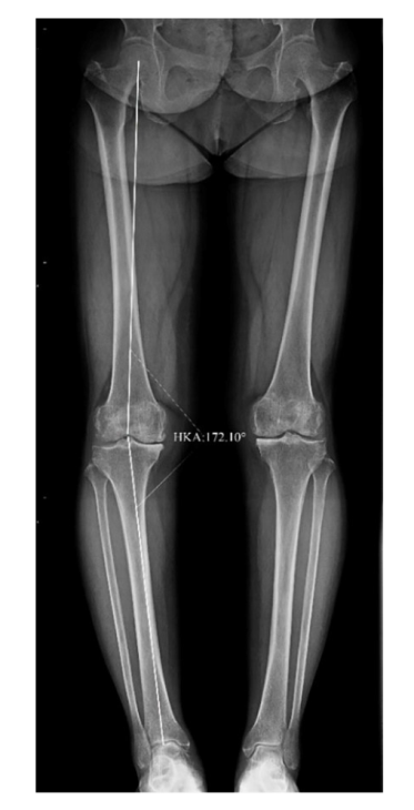
Fig.1 The weight-bearing full-length X-ray films of lower limbs
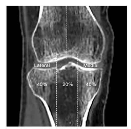
Fig.2 CT reconstruction of knee joint coronal area diagram
| 组别 | n | 股骨内髁/ (mg/cm3) | 股骨外髁/ (mg/cm3) | 胫骨内侧平台/ (mg/cm3) | 胫骨外侧平台/ (mg/cm3) | 胫骨内侧平台/ 胫骨外侧平台 | 股骨内髁/ 股骨外髁 |
|---|---|---|---|---|---|---|---|
| 力线正常组 | 131 | 162.0(130.6,195.4) | 166.1(136.4,199.9) | 125.1(102.0,143.7) | 117.7(92.8,137.6) | 1.07(1.01,1.15) | 0.96(0.93,1.00) |
| 膝内翻组 | 319 | 198.1(162.6,245.4) | 149.9(122.2,186.9) | 163.4(134.0,200.9) | 97.9(68.6,123.2) | 1.65(1.35,2.19) | 1.26(1.14,1.47) |
| Z | 7.085** | 2.885** | 8.718** | 4.337** | 14.425** | 13.808** |
Tab.1 Comparison of BMD in femoro-tibial medial and lateral compartment between the two groups
| 组别 | n | 股骨内髁/ (mg/cm3) | 股骨外髁/ (mg/cm3) | 胫骨内侧平台/ (mg/cm3) | 胫骨外侧平台/ (mg/cm3) | 胫骨内侧平台/ 胫骨外侧平台 | 股骨内髁/ 股骨外髁 |
|---|---|---|---|---|---|---|---|
| 力线正常组 | 131 | 162.0(130.6,195.4) | 166.1(136.4,199.9) | 125.1(102.0,143.7) | 117.7(92.8,137.6) | 1.07(1.01,1.15) | 0.96(0.93,1.00) |
| 膝内翻组 | 319 | 198.1(162.6,245.4) | 149.9(122.2,186.9) | 163.4(134.0,200.9) | 97.9(68.6,123.2) | 1.65(1.35,2.19) | 1.26(1.14,1.47) |
| Z | 7.085** | 2.885** | 8.718** | 4.337** | 14.425** | 13.808** |
| 组别 | n | 股骨内髁/ (mg/cm3) | 股骨外髁/ (mg/cm3) | 胫骨内侧平台/ (mg/cm3) | 胫骨外侧平台/ (mg/cm3) | 胫骨内侧平台/ 胫骨外侧平台 | 股骨内髁/ 股骨外髁 |
|---|---|---|---|---|---|---|---|
| 力线正常组 | 52 | 169.8(154.2,204.4) | 177.7(158.7,209.8) | 134.6(119.9,155.2) | 126.6(113.8,142.9) | 1.04(1.00,1.10) | 0.96(0.92,0.99) |
| 膝内翻组 | 106 | 232.5(189.6,257.1) | 187.3(148.4,205.1) | 182.8(151.5,206.0) | 119.9(102.4,141.6) | 1.42(1.25,1.78) | 1.23(1.12,1.37) |
| Z | 5.732** | 0.599 | 6.923** | 1.108 | 9.303** | 8.596** |
Tab.2 Comparison of BMD in femoro-tibial medial and lateral compartment between the two groups of male patients with KOA
| 组别 | n | 股骨内髁/ (mg/cm3) | 股骨外髁/ (mg/cm3) | 胫骨内侧平台/ (mg/cm3) | 胫骨外侧平台/ (mg/cm3) | 胫骨内侧平台/ 胫骨外侧平台 | 股骨内髁/ 股骨外髁 |
|---|---|---|---|---|---|---|---|
| 力线正常组 | 52 | 169.8(154.2,204.4) | 177.7(158.7,209.8) | 134.6(119.9,155.2) | 126.6(113.8,142.9) | 1.04(1.00,1.10) | 0.96(0.92,0.99) |
| 膝内翻组 | 106 | 232.5(189.6,257.1) | 187.3(148.4,205.1) | 182.8(151.5,206.0) | 119.9(102.4,141.6) | 1.42(1.25,1.78) | 1.23(1.12,1.37) |
| Z | 5.732** | 0.599 | 6.923** | 1.108 | 9.303** | 8.596** |
| 组别 | n | 股骨内髁/ (mg/cm3) | 股骨外髁/ (mg/cm3) | 胫骨内侧平台/ (mg/cm3) | 胫骨外侧平台/ (mg/cm3) | 胫骨内侧平台/ 胫骨外侧平台 | 股骨内髁/ 股骨外髁 |
|---|---|---|---|---|---|---|---|
| 力线正常组 | 79 | 156.8(120.9,182.5) | 153.1(125.7,181.1) | 117.9(99.1,140.4) | 105.0(87.5,130.6) | 1.08(1.03,1.20) | 0.96(0.93,1.01) |
| 膝内翻组 | 213 | 183.0(155.3,225.2) | 139.6(110.4,166.9) | 156.8(120.6,191.8) | 82.7(61.2,107.1) | 1.75(1.44,2.40) | 1.28(1.15,1.53) |
| Z | 5.429** | 2.710** | 6.480** | 4.553** | 11.151** | 10.750** |
Tab.3 Comparison of BMD in femoro-tibial medial and lateral compartment between the two groups of female patients with KOA
| 组别 | n | 股骨内髁/ (mg/cm3) | 股骨外髁/ (mg/cm3) | 胫骨内侧平台/ (mg/cm3) | 胫骨外侧平台/ (mg/cm3) | 胫骨内侧平台/ 胫骨外侧平台 | 股骨内髁/ 股骨外髁 |
|---|---|---|---|---|---|---|---|
| 力线正常组 | 79 | 156.8(120.9,182.5) | 153.1(125.7,181.1) | 117.9(99.1,140.4) | 105.0(87.5,130.6) | 1.08(1.03,1.20) | 0.96(0.93,1.01) |
| 膝内翻组 | 213 | 183.0(155.3,225.2) | 139.6(110.4,166.9) | 156.8(120.6,191.8) | 82.7(61.2,107.1) | 1.75(1.44,2.40) | 1.28(1.15,1.53) |
| Z | 5.429** | 2.710** | 6.480** | 4.553** | 11.151** | 10.750** |
| 组别 | n | 股骨内髁/ (mg/cm3) | 股骨外髁/ (mg/cm3) | 胫骨内侧平台/ (mg/cm3) | 胫骨外侧平台/ (mg/cm3) | 胫骨内侧平台/ 胫骨外侧平台 | 股骨内髁/ 股骨外髁 |
|---|---|---|---|---|---|---|---|
| 力线正常组 | 48 | 195.2(168.6,207.5) | 200.4(166.3,215.4) | 142.1(120.5,163.8) | 132.4(112.5,147.0) | 1.08(1.02,1.15) | 0.97(0.93,0.99) |
| 膝内翻组 | 137 | 215.0(182.0,254.8) | 171.1(147.3,201.0) | 182.7(151.4,216.5) | 114.2(89.7,139.2) | 1.51(1.29,1.85) | 1.22(1.11,1.40) |
| Z | 3.774** | 2.944** | 5.585** | 2.806** | 9.221** | 7.899** |
Tab.4 Comparison of BMD in femoro-tibial medial and lateral compartment between the two groups of KOA patients under 60 years of age
| 组别 | n | 股骨内髁/ (mg/cm3) | 股骨外髁/ (mg/cm3) | 胫骨内侧平台/ (mg/cm3) | 胫骨外侧平台/ (mg/cm3) | 胫骨内侧平台/ 胫骨外侧平台 | 股骨内髁/ 股骨外髁 |
|---|---|---|---|---|---|---|---|
| 力线正常组 | 48 | 195.2(168.6,207.5) | 200.4(166.3,215.4) | 142.1(120.5,163.8) | 132.4(112.5,147.0) | 1.08(1.02,1.15) | 0.97(0.93,0.99) |
| 膝内翻组 | 137 | 215.0(182.0,254.8) | 171.1(147.3,201.0) | 182.7(151.4,216.5) | 114.2(89.7,139.2) | 1.51(1.29,1.85) | 1.22(1.11,1.40) |
| Z | 3.774** | 2.944** | 5.585** | 2.806** | 9.221** | 7.899** |
| 组别 | n | 股骨内髁/ (mg/cm3) | 股骨外髁/ (mg/cm3) | 胫骨内侧平台/ (mg/cm3) | 胫骨外侧平台/ (mg/cm3) | 胫骨内侧平台/ 胫骨外侧平台 | 股骨内髁/ 股骨外髁 |
|---|---|---|---|---|---|---|---|
| 力线正常组 | 48 | 146.0(120.9,169.0) | 152.4(125.7,177.4) | 117.9(92.6,134.5) | 105.0(86.2,124.7) | 1.07(1.01,1.16) | 0.96(0.92,1.00) |
| 膝内翻组 | 137 | 178.2(154.6,235.3) | 135.8(109.4,164.7) | 153.6(118.9,185.7) | 83.2(59.8,109.2) | 1.74(1.42,2.36) | 1.30(1.17,1.55) |
| Z | 6.232** | 2.480** | 6.814** | 4.162** | 11.159** | 11.390** |
Tab.5 Comparison of BMD in femoro-tibial medial and lateral compartment between the two groups of KOA patients in aged ≥60 years
| 组别 | n | 股骨内髁/ (mg/cm3) | 股骨外髁/ (mg/cm3) | 胫骨内侧平台/ (mg/cm3) | 胫骨外侧平台/ (mg/cm3) | 胫骨内侧平台/ 胫骨外侧平台 | 股骨内髁/ 股骨外髁 |
|---|---|---|---|---|---|---|---|
| 力线正常组 | 48 | 146.0(120.9,169.0) | 152.4(125.7,177.4) | 117.9(92.6,134.5) | 105.0(86.2,124.7) | 1.07(1.01,1.16) | 0.96(0.92,1.00) |
| 膝内翻组 | 137 | 178.2(154.6,235.3) | 135.8(109.4,164.7) | 153.6(118.9,185.7) | 83.2(59.8,109.2) | 1.74(1.42,2.36) | 1.30(1.17,1.55) |
| Z | 6.232** | 2.480** | 6.814** | 4.162** | 11.159** | 11.390** |
| [1] | LEIFER V P, KATZ J N, LOSINA E. The burden of OA-health services and economics[J]. Osteoarthritis Cartilage, 2022, 30(1):10-16. doi:10.1016/j.joca.2021.05.007. |
| [2] | HUNTER D J, BIERMA-ZEINSTRA S. Osteoarthritis[J]. Lancet, 2019, 393(10182):1745-1759. doi:10.1016/S0140-6736(19)30417-9. |
| [3] | 马圣楠, 柯竟悦, 董洪铭, 等. 矫形器在内侧间室膝关节骨性关节炎中的应用[J]. 中国康复, 2023, 38(2):119-123. |
| MA S N, KE J Y, DONG H M, et al. Application of orthoses in medial compartment knee osteoarthritis[J]. Chinese Journal of Rehabilitation, 2023, 38(2):119-123. doi:10.3870/zgkf.2023.02.012. | |
| [4] | 史俊龙, 鹿战, 雷宏伟, 等. 下肢力线在盘状半月板损伤应用的研究进展[J]. 中国骨伤, 2020, 33(4):383-387. |
| SHI J L, LU Z, LEI H W, et al. Research progress on the application of lower limb alignment in discoid meniscus injury[J]. China J Orthop Trauma, 2020, 33(4):383-387. doi:10.12200/j.issn.1003-0034.2020.04.019. | |
| [5] | 李亚楠, 田军营, 杨洸, 等. 膝内翻矫形器械治疗膝关节骨性关节炎的临床观察[J]. 中国康复, 2024, 39(9):572-576. |
| LI Y N, TIAN J Y, YANG G, et al. Clinical observation of the knee varus orthosis in the treatmen of knee osteoarthritis patients[J]. Chinese Journal of Rehabilitation, 2024, 39(9):572-576. doi:10.3870/zgkf.2024.09.012. | |
| [6] | MESSIER S P, MIHALKO S L, BEAVERS D P, et al. Effect of high-intensity strength training on knee pain and knee joint compressive forces among adults with knee osteoarthritis:the start randomized clinical trial[J]. JAMA, 2021, 325(7):646-657. doi:10.1001/jama.2021.0411. |
| [7] | 陈川田, 张国如. 膝内翻全膝关节置换术下肢机械轴研究进展[J]. 国际骨科学杂志, 2023, 44(6):340-344. |
| CHEN C T, ZHANG G R. Research progress on the mechanical axis of the lower extremity in total knee arthroplasty for valgus knee[J]. Int J Orthop, 2023, 44(6):340-344. doi:10.3969/j.issn.1673-7083.2023.06.002. | |
| [8] | ROSSI R, COTTINO U, BRUZZONE M, et al. Total knee arthroplasty in the varus knee:tips and tricks[J]. Int Orthop, 2019, 43(1):151-158. doi:10.1007/s00264-018-4116-3. |
| [9] | BURNETT W D, KONTULAINEN S A, MCLENNAN C E, et al. Knee osteoarthritis patients with more subchondral cysts have altered tibial subchondral bone mineral density[J]. BMC Musculoskelet Disord, 2019, 20(1):14. doi:10.1186/s12891-018-2388-9. |
| [10] | BURNETT W D, KONTULAINEN S A, MCLENNAN C E, et al. Proximal tibial trabecular bone mineral density is related to pain in patients with osteoarthritis[J]. Arthritis Res Ther, 2017, 19(1):200. doi:10.1186/s13075-017-1415-9. |
| [11] | ISHII Y, NOGUCHI H, SATO J, et al. Association between bone mineral density distribution and various radiographic parameters in patients with advanced medial osteoarthritis of the knee[J]. J Orthop Sci, 2019, 24(4):686-692. doi:10.1016/j.jos.2018.12.019. |
| [12] | KAMATH A F, ISRAELITE C, HORNEFF J, et al. Editorial:What is varus or valgus knee alignment?:a call for a uniform radiographic classification[J]. Clin Orthop Relat Res, 2010, 468(6):1702-1704. doi:10.1007/s11999-010-1334-4. |
| [13] | BENNELL K L, CREABY M W, WRIGLEY T V, et al. Tibial subchondral trabecular volumetric bone density in medial knee joint osteoarthritis using peripheral quantitative computed tomography technology[J]. Arthritis Rheum, 2008, 58(9):2776-2785. doi:10.1002/art.23795. |
| [14] | MOCHIZUKI T, TANIFUJI O, KOGA Y, et al. Sex differences in femoral deformity determined using three-dimensional assessment for osteoarthritic knees[J]. Knee Surg Sports Traumatol Arthrosc, 2017, 25(2):468-476. doi:10.1007/s00167-016-4166-2. |
| [15] | OTA S, CHIBA D, SASAKI E, et al. Symptomatic bone marrow lesions induced by reduced bone mineral density in middle-aged women:a cross-sectional Japanese population study[J]. Arthritis Res Ther, 2019, 21(1):113. doi:10.1186/s13075-019-1900-4. |
| [16] | OMOUMI P, BABEL H, JOLLES B M, et al. Quantitative regional and sub-regional analysis of femoral and tibial subchondral bone mineral density(sBMD)using computed tomography(CT):comparison of non-osteoarthritic(OA)and severe OA knees[J]. Osteoarthritis Cartilage, 2017, 25(11):1850-1857. doi:10.1016/j.joca.2017.07.014. |
| [17] | ROBERTS B C, SOLOMON L B, MERCER G, et al. Relationships between in vivo dynamic knee joint loading, static alignment and tibial subchondral bone microarchitecture in end-stage knee osteoarthritis[J]. Osteoarthritis Cartilage, 2018, 26(4):547-556. doi:10.1016/j.joca.2018.01.014. |
| [18] | YOON C, CHANG M J, CHANG C B, et al. Bone mineral density around the knee joint:correlation with central bone mineral density and associated factors[J]. J Clin Densitom, 2020, 23(1):82-91. doi:10.1016/j.jocd.2018.07.005. |
| [19] | LO G H, MERCHANT M G, DRIBAN J B, et al. Knee alignment is quantitatively related to periarticular bone morphometry and density,especially in patients with osteoarthritis[J]. Arthritis Rheumatol, 2018, 70(2):212-221. doi:10.1002/art.40325. |
| Viewed | ||||||
|
Full text |
|
|||||
|
Abstract |
|
|||||
