Tianjin Medical Journal ›› 2022, Vol. 50 ›› Issue (9): 897-901.doi: 10.11958/20220323
• Cell and Molecular Biology • Next Articles
KANG Jingjing1( ), CAO Xiang1,2,△()
Received:2022-02-28
Revised:2022-03-30
Published:2022-09-15
Online:2022-09-05
Contact:
CAO Xiang
E-mail:kangjingjingkuaile@163.com;xiangcao1988@163.com
KANG Jingjing, CAO Xiang. Lycorine inhibited LPS-induced primary microglial inflammatory response via TLR4/NF-κB signaling pathway[J]. Tianjin Medical Journal, 2022, 50(9): 897-901.
CLC Number:
| 基因名称 | 引物序列(5′→3′) | 产物大小(bp) |
|---|---|---|
| IL-1β | 上游:AAGCCTCGTGCTGTCGGACC | 140 |
| 下游:TGAGGCCCAAGGCCACAGG | ||
| IL-6 | 上游:GCTGGTGACAACCACGGCCT | 107 |
| 下游:AGCCTCCGACTTGTGAAGTGGT | ||
| TNF-α | 上游:CAAGGGACAAGGCTGCCCCG | 109 |
| 下游:GCAGGGGCTCTTGACGGCAG | ||
| CD86 | 上游:GACCGTTGTGTGTGTTCTGG | 148 |
| 下游:GATGAGCAGCATCACAAGGA | ||
| CD206 | 上游:TTCGGTGGACTGTGGACGAGCA | 108 |
| 下游:ATAAGCCACCTGCCACTCCGGT | ||
| GAPDH | 上游:GCCAAGGCTGTGGGCAAGGT | 112 |
| 下游:TCTCCAGGCGGCACGTCAGA |
Tab.1 The sequences of the primers for qPCR
| 基因名称 | 引物序列(5′→3′) | 产物大小(bp) |
|---|---|---|
| IL-1β | 上游:AAGCCTCGTGCTGTCGGACC | 140 |
| 下游:TGAGGCCCAAGGCCACAGG | ||
| IL-6 | 上游:GCTGGTGACAACCACGGCCT | 107 |
| 下游:AGCCTCCGACTTGTGAAGTGGT | ||
| TNF-α | 上游:CAAGGGACAAGGCTGCCCCG | 109 |
| 下游:GCAGGGGCTCTTGACGGCAG | ||
| CD86 | 上游:GACCGTTGTGTGTGTTCTGG | 148 |
| 下游:GATGAGCAGCATCACAAGGA | ||
| CD206 | 上游:TTCGGTGGACTGTGGACGAGCA | 108 |
| 下游:ATAAGCCACCTGCCACTCCGGT | ||
| GAPDH | 上游:GCCAAGGCTGTGGGCAAGGT | 112 |
| 下游:TCTCCAGGCGGCACGTCAGA |
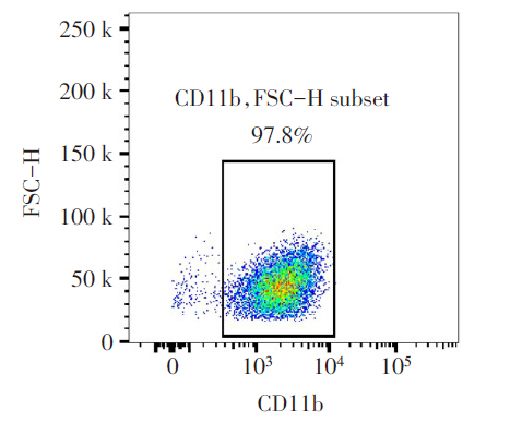
Fig.1 Purity verification of primary microglia cells
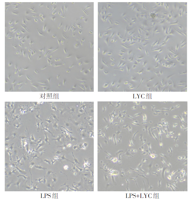
Fig.2 Effects of LYC on the morphology of primary microglia cells (×200)
| 组别 | IL-1β mRNA | IL-6 mRNA | ||
|---|---|---|---|---|
| 对照组 | 1.026±0.137 | 1.047±0.118 | ||
| LYC组 | 1.135±0.507 | 1.032±0.156 | ||
| LPS组 | 1 357.000±186.300a | 3 644.000±391.400a | ||
| LPS+LYC组 | 631.900±83.950ab | 1 549.000±267.200ab | ||
| F | 159.500** | 212.000** | ||
| 组别 | TNF-α mRNA | NO含量 | ||
| 对照组 | 1.036±0.128 | 1.185±0.366 | ||
| LYC组 | 1.136±0.508 | 2.238±0.949 | ||
| LPS组 | 397.700±62.410a | 28.770±2.472a | ||
| LPS+LYC组 | 248.800±45.230ab | 18.830±4.847ab | ||
| F | 103.100** | 93.640** | ||
Tab.2 Comparison of IL-1β, IL-6 and TNF-α mRNA expression level and NO content between the four groups (n=4,μmol/L,$\bar{x}±s$)
| 组别 | IL-1β mRNA | IL-6 mRNA | ||
|---|---|---|---|---|
| 对照组 | 1.026±0.137 | 1.047±0.118 | ||
| LYC组 | 1.135±0.507 | 1.032±0.156 | ||
| LPS组 | 1 357.000±186.300a | 3 644.000±391.400a | ||
| LPS+LYC组 | 631.900±83.950ab | 1 549.000±267.200ab | ||
| F | 159.500** | 212.000** | ||
| 组别 | TNF-α mRNA | NO含量 | ||
| 对照组 | 1.036±0.128 | 1.185±0.366 | ||
| LYC组 | 1.136±0.508 | 2.238±0.949 | ||
| LPS组 | 397.700±62.410a | 28.770±2.472a | ||
| LPS+LYC组 | 248.800±45.230ab | 18.830±4.847ab | ||
| F | 103.100** | 93.640** | ||
| 组别 | CD86 mRNA | CD206 mRNA | CD86(%) | CD206(%) |
|---|---|---|---|---|
| 对照组 | 0.921±0.062 | 1.190±0.419 | 9.150±0.387 | 8.463±0.697 |
| LYC组 | 0.741±0.207 | 0.972±0.278 | 9.020±0.486 | 8.850±0.265 |
| LPS组 | 6.003±1.094a | 3.682±0.584a | 36.150±2.525a | 16.680±1.396a |
| LPS+LYC组 | 3.427±0.542ab | 6.200±0.695ab | 22.400±2.754ab | 25.150±1.234ab |
| F | 63.910** | 89.730** | 186.600** | 246.500** |
Tab.3 Comparison of CD86 and CD206 mRNA expression levels and phenotypic ratio between the four groups (n=4,$\bar{x}±s$)
| 组别 | CD86 mRNA | CD206 mRNA | CD86(%) | CD206(%) |
|---|---|---|---|---|
| 对照组 | 0.921±0.062 | 1.190±0.419 | 9.150±0.387 | 8.463±0.697 |
| LYC组 | 0.741±0.207 | 0.972±0.278 | 9.020±0.486 | 8.850±0.265 |
| LPS组 | 6.003±1.094a | 3.682±0.584a | 36.150±2.525a | 16.680±1.396a |
| LPS+LYC组 | 3.427±0.542ab | 6.200±0.695ab | 22.400±2.754ab | 25.150±1.234ab |
| F | 63.910** | 89.730** | 186.600** | 246.500** |
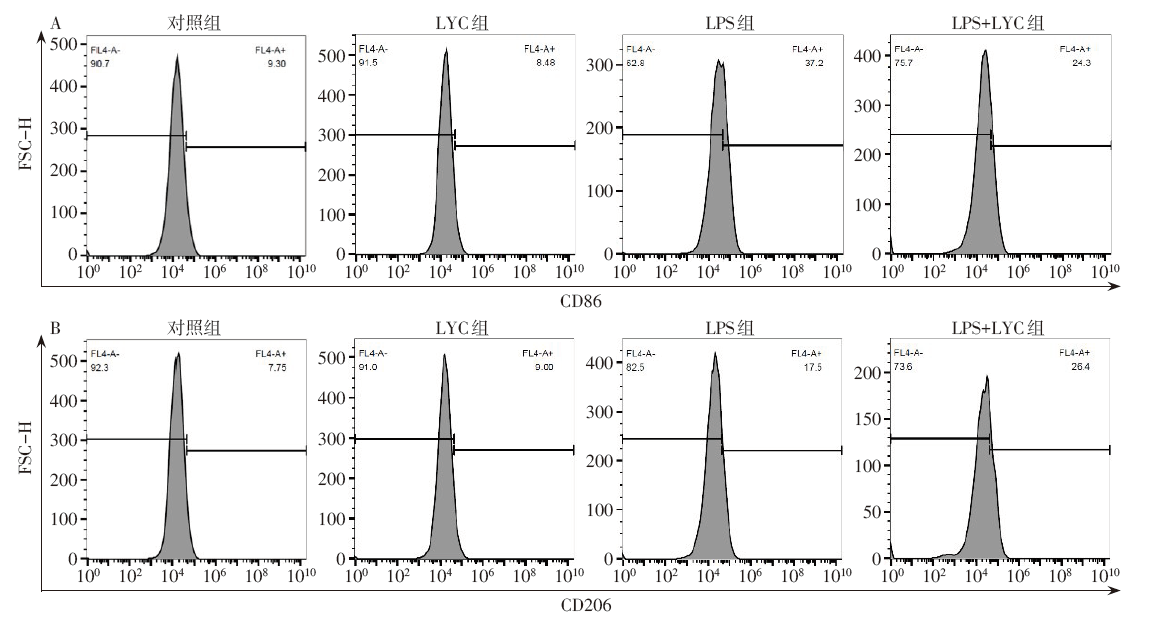
Fig.3 The flow cytometry results of LYC on different phenotypic primary microglia cells induced by LPS
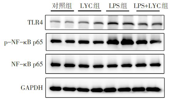
Fig.4 Effects of LYC on TLR4/NF-κB signaling pathway in LPS-induced primary microglia cells
| 组别 | TLR4 | p-NF-κB p65 |
|---|---|---|
| 对照组 | 0.992±0.153 | 1.000±0.064 |
| LYC组 | 1.391±0.180 | 1.139±0.063 |
| LPS组 | 2.547±0.016a | 2.214±0.242a |
| LPS+LYC组 | 1.665±0.207ab | 1.246±0.384b |
| F | 52.930** | 17.070** |
Tab.4 Comparison of TLR4/ NF-κB signaling pathway protein expression levels between the four groups (n=3,$\bar{x}±s$)
| 组别 | TLR4 | p-NF-κB p65 |
|---|---|---|
| 对照组 | 0.992±0.153 | 1.000±0.064 |
| LYC组 | 1.391±0.180 | 1.139±0.063 |
| LPS组 | 2.547±0.016a | 2.214±0.242a |
| LPS+LYC组 | 1.665±0.207ab | 1.246±0.384b |
| F | 52.930** | 17.070** |
| [1] | SALTER M W, STEVENS B. Microglia emerge as central players in brain disease[J]. Nat Med, 2017, 23(9):1018-1027. doi: 10.1038/nm.4397. |
| [2] | XU L, HE D, BAI Y. Microglia-mediated inflammation and neurodegenerative disease[J]. Mol Neurobiol, 2016, 53(10):6709-6715. doi: 10.1007/s12035-015-9593-4. |
| [3] | HU X, LEAK R K, SHI Y, et al. Microglial and macrophage polarization-new prospects for brain repair[J]. Nat Rev Neurol, 2015, 11(1):56-64. doi: 10.1038/nrneurol.2014.207. |
| [4] | WU S, QIU Y, SHAO Y, et al. Lycorine displays potent antitumor efficacy in colon carcinoma by targeting STAT3[J]. Front Pharmacol, 2018, 9:881. doi: 10.3389/fphar.2018.00881. |
| [5] | LV X, ZHANG M, YU S, et al. Antiviral and virucidal activities of lycorine on duck tembusu virus in vitro by blocking viral internalization and entry[J]. Poult Sci, 2021, 100(10):101404. doi: 10.1016/j.psj.2021.101404. |
| [6] | CAO Z, YANG P, ZHOU Q. Multiple biological functions and pharmacological effects of lycorine[J]. Sci China Chem, 2013, 56(10):1382-1391. doi: 10.1007/s11426-013-4967-9. |
| [7] | KANG J, ZHANG Y, CAO X, et al. Lycorine inhibits lipopolysaccharide- induced iNOS and COX-2 up-regulation in RAW264.7 cells through suppressing P38 and STATs activation and increases the survival rate of mice after LPS challenge[J]. Int Immunopharmacol, 2012, 12(1):249-256. doi: 10.1016/j.intimp.2011.11.018. |
| [8] | 果婷婷, 王辉强, 李玉环, 等. 石蒜碱及其衍生物的药理作用研究进展[J]. 中国医药生物技术, 2018, 13(5):463-466. |
| GUO T T, WANG H Q, LI Y H, et al. Progress in pharmacological action of lycorine and its derivatives[J]. Chinese Medicinal Biotechnology, 2018, 13(5):463-466. doi: 10.3969/j.issn.1673-713X.2018.05.014. | |
| [9] | SHADFAR S, HWANG C J, LIM M S, et al. Involvement of inflammation in Alzheimer's disease pathogenesis and therapeutic potential of anti-inflammatory agents[J]. Arch Pharm Res, 2015, 38(12):2106-2119. doi: 10.1007/s12272-015-0648-x. |
| [10] | ANDERSEN M S, BANDRES-CIGA S, REYNOLDS R H, et al. Heritability enrichment implicates microglia in Parkinson's Disease pathogenesis[J]. Ann Neurol, 2021, 89:942-951. doi: 10.1002/ana.26032. |
| [11] | WERNER Y, MASS E, ASHOK K P, et al. Cxcr4 distinguishes HSC-derived monocytes from microglia and reveals monocyte immune responses to experimental stroke[J]. Nat Neurosci, 2020, 23(3):351-362. doi: 10.1038/s41593-020-0585-y. |
| [12] | JOHNSON V E, STEWART J E, BEGBIE F D, et al. Inflammation and white matter degeneration persist for years after a single traumatic brain injury[J]. Brain, 2013, 136(Pt 1):28-42. doi: 10.1093/brain/aws322. |
| [13] | SHI S, LI C, ZHANG Y, et al. Lycorine hydrochloride inhibits melanoma cell proliferation,migration and invasion via down-regulating p21Cip1/WAF1[J]. Am J Cancer Res, 2021, 11(4):1391-1409. |
| [14] | ZHANG Y N, ZHANG Q Y, LI X D, et al. Gemcitabine,lycorine and oxysophoridine inhibit novel coronavirus(SARS-CoV-2)in cell culture[J]. Emerg Microbes Infect, 2020, 9(1):1170-1173. doi: 10.1080/22221751.2020.1772676. |
| [15] | PARK H J, GHOLAM-ZADEH M, SUH J H, et al. Lycorine attenuates autophagy in osteoclasts via an axis of mROS/TRPML1/TFEB to reduce LPS-induced bone loss[J]. Oxid Med Cell Longev, 2019, 2019:8982147. doi: 10.1155/2019/8982147. |
| [16] | LI X, KANG J, LV H, et al. CircPrkcsh,a circular RNA,contributes to the polarization of microglia towards the M1 phenotype induced by spinal cord injury and acts via the JNK/p38 MAPK pathway[J]. FASEB J, 2021, 35(12):e22014. doi: 10.1096/fj.202100993R. |
| [17] | LU L, WANG H, LIU X, et al. Pyruvate kinase isoform M2 impairs cognition in systemic lupus erythematosus by promoting microglial synaptic pruning via the beta-catenin signaling pathway[J]. J Neuroinflammation, 2021, 18(1):229. doi: 10.1186/s12974-021-02279-9. |
| [18] | ZAGHLOUL N, KUREPA D, BADER M Y, et al. Prophylactic inhibition of NF-kappaB expression in microglia leads to attenuation of hypoxic ischemic injury of the immature brain[J]. J Neuroinflammation, 2020, 17(1):365. doi: 10.1186/s12974-020-02031-9. |
| [19] | BIAN H J, XU S Y, LI H Q, et al. JLX001 ameliorates cerebral ischemia injury by modulating microglial polarization and compromising NLRP3 inflammasome activation via the NF-kappaB signaling pathway[J]. Int Immunopharmacol, 2021, 101(Pt A):108325. doi: 10.1016/j.intimp.2021.108325. |
| [1] | CHEN Jingjing, NONG Zhangsong, TAN Liangyuan, YANG Peipei, LIANG Yingye, TANG Hongliang, WANG Kailong. Research progress on the role of microglia polarization in neuropathic pain [J]. Tianjin Medical Journal, 2024, 52(9): 1000-1003. |
| [2] | FAN Huihui, REN Yumei, TIAN Xinlei, ZHANG Kai, LI Xiaoli. Effects of Zhike Pingchuan Formula on airway inflammation and TLR4/TRAF6/NF-κB pathway in bronchial asthma mice [J]. Tianjin Medical Journal, 2024, 52(9): 924-929. |
| [3] | JIA Weining, BAO Yaling, LEI Hui, YIN Xiaoning. The effect of prunella vulgaris extract on inflammatory response and peritoneal macrophages in septic mice [J]. Tianjin Medical Journal, 2024, 52(9): 930-935. |
| [4] | LIU Bin, YANG Long, LI Wenli, SHAO Ningning, DONG Jinrui. Mechanism of microglia ferroptosis in smoke inhalation-induced brain injury [J]. Tianjin Medical Journal, 2024, 52(8): 791-797. |
| [5] | YE Zhaoyang, MA Jianzhong, LI Houjun, WEI Kunpeng. Relationship between peripheral blood TLR4, IL-1β and NLR and the progression and prognosis of acute pancreatitis [J]. Tianjin Medical Journal, 2024, 52(6): 648-652. |
| [6] | JIA Xirui, LIU Lijie. The role and research progress of microglia in sepsis related encephalopathy [J]. Tianjin Medical Journal, 2024, 52(5): 557-560. |
| [7] | XIAO Yuqian, SUN Kexin, WAN Jun, CHEN Shuying, CHEN Limin, WANG Yan, BAI Yanjie. Research progress of RNA m6A methylation in post-stroke cognitive impairment [J]. Tianjin Medical Journal, 2024, 52(3): 331-336. |
| [8] | LIN Yao, LIU Congna, WANG Shixia, ZHANG Zhiyong. Effect of acacetin on lipopolysaccharide induced apoptosis of dental pulp cells by regulating the HMGB1/TLR4 signaling pathway [J]. Tianjin Medical Journal, 2024, 52(12): 1238-1243. |
| [9] | SHI Jingbo, LI Changnian, MENG Yuxuan, XIE Guangdong, XU Jie, RONG Baohai. Mechanism study of Qingchang mixture in the treatment of postoperative abdominal adhesions by regulating the expression of Th1 and Th2 cytokines [J]. Tianjin Medical Journal, 2024, 52(12): 1244-1250. |
| [10] | OUYANG Jie, ZHAO Haiqian, KONG Yun, NIU Qin, CHEN Ying, SI Yongyu. The effect of electroacupuncture on paclitaxel-induced neuropathic pain in rats [J]. Tianjin Medical Journal, 2024, 52(11): 1141-1145. |
| [11] | LONG Hua, CHEN Yifei, WANG Qingshu. Effect of remimazolam on apoptosis of intestinal epithelial cells in burned rats by regulating TLR4/MyD88/NF-κB signaling pathway [J]. Tianjin Medical Journal, 2024, 52(11): 1152-1157. |
| [12] | ZHANG Zhiqiang, WANG Weiwei, DONG Tengjing, LIAN Hongkai. The relationship between TLR4, JAK3 gene expression and Th17/Treg imbalance in peripheral blood of patients with ankylosing spondylitis [J]. Tianjin Medical Journal, 2024, 52(10): 1065-1068. |
| [13] | CHEN Hao, LI Rui, YI Fei, ZHOU Li, CHEN Jiaqi, ZHU Fan, GUAN Chengyan, WU Na. Construction of mouse intestinal organoid inflammation model [J]. Tianjin Medical Journal, 2024, 52(1): 16-21. |
| [14] | WANG Shengcheng, LI Qi, CAI Xiaoyang, TANG Yongjie. Effect and mechanism of tetrandrine on airway remodeling in bronchial asthma mice [J]. Tianjin Medical Journal, 2023, 51(9): 943-947. |
| [15] | CHEN Xilong, WANG Hongjun, SONG Zhengyu, WANG Jing. Arctigenin alleviates neuronal damage of acute cerebral infarction in rats by inhibiting the HMGB1/TLR4/NF-κB pathway [J]. Tianjin Medical Journal, 2023, 51(8): 825-829. |
| Viewed | ||||||
|
Full text |
|
|||||
|
Abstract |
|
|||||