Tianjin Medical Journal ›› 2022, Vol. 50 ›› Issue (11): 1121-1127.doi: 10.11958/20212787
• Cell and Molecular Biology • Next Articles
LI Wenwen( ), ZHU Zhixin, HAN Limin, JIAO Yanlin, WENG Zongqin, ZHAO Hailong△()
Received:2022-01-04
Revised:2022-04-19
Published:2022-11-15
Online:2022-11-11
Contact:
ZHAO Hailong
E-mail:lw20200701@163.com;Hailongzhao@zmu.edu.cn
LI Wenwen, ZHU Zhixin, HAN Limin, JIAO Yanlin, WENG Zongqin, ZHAO Hailong. Molecular mechanism study of melanoma cell proliferation regulated by miR-101-3p-E2F2 targeting pathway[J]. Tianjin Medical Journal, 2022, 50(11): 1121-1127.
CLC Number:
| 基因名称 | 引物序列(5′→3′) | 产物大小(bp) |
|---|---|---|
| miR-101-3p | 上游:TACAGTACTGTGATAACTGAA 下游:GGAGTAG ATGATGGTTAGC | 58 |
| U6 | 上游:CTCGCTTCGGCAGCACA 下游:AACGCTTCACGAATTTGCGT | 94 |
| E2F2 | 上游:CTCTCTGAGCTTCAAGCACCTG 下游:CTTGACGGCAATCACT GTCTGC | 118 |
| GAPDH | 上游:ACCACAGTCCATGCCATCAC 下游:TCCACCACCCTGTTGCTGTA | 452 |
Tab.1 Primer sequence for qPCR
| 基因名称 | 引物序列(5′→3′) | 产物大小(bp) |
|---|---|---|
| miR-101-3p | 上游:TACAGTACTGTGATAACTGAA 下游:GGAGTAG ATGATGGTTAGC | 58 |
| U6 | 上游:CTCGCTTCGGCAGCACA 下游:AACGCTTCACGAATTTGCGT | 94 |
| E2F2 | 上游:CTCTCTGAGCTTCAAGCACCTG 下游:CTTGACGGCAATCACT GTCTGC | 118 |
| GAPDH | 上游:ACCACAGTCCATGCCATCAC 下游:TCCACCACCCTGTTGCTGTA | 452 |
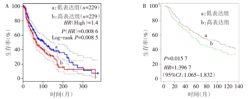
Fig.1 Survival curves of melanoma patients with different expression levels of E2F2
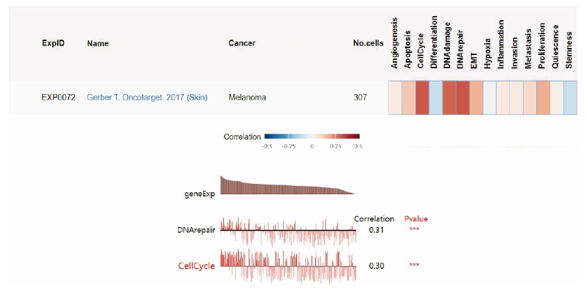
Fig.2 CancerSEA database analyzes the functional diagram of E2F2 in melanoma
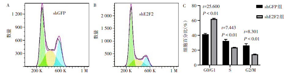
Fig.3 The changes of cell cycle detected by PI staining flow cytometry after shGFP or shE2F2 treatment
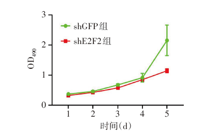
Fig.4 The effect shGFP or shE2F2 on cell viability of MV3 cells detected by MTT
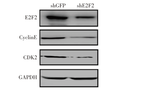
Fig.5 Expression of cycle-related proteins in MV3 cells treated with shGFP or shE2F2
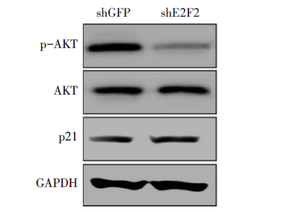
Fig.6 Expression changes of p-AKT, AKT and p21 in MV3 cells treated with shGFP or shE2F2
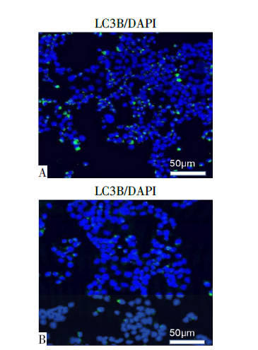
Fig.7 The changes of LC3B fluorescence signal of MV3 cells after shGFP or shE2F2 treatment
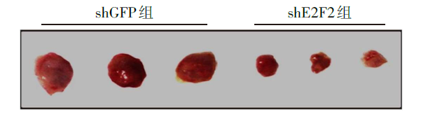
Fig.8 The effect of shE2F2 on the growth of melanoma in vivo
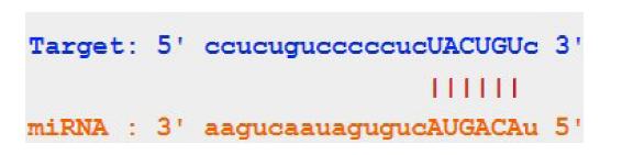
Fig.9 The binding sequence diagram between miR-101 and the 3'-UTR sequence of E2F2 predicted by StarBase
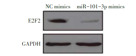
Fig.10 Protein expression levels of E2F2 in MV3 cells treated with NC or miR-101-3p mimics
| 组别 | 1 d | 2 d | 3 d | 4 d | 5 d |
|---|---|---|---|---|---|
| NC mimics组 | 0.18±0.02 | 0.38±0.03 | 0.74±0.05 | 1.11±0.16 | 1.90±0.05 |
| miR-101-3p mimics组 | 0.19±0.02 | 0.36±0.03 | 0.68±0.04 | 0.87±0.08 | 1.26±0.12 |
| t | 0.692 | 0.711 | 1.779 | 2.336 | 8.784** |
Tab.2 Comparison of MV3 cell viability after treatment with NC mimics and miR-101-3p mimics between the two groups
| 组别 | 1 d | 2 d | 3 d | 4 d | 5 d |
|---|---|---|---|---|---|
| NC mimics组 | 0.18±0.02 | 0.38±0.03 | 0.74±0.05 | 1.11±0.16 | 1.90±0.05 |
| miR-101-3p mimics组 | 0.19±0.02 | 0.36±0.03 | 0.68±0.04 | 0.87±0.08 | 1.26±0.12 |
| t | 0.692 | 0.711 | 1.779 | 2.336 | 8.784** |
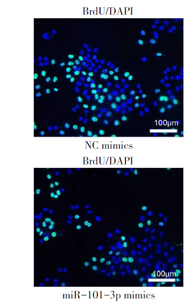
Fig.11 BrdU positive staining in MV3 cells treated with NC mimics or miR-101-3p mimics
| [1] | SCHADENDORF D, VAN AKKOOI A C J, BERKING C, et al. Melanoma[J]. Lancet, 2018, 392(10151):971-984. doi:10.1016/S0140-6736(18)31559-9. |
| [2] | AUDRITO V, MANAGÒ A, GAUDINO F, et al. Targeting metabolic reprogramming in metastatic melanoma: The key role of nicotinamide phosphoribosyltransferase (NAMPT)[J]. Semin Cell Dev Biol, 2020, 98:192-201. doi:10.1016/j.semcdb.2019.05.001. |
| [3] | ZHAO G, GREEN C F, HUI Y H, et al. Discovery of a highly selective NAMPT inhibitor that demonstrates robust efficacy and improved retinal toxicity with nicotinic acid coadministration[J]. Mol Cancer Ther, 2017, 16(12):2677-2688. doi:10.1158/1535-7163.MCT-16-0674. |
| [4] | LIANG B, ZHAO J, WANG X. Clinical performance of E2Fs1-3 in kidney clear cell renal cancer,evidence from bioinformatics analysis[J]. Genes Cancer, 2017, 8(5/6):600-607. doi:10.18632/genes andcancer.143. |
| [5] | AN X, MA H, LIU Y, et al. Effects of miR-101-3p on goat granulosa cells in vitro and ovarian development in vivo via STC1[J]. J Anim Sci Biotechnol, 2020, 11:102. doi:10.1186/s40104-020-00506-6. |
| [6] | VAN ZEIJL M C, WAN DEN EERTWEGH A J, HAANEN J B, et al. (Neo)adjuvant systemic therapy for melanoma[J]. Eur J Surg Oncol, 2017, 43(3):534-543. doi:10.1016/j.ejso.2016.07.001. |
| [7] | GOODY D, GUPTA S K, ENGELMANN D, et al. Drug repositioning inferred from E2F1-coregulator interactions studies for the prevention and treatment of metastatic cancers[J]. Theranostics, 2019, 9(5):1490-1509. doi:10.7150/thno.29546. |
| [8] | ZHAO H, TANG W, CHEN X, et al. The NAMPT/E2F2/SIRT1 axis promotes proliferation and inhibits p53-dependent apoptosis in human melanoma cells[J]. Biochem Biophys Res Commun, 2017, 493(1):77-84. doi:10.1016/j.bbrc.2017.09.071. |
| [9] | ZENG Z, CAO Z, TANG Y. Increased E2F2 predicts poor prognosis in patients with HCC based on TCGA data[J]. BMC Cancer, 2020, 20(1):1037. doi:10.1186/s12885-020-07529-2. |
| [10] | HUANG Y L, NING G, CHEN L B, et al. Promising diagnostic and prognostic value of E2Fs in human hepatocellular carcinoma[J]. Cancer Manag Res, 2019, 11:1725-1740. doi:10.2147/CMAR.S182001. |
| [11] | SUN C C, LI S J, HU W, et al. Comprehensive analysis of the expression and prognosis for E2Fs in human breast cancer[J]. Mol Ther, 2019, 27(6):1153-1165. doi:10.1016/j.ymthe.2019.03.019. |
| [12] | YANG C, ZHANG Z C, LIU T B, et al. E2F1/2/7/8 as independent indicators of survival in patients with cervical squamous cell carcinoma[J]. Cancer Cell Int, 2020, 20:500. doi:10.1186/s12935-020- 01594-0. |
| [13] | LIU X, HU C. Novel potential therapeutic target for E2F1 and prognostic factors of E2F1/2/3 /5/7/8 in human gastric cancer[J]. Mol Ther Methods Clin Dev, 2020, 18:824-838. doi:10.1016/j.omtm.2020.07.017. |
| [14] | GAO J, CHEN X, SHAN C, et al. Autophagy in cardiovascular diseases: role of noncoding RNAs[J]. Mol Ther Nucleic Acids, 2020, 23:101-118. doi:10.1016/j.omtn.2020.10.039. |
| [15] | YAN X, ZHOU R, MA Z. Autophagy-cell survival and death[J]. Adv Exp Med Biol, 2019, 1206:667-696. doi:10.1007/978-981-15-0602-4_29. |
| [16] | CUI G, WANG H, LIU W, et al. Glycogen phosphorylase B is regulated by miR101-3p and promotes hepatocellular carcinoma tumorigenesis[J]. Front Cell Dev Biol, 2020, 8:566494. doi:10.3389/fcell.2020.566494. |
| [17] | HUANG Z, WU X, LI J. miR-101 suppresses colon cancer cell migration through regulation of EZH2[J]. Rev Esp Enferm Dig, 2021, 113(4):255-260. doi:10.17235/reed.2020.6800/2019. |
| [18] | WU R S, QIU E H, ZHU J J, et al. MiR-101 promotes nasopharyngeal carcinoma cell apoptosis through inhibiting Ras/Raf/MEK/ERK signaling pathway[J]. Eur Rev Med Pharmacol Sci, 2020, 24(16):8240. doi:10.26355/eurrev_202008_22580. |
| [19] | HUANG Y, ZOU Y, LIN L, et al. miR-101 regulates cell proliferation and apoptosis by targeting KDM1A in diffuse large B cell lymphoma[J]. Cancer Manag Res, 2019, 11:2739-2746. doi:10.2147/CMAR.S197744. |
| [20] | WANDLER A, RIBER-HANSEN R, HAGER H, et al. Quantification of microRNA-21 and microRNA- 125b in melanoma tissue[J]. Melanoma Res, 2017, 27(5):417-428. doi:10.1097/CMR.0000000000000374. |
| Viewed | ||||||
|
Full text |
|
|||||
|
Abstract |
|
|||||