Tianjin Medical Journal ›› 2025, Vol. 53 ›› Issue (5): 537-541.doi: 10.11958/20250046
• Applied Research • Previous Articles Next Articles
MA Ping1( ), XU Xiaoming2, YE Degang1
Received:2025-01-07
Revised:2025-03-10
Published:2025-05-15
Online:2025-05-28
MA Ping, XU Xiaoming, YE Degang. Application of DWI and ADC values in differential diagnosis of cervical lymph nodes in patients with nasopharyngeal carcinoma[J]. Tianjin Medical Journal, 2025, 53(5): 537-541.
CLC Number:
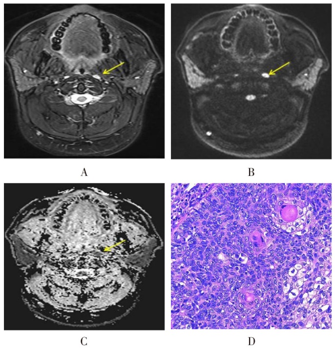
Fig.1 MRI and pathological findings of malignant lymph nodes
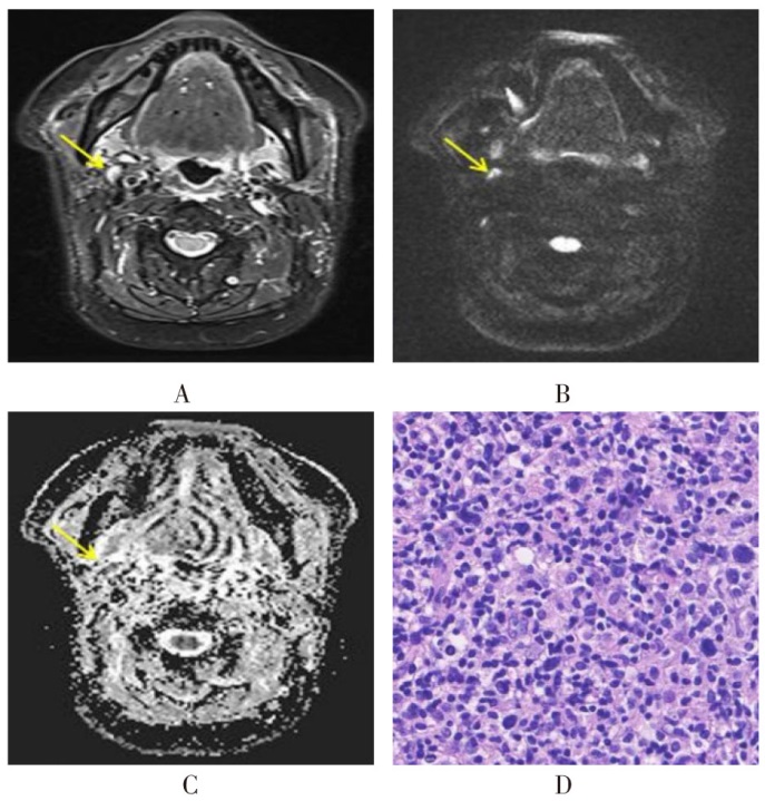
Fig.2 MRI and pathological findings of benign lymph nodes
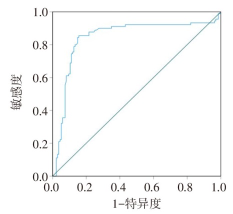
Fig.3 ROC curve of ADC for differential diagnosis of benign and malignant cervical lymph nodes in nasopharyngeal carcinoma
| 方法 | 病理组织学诊断结果 | 合计 | |
|---|---|---|---|
| 恶性 | 良性 | ||
| MRI平扫 | |||
| 恶性 | 48 | 13 | 61 |
| 良性 | 17 | 20 | 37 |
| ADC | |||
| 恶性 | 58 | 5 | 63 |
| 良性 | 7 | 28 | 35 |
| 合计 | 65 | 33 | 98 |
Tab.1 Qualitative diagnosis and histopathological diagnosis of different examination methods
| 方法 | 病理组织学诊断结果 | 合计 | |
|---|---|---|---|
| 恶性 | 良性 | ||
| MRI平扫 | |||
| 恶性 | 48 | 13 | 61 |
| 良性 | 17 | 20 | 37 |
| ADC | |||
| 恶性 | 58 | 5 | 63 |
| 良性 | 7 | 28 | 35 |
| 合计 | 65 | 33 | 98 |
| [1] | 王璐萍, 张祥意, 周明辉, 等. 精确放疗时代T1期鼻咽癌患者不同颈淋巴结转移状态的预后情况分析[J]. 实用癌症杂志, 2024, 39(4):551-553,565. |
| WANG L P, ZHANG X Y, ZHOU M H, et al. Prognostic analysis of different cervical lymph node metastasis transitions in T1 stage nasopharyngeal carcinoma patients during precise radiotherapy[J]. The Practical Journal of Cancer, 2024, 39(4):551-553,565. doi:10.3969/j.issn.1001-5930.2024.04.007. | |
| [2] | ALONTO A, MONDIA M, PLANILLA C, et al. Rare case of intramedullary spinal cord metastasis from nasopharyngeal carcinoma[J]. Curr Probl Cancer, 2021, 45(6):100713. doi:10.1016/j.currproblcancer.2021.100713. |
| [3] | 刘懿炜, 王钢, 周子博, 等. 影像学方法在鼻咽癌颈淋巴结转移诊断中的应用进展[J]. 分子影像学杂志, 2024, 47(7):764-768. |
| LIU Y W, WANG G, ZHOU Z B, et al. Advances in the application of medical imaging methods in the diagnosis of cervical lymph node metastasis in nasopharyngeal carcinoma[J]. Journal of Molecular Imaging, 2024, 47(7):764-768. doi:10.12122/j.issn.1674-4500.2024.07.17. | |
| [4] | 邓宇辰, 毕秋, 吴昆华. 不同扩散成像模型在子宫内膜癌中的研究进展[J]. 国际医学放射学杂志, 2024, 47(5):599-604. |
| DENG Y C, BI Q, WU K H. Research progress on different diffusion imaging models in endometrial carcinoma[J]. International Journal of Medical Radiology, 2024, 47(5):599-604. doi:10.19300/j.2024.Z21223. | |
| [5] | 丛榕, 马霄虹. 肝细胞癌放射治疗的疗效预测及评估的MRI应用进展[J]. 国际医学放射学杂志, 2023, 46(6):696-700. |
| CONG R, MA X H. Advances in the application of MRI in predicting and evaluating the efficacy of radiotherapy for hepatocellular carcinoma[J]. International Journal of Medical Radiology, 2023, 46(6):696-700. doi:10.19300/j.2023.Z20688. | |
| [6] | 顾大勇, 黄生富, 宗丹, 等. 磁共振弥散加权成像对鼻咽癌放疗后颈部淋巴结复发早期诊疗的临床价值[J]. 中华放射肿瘤学杂志, 2019, 28(8):571-574. |
| GU D Y, HUANG S F, ZONG D, et al. Clinical value of magnetic resonance diffusion-weighted imaging in early diagnosis and treatment of cervical lymph node recurrence after radiotherapy of nasopharyngeal carcinoma[J]. Chin J Radiat Oncol, 2019, 28(8):571-574. doi:10.3760/cma.j.issn.1004-4221.2019.08.003. | |
| [7] | 武敬君, 张旼旼, 戴慧, 等. 基于MRI-DWI的影像组学在预测鼻咽癌淋巴结转移中的价值[J]. 中国医学计算机成像杂志, 2024, 30(3):305-311. |
| WU J J, ZHANG M M, DAI H, et al. The value of MRI-DWI based radiomics in predicting lymph node metastasis of nasopharyngeal carcinoma[J]. Chin Comput Med Imag, 2024, 30(3):305-311. doi:10.3969/j.issn.1006-5741.2024.03.008. | |
| [8] | 张伟. 18F-NaF PET/CT、18F-FDG PET/CT及MRI对鼻咽癌患者颅底骨质受侵及骨转移的评估价值比较[J]. 中国CT和MRI杂志, 2022, 20(4):28-31. |
| ZHANG W. Comparison of evaluated value of 18F-NaF PET/CT,18F-FDG PET/CT,and MRI on skull base invasion and bone metastasis in patients with nasopharyngeal carcinoma[J]. Chinese Journal of CT and MRI, 2022, 20(4):28-31. doi:10.3969/j.issn.1672-5131.2022.04.010. | |
| [9] | GAO Y, REIG B, HEACOCK L, et al. Magnetic resonance imaging in screening of breast cancer[J]. Radiol Clin North Am, 2021, 59(1):85-98. doi:10.1016/j.rcl.2020.09.004. |
| [10] | 包丹, 张娅, 胡镭, 等. 表观扩散系数值对鼻咽癌疾病进展风险预测价值的研究[J]. 实用放射学杂志, 2022, 38(4):538-542. |
| BAO D, ZHANG Y, HU L, et al. Predictive value of apparent diffusion coefficient on the risk of disease progression with nasopharyngeal carcinoma[J]. J Prac Radiol, 2022, 38(4):538-542. doi:10.3969/j.issn.1002-1671.2022.04.006. | |
| [11] | 黄梓杰, 胡玉芳, 覃宝仪, 等. 多重灵敏度编码扩散加权成像在提高鼻咽癌图像质量的应用研究[J]. 实用放射学杂志, 2023, 39(7):1179-1182. |
| HUANG Z J, HU Y F, QIN B Y, et al. Application of multiple sensitivity encoding diffusion weighted imaging in improving the image quality of nasopharyngeal carcinoma[J]. J Prac Radiol, 2023, 39(7):1179-1182. doi:10.3969/j.issn.1002-1671.2023.07.032. | |
| [12] | 赵灿灿, 李淑华, 王雪莲, 等. 基于多模态MRI影像组学模型预测鼻咽癌放化疗疗效[J]. 蚌埠医学院学报, 2024, 49(6):771-775. |
| ZHAO C C, LI S H, WANG X L, et al. Prediction value of multi-modal MRI radiomics model in the efficacy of chemoradiotherapy for nasopharyngeal carcinoma[J]. J Bengbu Med Coll, 2024, 49(6):771-775. doi:10.13898/j.cnki.issn.1000-2200.2024.06.016. | |
| [13] | 杨栋, 张宏岩, 张邵婕, 等. IVIM-DWI联合血清Apo-1/Fas评估鼻咽癌患者诱导化疗+同步放化疗近期疗效的价值[J]. 临床和实验医学杂志, 2022, 21(19):2107-2111. |
| YANG D, ZHANG H Y, ZHANG S J, et al. Value of IVIM-DWI combined with serum Apo-1/Fas in evaluating the short-term efficacy of induction chemotherapy+concurrent chemoradiotherapy in patients with nasopharyngeal carcinoma[J]. Journal of Clinical and Experimental Medicine, 2022, 21(19):2107-2111. doi:10.3969/j.issn.1671-4695.2022.19.025. | |
| [14] | 周霖, 曾蕾. 不同表观扩散系数对前列腺癌盆腔转移性淋巴结的定性诊断价值[J]. 肿瘤防治研究, 2019, 46(3):248-252. |
| ZHOU L, ZENG L. Qualitative diagnostic value of different apparent diffusion coefficients on metastatic pelvic lymph nodes in prostate cancer[J]. Cancer Res Prev Treat, 2019, 46(3):248-252. doi:10.3971/j.issn.1000-8578.2019.18.1393. | |
| [15] | 周镇源, 刘志锋, 蔡金辉, 等. 表观扩散系数鉴别颈部淋巴结病变诊断价值[J]. 实用放射学杂志, 2019, 35(8):1220-1224. |
| ZHOU Z Y, LIU Z F, CAI J H, et al. The diagnostic value of ADC in differential diagnosis of cervical lymph node lesions[J]. J Pract Radiol, 2019, 35(8):1220-1224. doi:10.3969/j.issn.1002-1671.2019.07.005. | |
| [16] | 黄涛, 蒋平平, 樊蓝振, 等. 3.0T MR多b值DWI对鼻咽癌患者颈部良恶性淋巴结的鉴别诊断价值[J]. 医学影像学杂志, 2020, 30(3):367-374. |
| HUANG T, JIANG P P, FAN L Z, et al. Value of multi b-value DWI of 3.0T MR in differential diagnosis of benign and malignant cervical lymph nodes in patients with nasopharyngeal carcinoma[J]. Journal of Medical Imaging, 2020, 30(3):367-374. | |
| [17] | 满育平, 马隆佰, 吴春梅, 等. 3.0T磁共振ADC值及DCE-MRI定量分析对颈部良恶性淋巴结鉴别诊断[J]. 放射学实践, 2019, 34(6):619-623. |
| MAN Y P, MA L B, WU C M, et al. The differential value of ADC and quantitative parameters of DCE-MRI at 3.0T in diagnosing benign and malignant cervical lymph nodes[J]. Radiol Practice, 2019, 34(6):619-623. doi:10.13609/j.cnki.1000-0313.2019.06.005. | |
| [18] | AHN J H, KIM S H, SON J H, et al. Added value of diffusion-weighted imaging for evaluation of extramural venous invasion in patients with primary rectal cancer[J]. Br J Radiol, 2019, 92(1096):20180821. doi:10.1259/bjr.20180821. |
| [1] | WANG Lili, WANG Xuyan, ZHANG Pei, HAN Mingming, ZHANG Jing, ZHAO Mingxin. Expression and clinical significance of CLDN6, TRIM59 and CMTM6 in nasopharyngeal carcinoma [J]. Tianjin Medical Journal, 2025, 53(3): 272-276. |
| [2] | TIAN Youjun, TAN Zhengwu, YANG Ke, PENG Jianmin, CHEN Hongtao, HUANG Zhiping. The predictive value of multi-sequence MRI radiomics in the therapeutic effect of concurrent chemoradiotherapy on locally advanced cervical squamous cell carcinoma [J]. Tianjin Medical Journal, 2025, 53(2): 213-218. |
| [3] | WU Caixin, YAN Yan, DENG Yuanlin, DU Yamin, YANG Zhenwen, PAN Qing, YANG Fan. The value of cardiac magnetic resonance in evaluating severe pulmonary hypertension associated with connective tissue disease [J]. Tianjin Medical Journal, 2024, 52(7): 691-694. |
| [4] | ZHU Gangming, DONG Yongde, ZHU Ruiting, TAN Yuanman, TAO Juan, LIU Xiao, CHEN Decheng, YANG Gai. The value of magnetic resonance relaxation time quantitative imaging in predicting molecular subtypes of invasive ductal carcinoma [J]. Tianjin Medical Journal, 2024, 52(7): 770-774. |
| [5] | GU Cheng, SHEN Xinyu, SUN Jinghua, YAN Saike, WANG Haiping. The evaluation value of dynamic MRI imaging technology for LARS after anorectal preservation surgery in low rectal cancer [J]. Tianjin Medical Journal, 2024, 52(6): 653-657. |
| [6] | WU Qian, WANG Yi, CHEN Nian, ZHOU Kai, TIAN Xin, XU Hui, GOU Xiaoxia. Effect of induction chemotherapy on immune function and inflammatory indicators in patients with nasopharyngeal carcinoma [J]. Tianjin Medical Journal, 2024, 52(4): 397-402. |
| [7] | GAO Guangren, FENG Lianrong, FU Jinguo, GUO Run, NIU Heping, LI Fengpeng, ZHANG Qianyu, ZHANG Jun. Characteristics of myocardial injury in patients with acute myocardial infarction complicated with pleural effusion and its influence on long-term prognosis [J]. Tianjin Medical Journal, 2024, 52(2): 197-200. |
| [8] | XIE Haoran, LI Yihao, LIU Cheng, XIA Yuting, QIU Shenglei, XIONG Bin, FENG Qizhen. Research of predictive factors of axillary lymph node metastasis in breast cancer under the context of DIP payment of medical insurance [J]. Tianjin Medical Journal, 2024, 52(11): 1193-1196. |
| [9] | WANG Jianlin, WANG Yanhua, SHI Lei. Relationship between evaluation results of LGE-CMR and serum myocardial markers in patients with acute myocardial infarction [J]. Tianjin Medical Journal, 2022, 50(4): 393-938. |
| [10] | LIAO Zonghui , YUAN Yi, PU Hong, LI Hang. The application value of region of interest selection for intravoxel incoherent motion diffusion-weighted imaging parameters in the evaluation of tumor angiogenesis and lymphovascular invasion in rectal cancer [J]. Tianjin Medical Journal, 2022, 50(2): 200-205. |
| [11] | GUO Jun, CAO Jing, LI Cheng, LI Ze-kui, GAO Si-wen, ZHANG Juan. Imaging analysis of patients with temporomandibular joint disorder before and after repositioning splint treatment [J]. Tianjin Medical Journal, 2021, 49(7): 735-741. |
| [12] | LI Hui-xia, ZHAO Yu-long, WANG Qi, LI Xiao-hui, FENG Lin, ZHANG Yong-ting . Analysis of surgical outcomes of lumbarspinal stenosis with redundant nerve roots [J]. Tianjin Medical Journal, 2021, 49(10): 1085-1088. |
| [13] | WANG Yue-bo, LIAO Zong-hui, LI Hang△. A comparative study of diffusion-weighted imaging region of interest selection in cervical cancer differentiation, lymph node metastasis and lymphovascular invasion [J]. Tianjin Medical Journal, 2021, 49(1): 79-84. |
| [14] | MENG Xiang-lu, , SUN Ji-wei , WANG Huan , DAI Li-mei , ZHAO Shao-li , ZHAO Yu-meng , FENG Ling-ling , WANG Wen-hong△. The diagnostic efficiency of MRC combined with fecal calprotectin in the activity of ulcerative colitis [J]. Tianjin Medical Journal, 2020, 48(7): 635-641. |
| [15] | XIE Zong-yuan, YU Xiang-yang, TAN Zhi-bin, LI Hui , WANG Ya-jing, WANG Zhi-qiang, LIU Tao. The diagnostic value of DWI and DCE-MRI for benign and malignant regional lymph nodes of rectal cancer [J]. Tianjin Medical Journal, 2019, 47(6): 575-579. |
| Viewed | ||||||
|
Full text |
|
|||||
|
Abstract |
|
|||||