Tianjin Medical Journal ›› 2024, Vol. 52 ›› Issue (7): 691-694.doi: 10.11958/20231770
• Monograph·Connective Tissue Diseases-Interstitial Lung Disease/Pulmo-nary Arterial Hypertension • Previous Articles Next Articles
WU Caixin1( ), YAN Yan1, DENG Yuanlin1, DU Yamin1, YANG Zhenwen2, PAN Qing3, YANG Fan1,∆()
Received:2023-11-15
Revised:2024-01-03
Published:2024-07-15
Online:2024-07-11
Contact:
∆E-mail:dr_yangfan0201@163.com
WU Caixin, YAN Yan, DENG Yuanlin, DU Yamin, YANG Zhenwen, PAN Qing, YANG Fan. The value of cardiac magnetic resonance in evaluating severe pulmonary hypertension associated with connective tissue disease[J]. Tianjin Medical Journal, 2024, 52(7): 691-694.
CLC Number:
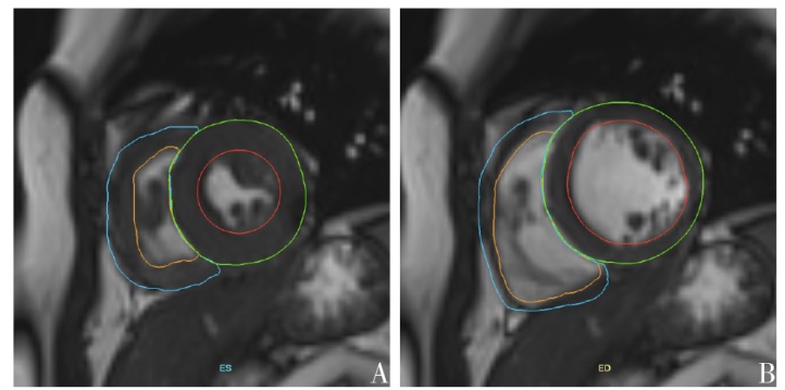
Fig.1 Short axis film sequence image sketch diagram
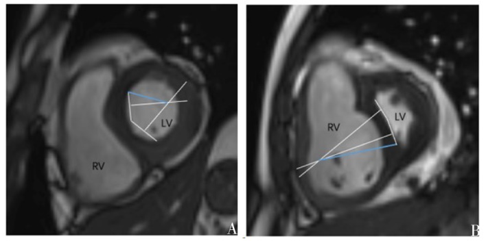
Fig.2 Example of delineations of CIVS on cine images
| 组别 | n | 女性 | 年龄/岁 | 身高/cm | |
|---|---|---|---|---|---|
| 轻中度组 | 34 | 32(94.12) | 40.97±11.20 | 160.15±5.41 | |
| 重度组 | 14 | 14(100.00) | 44.86±12.99 | 159.71±4.89 | |
| t或Z | 1.000▲ | 1.043 | 0.259 | ||
| 组别 | 体质量/kg | BNP/(ng/L) | |||
| 轻中度组 | 55.06±10.29 | 137.20(40.60,350.34) | |||
| 重度组 | 57.93±10.00 | 280.50(138.50,402.25) | |||
| t或Z | 0.885 | 1.463 | |||
Tab.1 Comparison of clinical parameters between two groups of patients
| 组别 | n | 女性 | 年龄/岁 | 身高/cm | |
|---|---|---|---|---|---|
| 轻中度组 | 34 | 32(94.12) | 40.97±11.20 | 160.15±5.41 | |
| 重度组 | 14 | 14(100.00) | 44.86±12.99 | 159.71±4.89 | |
| t或Z | 1.000▲ | 1.043 | 0.259 | ||
| 组别 | 体质量/kg | BNP/(ng/L) | |||
| 轻中度组 | 55.06±10.29 | 137.20(40.60,350.34) | |||
| 重度组 | 57.93±10.00 | 280.50(138.50,402.25) | |||
| t或Z | 0.885 | 1.463 | |||
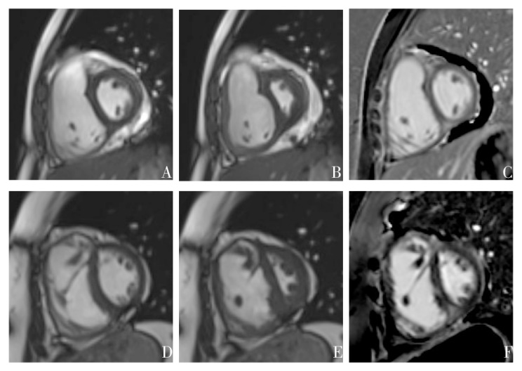
Fig.3 CMR short-axial images of end-systole, end-diastole and LGE in CTD-PAH patients
| 组别 | LV MM/g | CIVS | IVS形变时间占比/% | LGE MM/g | LGE MM占比/% | ||||||
|---|---|---|---|---|---|---|---|---|---|---|---|
| 轻中度组 | 75.42±17.66 | 0.41(0.27,0.55) | 66.41±18.36 | 2.92(0.12,5.52) | 3.86(0.20,7.77) | ||||||
| 重度组 | 73.70±21.25 | 0.02(-0.28,0.48) | 83.50±15.24 | 2.69(2.05,4.98) | 5.01(2.62,7.70) | ||||||
| t或Z | 0.289 | 2.100* | 3.069** | 0.455 | 0.910 | ||||||
| 组别 | n | RV EDVI/(mL/m2) | RV ESVI/(mL/m2) | RV SVI/(mL/m2) | RV EF/% | RV MM/g | |||||
| 轻中度组 | 34 | 91.96±24.68 | 57.60±21.19 | 34.09±11.15 | 38.23±10.63 | 55.11±17.09 | |||||
| 重度组 | 14 | 109.68±27.73 | 75.31±22.30 | 34.38±11.44 | 31.56±8.62 | 72.16±14.12 | |||||
| t或Z | 2.183* | 2.593* | 0.081 | 2.078* | 3.292** | ||||||
Tab.2 Comparison of CMR parameters between two groups of patients
| 组别 | LV MM/g | CIVS | IVS形变时间占比/% | LGE MM/g | LGE MM占比/% | ||||||
|---|---|---|---|---|---|---|---|---|---|---|---|
| 轻中度组 | 75.42±17.66 | 0.41(0.27,0.55) | 66.41±18.36 | 2.92(0.12,5.52) | 3.86(0.20,7.77) | ||||||
| 重度组 | 73.70±21.25 | 0.02(-0.28,0.48) | 83.50±15.24 | 2.69(2.05,4.98) | 5.01(2.62,7.70) | ||||||
| t或Z | 0.289 | 2.100* | 3.069** | 0.455 | 0.910 | ||||||
| 组别 | n | RV EDVI/(mL/m2) | RV ESVI/(mL/m2) | RV SVI/(mL/m2) | RV EF/% | RV MM/g | |||||
| 轻中度组 | 34 | 91.96±24.68 | 57.60±21.19 | 34.09±11.15 | 38.23±10.63 | 55.11±17.09 | |||||
| 重度组 | 14 | 109.68±27.73 | 75.31±22.30 | 34.38±11.44 | 31.56±8.62 | 72.16±14.12 | |||||
| t或Z | 2.183* | 2.593* | 0.081 | 2.078* | 3.292** | ||||||
| CMR参数 | 截断值 | 约登 指数 | AUC(95%CI) | 敏感度 | 特异度 |
|---|---|---|---|---|---|
| RV EDVI | 91.25 mL/m2 | 0.404 | 0.686(0.527~0.845) | 0.618 | 0.786 |
| RV ESVI | 54.30 mL/m2 | 0.429 | 0.731(0.589~0.873) | 0.500 | 0.929 |
| RV EF | 24.55% | 0.298 | 0.659(0.489~0.828) | 0.941 | 0.357 |
| RV MM | 62.05 g | 0.492 | 0.792(0.653~0.931) | 0.706 | 0.786 |
| CIVS | 0.265 | 0.408 | 0.694(0.504~0.885) | 0.765 | 0.643 |
| IVS形变 时间占比 | 0.82% | 0.467 | 0.766(0.630~0.901) | 0.824 | 0.643 |
| 联合指标 | 0.651 | 0.840(0.709~0.972) | 0.794 | 0.857 |
Tab.3 Predictive values of parameters and the combined parameter for severe CTD-PAH patients
| CMR参数 | 截断值 | 约登 指数 | AUC(95%CI) | 敏感度 | 特异度 |
|---|---|---|---|---|---|
| RV EDVI | 91.25 mL/m2 | 0.404 | 0.686(0.527~0.845) | 0.618 | 0.786 |
| RV ESVI | 54.30 mL/m2 | 0.429 | 0.731(0.589~0.873) | 0.500 | 0.929 |
| RV EF | 24.55% | 0.298 | 0.659(0.489~0.828) | 0.941 | 0.357 |
| RV MM | 62.05 g | 0.492 | 0.792(0.653~0.931) | 0.706 | 0.786 |
| CIVS | 0.265 | 0.408 | 0.694(0.504~0.885) | 0.765 | 0.643 |
| IVS形变 时间占比 | 0.82% | 0.467 | 0.766(0.630~0.901) | 0.824 | 0.643 |
| 联合指标 | 0.651 | 0.840(0.709~0.972) | 0.794 | 0.857 |
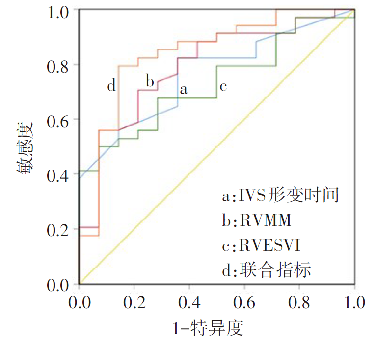
Fig.4 ROC of severe of CTD-PAH patients evaluated by CMR parameters
| [1] | WANG Z, LI J, WANG X, et al. Evaluation of pulmonary arterial pressure in patients with connective tissue disease-associated pulmonary arterial hypertension by myocardial perfusion imaging[J]. Ann Noninvasive Electrocardiol, 2022, 27(2):e12927. doi:10.1111/anec.12927. |
| [2] | 范倩, 巩路, 魏蔚, 等. 原发性干燥综合征合并肺动脉高压的临床研究[J]. 天津医药, 2013, 41(1):9-11. |
| FAN Q, GONG L, WEI W, et al. Clinical study of pulmonary hypertension in primary Sjogren’s syndrome[J]. Tianjin Med J, 2013, 41(1):9-11. doi:10.3969/j.issn.0253-9896.2013.01.003. | |
| [3] | KUMAR A, NEEMA P K. Severe pulmonary hypertension and right ventricular failure[J]. Indian J Anaesth, 2017, 61(9):753-759. doi:10.4103/ija.IJA_420_17. |
| [4] | ZHANG Z, WANG M, YANG Z, et al. Noninvasive prediction of pulmonary artery pressure and vascular resistance by using cardiac magnetic resonance indices[J]. Int J Cardiol, 2017, 227:915-922. doi:10.1016/j.ijcard.2016.10.068. |
| [5] | BRONCANO J, BHALLA S, GUTIERREZ F R, et al. Cardiac MRI in pulmonary hypertension:from magnet to bedside[J]. Radiographics, 2020, 40(4):982-1002. doi:10.1148/rg.2020190179. |
| [6] | DELLEGROTTAGLIE S, SANZ J, POON M, et al. Pulmonary hypertension: accuracy of detection with left ventricular septal-to-free wall curvature ratio measured at cardiac MR[J]. Radiology, 2007, 243(1):63-69. doi:10.1148/radiol.2431060067. |
| [7] | MAVROGENI S, BRATIS K, MARKUSSIS V, et al. The diagnostic role of cardiac magnetic resonance imaging in detecting myocardial inflammation in systemic lupus erythematosus. Differentiation from viral myocarditis[J]. Lupus, 2013, 22(1):34-43. doi:10.1177/0961203312462265. |
| [8] | PADERVINSKIENE L, KRIVICKIENE A, HOPPENOT D, et al. Prognostic value of left ventricular function and mechanics in pulmonary hypertension:a pilot cardiovascular magnetic resonance feature tracking study[J]. Medicina, 2019,55:73. doi:10.3390/medicina55030073. |
| [9] | MAVROGENI S, MARKOUSIS-MAVROGENIS G, KOUTSOGEORGOPOULOU L, et al. Cardiovascular magnetic resonance imaging:clinical implications in the evaluation of connective tissue diseases[J]. J Inflamm Res, 2017, 10:55-61. doi:10.2147/JIR.S115508. |
| [10] | 杨帆, 王丹, 张璋, 等. 超声心动图和CMR评估肺动脉高压病人心功能的研究进展[J]. 国际医学放射学杂志, 2018, 41(2):164-169. |
| YANG F, WANG D, ZHANG Z, et al. Progress of TTE and CMR in evaluating cardiac function in patients with pulmonary arterial hypertension[J]. Int J Med Radiol, 2018, 41(2):164-169. doi:10.19300/j.2018.Z5149. | |
| [11] | 马珂凡, 祝因苏, 徐怡. 心脏磁共振评估肺动脉高压的研究进展[J]. 国际医学放射学杂志, 2022, 45(6)694-699. |
| MA K F, ZHU Y S, XU Y. Progress of cardiac magnetic resonance in the evaluation of pulmonary hypertension[J]. Int J Med Radiol, 2022, 45(6):694-699. doi:10.19300/j.2022.Z19780. | |
| [12] | REN W, GUO J J, YANG F, et al. Indication of the prognosis of pulmonary hypertension by using CMR function parameters[J]. Eur Radiol, 2021, 31(9):7121-7131. doi:10.1007/s00330-021-07835-8. |
| [13] | HUMBERT M, KOVACS G, HOEPER M M, et al. 2022ESC/ERS Guidelines for the diagnosis and treatment of pulmonary hypertension[J]. Eur Heart J, 2022, 43(38):3618-3731. doi:10.1093/eurheartj/ehac237. |
| [14] | KJELLSTROM B, LINDHOLM A, OSTENFELD E. Correction to:cardiac magnetic resonance imaging in pulmonary arterial hypertension:ready for lienical practice and guidelines?[J]. Cur Heart Fail Rep, 2020,17:457. doi:10.1007/s11897-020-00492-w. |
| [15] | JOHNS C S, KIELY D G, RAJARAM S, et al. Diagnosis of pulmonary hypertension with cardiac MRI:derivation and validation of regression models[J]. Radiology, 2019, 290(1):61-68. doi:10.1148/radiol.2018180603. |
| [16] | 王丹, 张璋, 杨帆, 等. 不同室间隔形态肺高血压患者的心室功能特点:CMR初步研究[J]. 中国肺癌杂志, 2018, 21(5):397-402. |
| WANG D, ZHANG Z, YANG F, et al. Characteristics of ventricular function in pulmonary hypertension patients with different shape of interventricular septum:preliminary study with cardiac magnetic resonance imaging[J]. Chinese Journal of Lung Cancer, 2018, 21(5):397-402. doi:10.3779/j.issn.1009-3419.2018.05.07. |
| Viewed | ||||||
|
Full text |
|
|||||
|
Abstract |
|
|||||