天津医药 ›› 2023, Vol. 51 ›› Issue (12): 1326-1331.doi: 10.11958/20230708
聂进1( ), 刘代顺2, 张建勇1, 刘楚1, 周亮1,△()
收稿日期:2023-05-16
修回日期:2023-06-15
出版日期:2023-12-15
发布日期:2023-12-22
通讯作者:
△ E-mail:作者简介:聂进(1988),男,副主任医师,主要从事慢性阻塞性肺疾病与哮喘的临床与基础方面研究。E-mail:基金资助:
NIE Jin1(), LIU Daishun2, ZHANG Jianyong1, LIU Chu1, ZHOU Liang1,△()
Received:2023-05-16
Revised:2023-06-15
Published:2023-12-15
Online:2023-12-22
Contact:
△ E-mail:聂进, 刘代顺, 张建勇, 刘楚, 周亮. 脐带间充质干细胞外泌体对慢性阻塞性肺疾病大鼠肺部炎症的作用机制探讨[J]. 天津医药, 2023, 51(12): 1326-1331.
NIE Jin, LIU Daishun, ZHANG Jianyong, LIU Chu, ZHOU Liang. The effect and mechanism of exosomes from umbilical cord mesenchymal stem cells on pulmonary inflammation in chronic obstructive pulmonary disease rats[J]. Tianjin Medical Journal, 2023, 51(12): 1326-1331.
摘要:
目的 探讨人脐带间充质干细胞外泌体对慢性阻塞性肺疾病(COPD)大鼠肺部炎症的作用及机制。方法 将18只SD雄性大鼠随机分为Control组、COPD组、COPD+Exo组,每组6只;COPD组、COPD+Exo组大鼠采用香烟烟熏+脂多糖(LPS)雾化吸入的方法建立COPD大鼠模型,COPD+Exo组经尾静脉注射100 μL人脐带间充质干细胞外泌体稀释液(含2×10⁷个外泌体),Control组和COPD组注射等量的磷酸盐缓冲液。干预后1周处死大鼠,收集外周血、肺泡灌洗液(BALF)和肺组织。HE染色观察大鼠肺组织病理改变,全自动血细胞分析仪计数外周血炎性细胞数目,酶联免疫吸附试验(ELISA)检测BALF和血清中白细胞介素(IL)-1β、IL-6、肿瘤坏死因子(TNF)-α水平,qRT-PCR检测肺组织中IL-1β、IL-6、TNF-α mRNA的表达,Western blot检测肺组织中Toll样受体4(TLR4)、磷酸化核因子κB(p-NF-κB p65)和核因子抑制蛋白(IκBα)的表达。结果 Control组大鼠肺泡结构完整、形态规则;COPD组大鼠肺泡结构紊乱,部分肺泡不规则扩张、融合成肺大疱甚至破裂,肺泡间隔增宽且可见较多炎性细胞浸润。与COPD组比较,COPD+Exo组大鼠肺泡结构较完整,肺泡间隔炎性细胞浸润减少,外周血中炎性细胞减少,BALF、血清及肺组织中IL-1β、IL-6、TNF-α表达水平降低(P<0.05);肺组织TLR4、IκBα蛋白表达水平升高,p-NF-κB p65表达水平降低(P<0.05)。结论 人脐带间充质干细胞外泌体通过调控TLR4/核因子κB(NF-κB)信号通路减轻COPD大鼠的肺部炎症,从而发挥肺保护作用。
中图分类号:
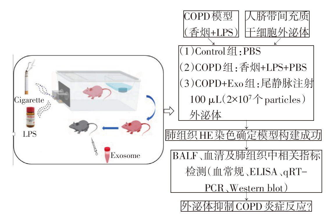
图1 人脐带间充质干细胞外泌体抑制COPD大鼠肺部炎症的实验流程图
Fig.1 Flow chart of experimental study on inhibition of lung inflammation by human umbilical mesenchymal stem cell exosomes in COPD rats
| 基因名称 | 引物(5′→3′) | 产物大小/bp |
|---|---|---|
| IL-1β | 上游:GCACAGTTCCCCAACTGGTA | 109 |
| 下游:ACACGGGTTCCATGGTGAAG | ||
| IL-6 | 上游:CTCTCCGCAAGAGACTTCCA | 92 |
| 下游:TCTCCTCTCCGGACTTGTGAA | ||
| TNF-α | 上游:CCACGCTCTTCTGTCTACTG | 145 |
| 下游:GCTACGGGCTTGTCACTC | ||
| GAPDH | 上游:CAGGAGGCATTGCTGATGAT | 138 |
| 下游:GAAGGCTGGGGCTCATTT |
表1 qRT-PCR引物序列
Tab.1 Primer sequence for qRT-PCR
| 基因名称 | 引物(5′→3′) | 产物大小/bp |
|---|---|---|
| IL-1β | 上游:GCACAGTTCCCCAACTGGTA | 109 |
| 下游:ACACGGGTTCCATGGTGAAG | ||
| IL-6 | 上游:CTCTCCGCAAGAGACTTCCA | 92 |
| 下游:TCTCCTCTCCGGACTTGTGAA | ||
| TNF-α | 上游:CCACGCTCTTCTGTCTACTG | 145 |
| 下游:GCTACGGGCTTGTCACTC | ||
| GAPDH | 上游:CAGGAGGCATTGCTGATGAT | 138 |
| 下游:GAAGGCTGGGGCTCATTT |
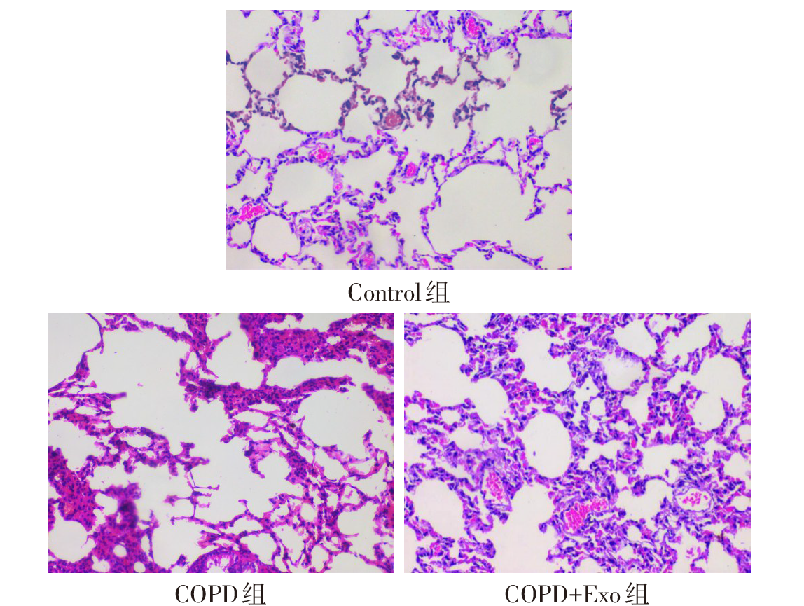
图2 各组大鼠肺组织HE染色情况(×200)
Fig.2 Representative images of HE staining of lung tissue in each group of rats (×200)
| 组别 | WBC | NEU | MON | LYM |
|---|---|---|---|---|
| Control组 | 0.48±0.07 | 0.77±0.24 | 0.03±0.00 | 0.25±0.00 |
| COPD组 | 3.10±0.27a | 2.01±0.20a | 0.15±0.01a | 2.63±0.19a |
| COPD+Exo组 | 1.59±0.55ab | 0.35±0.15b | 0.06±0.02b | 1.20±0.47ab |
| F | 27.197* | 39.738* | 51.100* | 33.116* |
表2 各组大鼠外周血炎性细胞计数
Tab.2 Number of inflammatory cells in peripheral blood of rats in each group (n=3,×109/L,$\bar{x}±s$)
| 组别 | WBC | NEU | MON | LYM |
|---|---|---|---|---|
| Control组 | 0.48±0.07 | 0.77±0.24 | 0.03±0.00 | 0.25±0.00 |
| COPD组 | 3.10±0.27a | 2.01±0.20a | 0.15±0.01a | 2.63±0.19a |
| COPD+Exo组 | 1.59±0.55ab | 0.35±0.15b | 0.06±0.02b | 1.20±0.47ab |
| F | 27.197* | 39.738* | 51.100* | 33.116* |
| 组别 | BALF | |||||
|---|---|---|---|---|---|---|
| IL-1β | IL-6 | TNF-α | ||||
| Control组 | 55.05±11.49 | 71.58±8.77 | 290.51±28.28 | |||
| COPD组 | 85.23±13.44a | 104.16±7.92a | 513.71±59.00a | |||
| COPD+Exo组 | 61.02±6.42b | 84.66±12.84b | 406.74±52.32ab | |||
| F | 10.827* | 13.234* | 26.634* | |||
| 组别 | 血清 | |||||
| IL-1β | IL-6 | TNF-α | ||||
| Control组 | 64.02±6.84 | 72.00±4.45 | 309.18±5.25 | |||
| COPD组 | 88.54±8.44a | 90.99±0.97a | 483.74±9.74a | |||
| COPD+Exo组 | 73.22±4.92b | 73.41±7.93b | 364.64±2.40b | |||
| F | 9.708* | 8.036* | 11.869* | |||
表3 各组大鼠BALF及血清中IL-1β、IL-6、TNF-α的表达水平
Tab.3 Expression levels of IL-1β, IL-6 and TNF-α in BALF and serum of rats in each group (n=6,ng/L,$\bar{x}±s$)
| 组别 | BALF | |||||
|---|---|---|---|---|---|---|
| IL-1β | IL-6 | TNF-α | ||||
| Control组 | 55.05±11.49 | 71.58±8.77 | 290.51±28.28 | |||
| COPD组 | 85.23±13.44a | 104.16±7.92a | 513.71±59.00a | |||
| COPD+Exo组 | 61.02±6.42b | 84.66±12.84b | 406.74±52.32ab | |||
| F | 10.827* | 13.234* | 26.634* | |||
| 组别 | 血清 | |||||
| IL-1β | IL-6 | TNF-α | ||||
| Control组 | 64.02±6.84 | 72.00±4.45 | 309.18±5.25 | |||
| COPD组 | 88.54±8.44a | 90.99±0.97a | 483.74±9.74a | |||
| COPD+Exo组 | 73.22±4.92b | 73.41±7.93b | 364.64±2.40b | |||
| F | 9.708* | 8.036* | 11.869* | |||
| 组别 | IL-1β | IL-6 | TNF-α |
|---|---|---|---|
| Control组 | 1.00±0.02 | 1.00±0.10 | 1.02±0.18 |
| COPD组 | 3.10±0.31a | 6.58±1.03a | 5.17±0.35a |
| COPD+Exo组 | 1.56±0.12ab | 1.65±0.02ab | 2.52±0.31ab |
| F | 62.411* | 51.588* | 104.294* |
表4 各组大鼠肺组织中IL-1β、IL-6、TNF-α mRNA的表达水平
Tab.4 Expression levels of IL-1β, IL-6 and TNF-α in lung tissue of rats in each group (n=6, $\bar{x}±s$)
| 组别 | IL-1β | IL-6 | TNF-α |
|---|---|---|---|
| Control组 | 1.00±0.02 | 1.00±0.10 | 1.02±0.18 |
| COPD组 | 3.10±0.31a | 6.58±1.03a | 5.17±0.35a |
| COPD+Exo组 | 1.56±0.12ab | 1.65±0.02ab | 2.52±0.31ab |
| F | 62.411* | 51.588* | 104.294* |
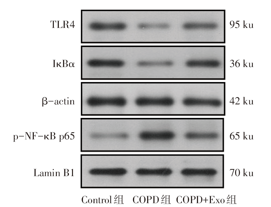
图3 Western blot检测各组大鼠肺组织中TLR4、IκBα、p-NF-κB p65蛋白的表达
Fig.3 Expression levels of TLR4, IκBα and p-NF-κB p65 protein in lung tissue of rats in each group detected by Western blot assay
| 组别 | TLR4 | IκBα | p-NF-κB p65 |
|---|---|---|---|
| Control组 | 0.73±0.01 | 0.50±0.03 | 0.31±0.02 |
| COPD组 | 0.30±0.00a | 0.23±0.01a | 0.92±0.10a |
| COPD+Exo组 | 0.56±0.02ab | 0.46±0.02b | 0.58±0.06ab |
| F | 824.637* | 130.246* | 59.341* |
表5 各组大鼠肺组织中TLR4、IκBα、p-NF-κB p65蛋白表达水平的比较
Tab.5 Comparison of TLR4, IκBα and p-NF-κB p65 protein levels in lung tissue between the three groups of rats (n=4,$\bar{x}±s$)
| 组别 | TLR4 | IκBα | p-NF-κB p65 |
|---|---|---|---|
| Control组 | 0.73±0.01 | 0.50±0.03 | 0.31±0.02 |
| COPD组 | 0.30±0.00a | 0.23±0.01a | 0.92±0.10a |
| COPD+Exo组 | 0.56±0.02ab | 0.46±0.02b | 0.58±0.06ab |
| F | 824.637* | 130.246* | 59.341* |
| [1] | CHRISTENSON S A, SMITH B M, BAFADHEL M, et al. Chronic obstructive pulmonary disease[J]. Lancet, 2022, 399(10342):2227-2242. doi:10.1016/S0140-6736(22)00470-6. |
| [2] | SONG Q, CHEN P, LIU X M. The role of cigarette smoke-induced pulmonary vascular endothelial cell apoptosis in COPD[J]. Respir Res, 2021, 22(1):39. doi:10.1186/s12931-021-01630-1. |
| [3] | 中华医学会呼吸病学分会慢性阻塞性肺疾病学组, 中国医师协会呼吸医师分会慢性阻塞性肺疾病工作委员会, 陈荣昌, 等. 慢性阻塞性肺疾病诊治指南(2021年修订版)[J]. 中华结核和呼吸杂志, 2021, 44(3):170-205. |
| Group of Chronic Obstructive Pulmonary Disease, Respiratory Branch of Chinese Medical Association, Working Committee of Chronic Obstructive Pulmonary Disease, Respiratory Branch of Chinese Medical Association, CHEN R C, et al. Guidelines for the diagnosis and treatment of chronic obstructive pulmonary disease (2021 revision)[J]. Chinese Journal of Tuberculosis and Respiration, 2021, 44(3):170-205. doi:10.3760/cma.j.cn112147-20210109-00031. | |
| [4] | LIU J, GAO J, LIANG Z, et al. Mesenchymal stem cells and their microenvironment[J]. Stem Cell Res Ther, 2022, 13(1):429. doi:10.1186/s13287-022-02985-y. |
| [5] | ZOU J X, YANG W N, CUI W S, et al. Therapeutic potential and mechanisms of mesenchymal stem cell-derived exosomes as bioactive materials in tendon-bone healing[J]. J Nanobiotechnology, 2023, 21(1):14. doi:10.1186/s12951-023-01778-6. |
| [6] | MOHAN A, AGARWAL S, CLAUSS M, et al. Extracellular vesicles:novel communicators in lung diseases[J]. Respir Res, 2020, 21(1):175. doi:10.1186/s12931-020-01423-y. |
| [7] | HARRELL C R, JOVICIC N, DJONOV V, et al. Mesenchymal stem cell-derived exosomes and other extracellular vesicles as new remedies in the therapy of inflammatory diseases[J]. Cells, 2019, 8(12):1605. doi:10.3390/cells8121605. |
| [8] | SHEN Z W, HUANG W, LIU J, et al. Effects of mesenchymal stem cell-derived exosomes on autoimmune diseases[J]. Front Immunol, 2021, 12:749192. doi:10.3389/fimmu.2021.749192. |
| [9] | LIN Z J, WU Y L, XU Y T, et al. Mesenchymal stem cell-derived exosomes in cancer therapy resistance:recent advances and therapeutic potential[J]. Mol Cancer, 2022, 21(1):179. doi:10.1186/s12943-022-01650-5. |
| [10] | ZHOU L, LUO H, LEE J W. Role of extracellular vesicles in lung diseases[J]. Chin Med J, 2022, 135(15):1765-1780. doi:10.1097/CM9.0000000000002118. |
| [11] | ABBASZADEH H, GHORBANI F, ABBASPOUR-AGHDAM S, et al. Chronic obstructive pulmonary disease and asthma:mesenchymal stem cells and their extracellular vesicles as potential therapeutic tools[J]. Stem Cell Res Ther, 2022, 13(1):262. doi:10.1186/s13287-022-02938-5. |
| [12] | SHU J Z, LI D F, OUYANG H P, et al. Comparison and evaluation of two different methods to establish the cigarette smoke exposure mouse model of COPD[J]. Sci Rep, 2017, 7(1):15454. doi:10.1038/s41598-017-15685-y. |
| [13] | KORSGREN M, LINDEN M, ENTWISTLE N, et al. Inhalation of LPS induces inflammatory airway responses mimicking characteristics of chronic obstructive pulmonary disease[J]. Clin Physiol Funct Imaging, 2012, 32(1):71-79. doi:10.1111/j.1475-097X.2011.01058.x. |
| [14] | SHI M M, YANG Q Y, MONSEL A, et al. Preclinical efficacy and clinical safety of clinical-grade nebulized allogenic adipose mesenchymal stromal cells-derived extracellular vesicles[J]. J Extracell Vesicles, 2021, 10(10):e12134. doi:10.1002/jev2.12134. |
| [15] | CHURG A, COSIO M, WRIGHT J L. Mechanisms of cigarette smoke-induced COPD:insights from animal models[J]. Am J Physiol Lung Cell Mol Physiol, 2008, 294(4):L612-L631. doi:10.1152/ajplung.00390.2007. |
| [16] | AGHAPOUR M, RAEE P, MOGHADDAM S J, et al. Airway epithelial barrier dysfunction in chronic obstructive pulmonary disease:role of cigarette smoke exposure[J]. Am J Respir Cell Mol Biol, 2018, 58(2):157-169. doi:10.1165/rcmb.2017-0200TR. |
| [17] | JONES B, DONOVAN C, LIU G, et al. Animal models of COPD:what do they tell us?[J]. Respirology, 2017, 22(1):21-32. doi:10.1111/resp.12908. |
| [18] | LIANG G B, HE Z H. Animal models of emphysema[J]. Chin Med J(Engl), 2019, 132(20):2465-2475. doi:10.1097/CM9.0000000000000469. |
| [19] | SMITH K R, LEONARD D, MCDONALD J D, et al. Inflammation,mucous cell metaplasia,and Bcl-2 expression in response to inhaled lipopolysaccharide aerosol and effect of rolipram[J]. Toxicol Appl Pharmacol, 2011, 253(3):253-260. doi:10.1016/j.taap.2011.04.001. |
| [20] | DE SOUZA XAVIER COSTA N, RIBEIRO JÚNIOR G,DOS SANTOS ALEMANY A A, et al. Early and late pulmonary effects of nebulized LPS in mice:An acute lung injury model[J]. PLoS One, 2017, 12(9):e0185474. doi:10.1371/journal.pone.0185474. |
| [21] | FONCECA A M, ZOSKY G R, BOZANICH E M, et al. Accumulation mode particles and LPS exposure induce TLR-4 dependent and independent inflammatory responses in the lung[J]. Respir Res, 2018, 19(1):15. doi:10.1186/s12931-017-0701-z. |
| [22] | 何永鸿, 强丽, 王宋平. 阿托伐他汀钙对COPD模型大鼠肺血管重塑的影响及机制探讨[J]. 天津医药, 2021, 49(6):598-602. |
| HE Y H, QIANG L, WANG S P. The effect of atorvastatin calcium on pulmonary vascular remodeling in chronic obstructive pulmonary disease rats[J]. Tianjin Med J, 2021, 49(6):598-602. doi:10.11958/202013011. | |
| [23] | ZHU W T, LI C H, DAI T T, et al. Effect of allyl isothiocyanate on oxidative stress in COPD via the AhR/CYP1A1 and Nrf2/NQO1 pathways and the underlying mechanism[J]. Phytomedicine, 2023, 114:154774. doi:10.1016/j.phymed.2023.154774. |
| [24] | JU J, LI Z, SHI Q. Baicalin inhibits inflammation in rats with chronic obstructive pulmonary disease by the TLR2/MYD88/NF-κBp65 signaling pathway[J]. Evid Based Complement Alternat Med, 2022, 2022:7273387. doi:10.1155/2022/7273387. |
| [25] | ARMITAGE J, TAN D, MOODLEY Y, et al. Mesenchymal stem cell infusion modulates systemic inflammation in patients with chronic obstructive pulmonary disease(COPD)[J]. Respirology, 2016, 21(Suppl 2):133. |
| [26] | GAO J, LIANG Y, CHEN J, et al. CXCR4 enhances the inhibitory effects of bone mesenchymal stem cells on lung cell apoptosis in a rat model of smoking-induced COPD[J]. Apoptosis, 2023, 28(3/4):639-652. doi:10.1007/s10495-022-01800-6. |
| [27] | WEISS D J, SEGAL K, CASABURI R, et al. Effect of mesenchymal stromal cell infusions on lung function in COPD patients with high CRP levels[J]. Respir Res, 2021, 22(1):142. doi:10.1186/s12931-021-01734-8. |
| [28] | ALVARENGA-NASCIMENTO C R, AADB LEIA, SANTOS T G, et al. Immunotherapeutic strategy with mesenchymal stem cells modulating inflammation in an experimental model of COPD[J]. European Respiratory Journal, 2020, 56(Suppl 64):314. |
| [29] | VOLAREVIC V, MARKOVIC B S, GAZDIC M, et al. Ethical and safety issues of stem cell-based therapy[J]. Int J Med Sci, 2018, 15(1):36-45. doi:10.7150/ijms.21666. |
| [30] | TANG Y Y, ZHOU Y, LI H J. Advances in mesenchymal stem cell exosomes:a review[J]. Stem Cell Res Ther, 2021, 12(1):71. doi:10.1186/s13287-021-02138-7. |
| [31] | CHAN A M L, SAMPASIVAM Y, LOKANATHAN Y. Biodistribution of mesenchymal stem cells (MSCs) in animal models and implied role of exosomes following systemic delivery of MSCs:a systematic review[J]. Am J Transl Res, 2022, 14(4):2147-2161. |
| [32] | XIA L J, ZHANG C L, LV N Y, et al. AdMSC-derived exosomes alleviate acute lung injury via transferring mitochondrial component to improve homeostasis of alveolar macrophages[J]. Theranostics, 2022, 12(6):2928-2947. doi:10.7150/thno.69533. |
| [33] | WAN X, CHEN S, FANG Y, et al. Mesenchymal stem cell-derived extracellular vesicles suppress the fibroblast proliferation by downregulating FZD6 expression in fibroblasts via micrRNA-29b-3p in idiopathic pulmonary fibrosis[J]. J Cell Physiol, 2020, 235(11):8613-8625. doi:10.1002/jcp.29706. |
| [34] | CHEN Q, LIN J, DENG Z Q, et al. Exosomes derived from human umbilical cord mesenchymal stem cells protect against papain-induced emphysema by preventing apoptosis through activating VEGF-VEGFR2-mediated AKT and MEK/ERK pathways in rats[J]. Regen Ther, 2022, 21:216-224. doi:10.1016/j.reth.2022.07.002. |
| [35] | YANG L, WEN M, LIU X, et al. Feikang granules ameliorate pulmonary inflammation in the rat model of chronic obstructive pulmonary disease via TLR2/4-mediated NF-κB pathway[J]. BMC Complement Med Ther, 2020, 20(1):170. doi:10.1186/s12906-020-02964-x. |
| [36] | ROY A, SRIVASTAVA M, SAQIB U, et al. Potential therapeutic targets for inflammation in toll-like receptor 4 (TLR4)-mediated signaling pathways[J]. Int Immunopharmacol, 2016, 40:79-89. doi:10.1016/j.intimp.2016.08.026. |
| [37] | LI Y, ZHAO J, SHAO H, et al. Preventive effect of total flavonoids of Trollius altaicus on a chronic obstructive pulmonary disease rat model based on the TLR4/NF-κB pathway[J]. Ann Transl Med, 2022, 10(4):222. doi:10.21037/atm-22-331. |
| [1] | 姜天佑, 李敏, 孙碧文, 李越洋, 邢丽静, 田晨. Let-7b诱导白血病相关巨噬细胞复极抑制AML的发展[J]. 天津医药, 2026, 54(3): 225-231. |
| [2] | 杨晓芳, 贾新燕, 丰文君. miR-181a-5p通过HMGB1/NF-κB信号通路调控狼疮性肾炎小鼠肾小球系膜细胞增殖和凋亡[J]. 天津医药, 2026, 54(3): 232-237. |
| [3] | 张婧, 魏玉英, 宁海虹, 韦红梅, 王嘉玮, 曹薇, 吴宾. DUSP9在2型糖尿病心肌病小鼠心肌损伤中的保护作用及其机制[J]. 天津医药, 2026, 54(3): 238-244. |
| [4] | 陈丽, 陈楠. COPD进展为Ⅱ型呼吸衰竭预警模型的建立与验证[J]. 天津医药, 2026, 54(2): 189-195. |
| [5] | 王文文, 董垚, 赵璐, 任鲁宁, 杜红阳. 火把花根片对中度特应性皮炎患者的临床疗效研究[J]. 天津医药, 2026, 54(1): 92-96. |
| [6] | 孔翠文, 路延双, 孙丽萍, 于芬芬. LncRNA SNHG14靶向miR-30a-5p对高糖诱导的足细胞损伤的影响[J]. 天津医药, 2025, 53(9): 903-909. |
| [7] | 周玉科, 孙伟, 高庆, 贺军. 七氟醚调节ROS/TXNIP/NLRP3通路对脊髓损伤大鼠神经细胞炎症的影响[J]. 天津医药, 2025, 53(9): 910-915. |
| [8] | 李硕, 张云鹏, 黄艳, 曹美然, 贾兰芳, 胡桂才, 黄兰, 段书众. 系统性免疫炎症指数与IgA肾病患者临床病理特征的关系[J]. 天津医药, 2025, 53(9): 932-936. |
| [9] | 于晓猛, 索睿, 杜昕桃, 索莹, 阿亚拉·阿斯哈尔, 郝天旭, 赵晓赟. 人脐带来源的间充质干细胞对慢性间歇低氧小鼠的影响[J]. 天津医药, 2025, 53(8): 814-819. |
| [10] | 陈慧, 赵凯, 刘振国, 常瑛, 巨康璐. 金丝桃素通过调控NLRP3炎症小体改善小鼠急性胰腺炎的机制探讨[J]. 天津医药, 2025, 53(8): 820-825. |
| [11] | 李迎杰, 张航, 郝少婷, 许菲, 郭魏超, 宋惠, 王艳芬. 生脉散合加味升陷汤对气虚血瘀型射血分数降低的心力衰竭的疗效观察[J]. 天津医药, 2025, 53(8): 860-864. |
| [12] | 李诚诚, 李龙骧, 于立恒. 灯盏细辛注射液联合阿托伐他汀钙治疗急性缺血性脑卒中的疗效观察[J]. 天津医药, 2025, 53(8): 865-869. |
| [13] | 蒋苏, 李东霞, 吕新翔, 崔艳红, 吕李婷. 基于PI3K/Akt/NF-κB信号通路探讨黄连素对特应性皮炎大鼠皮肤病理变化的治疗作用[J]. 天津医药, 2025, 53(7): 679-683. |
| [14] | 李林森, 冯玉梅. 卵巢切除小鼠的骨重塑特征研究[J]. 天津医药, 2025, 53(6): 566-570. |
| [15] | 马春梅, 于鹏, 张其程, 杨磊, 李棣华, 谭建, 孟召伟. 异硫氰酸苄酯联合索拉非尼治疗未分化甲状腺癌机制探讨[J]. 天津医药, 2025, 53(5): 449-455. |
| 阅读次数 | ||||||
|
全文 |
|
|||||
|
摘要 |
|
|||||