天津医药 ›› 2024, Vol. 52 ›› Issue (1): 50-55.doi: 10.11958/20231143
张琳琳( ), 赵唐明, 黄婵, 李善文, 甘卫华()
收稿日期:2023-08-29
出版日期:2024-01-15
发布日期:2024-01-18
通讯作者:
△E-mail:作者简介:张琳琳(1996),女,硕士在读,主要从事小儿肾脏疾病诊治及机制方面研究。E-mail:基金资助:
ZHANG Linlin(), ZHAO Tangming, HUANG Chan, LI Shanwen, GAN Weihua()
Received:2023-08-29
Published:2024-01-15
Online:2024-01-18
Contact:
△E-mail:张琳琳, 赵唐明, 黄婵, 李善文, 甘卫华. 新型多肽AMPP2在TGF-β1诱导的系膜细胞增殖中的作用及其机制[J]. 天津医药, 2024, 52(1): 50-55.
ZHANG Linlin, ZHAO Tangming, HUANG Chan, LI Shanwen, GAN Weihua. Effects and mechanism of AMPP2 on mesangial cell proliferation induced by TGF-β1[J]. Tianjin Medical Journal, 2024, 52(1): 50-55.
摘要:
目的 探究新型多肽AMPP2在转化生长因子β1(TGF-β1)诱导的小鼠系膜细胞增殖中的作用及相关机制。方法 体外用TGF-β1(10 μg/L)及AMPP2(10 ng/L)干预系膜细胞,分为Control组、AMPP2组、TGF-β1组及TGF-β1+AMPP2组。CCK-8法检测各组系膜细胞增殖活力;Western blot法检测各组细胞周期蛋白依赖性激酶(CDK)-4、CDK-6、细胞增殖核抗原(PCNA)、α平滑肌肌动蛋白(α-SMA)、Ⅰ型胶原蛋白(COL-Ⅰ)、纤维连接蛋白(FN)及磷酸化的SMAD同源物3/SMAD同源物3(p-SMAD3/SMAD3)的蛋白表达水平;qPCR法检测各组α-SMA、COL-Ⅰ、FN的mRNA表达水平。结果 与Control组相比,TGF-β1组系膜细胞增殖活力增加(P<0.05),与TGF-β1组相比,TGF-β1+AMPP2组系膜细胞增殖活力下降(P<0.05);与Control组相比,TGF-β1组CDK-4、CDK-6、PCNA蛋白以及α-SMA、COL-Ⅰ、FN蛋白和mRNA表达水平升高(P<0.05),与TGF-β1组相比,TGF-β1+AMPP2组上述蛋白及mRNA表达水平均下降(P<0.05);与Control组相比,TGF-β1组p-SMAD3/SMAD3水平显著升高(P<0.05),与TGF-β1组相比,TGF-β1+AMPP2组p-SMAD3/SMAD3的蛋白表达量显著降低(P<0.05)。结论 AMPP2可能通过调控TGF-β1/SMAD3通路抑制系膜细胞增殖。
中图分类号:
| 基因 | 引物序列(5′→3′) | 产物大小/bp |
|---|---|---|
| α-SMA | 上游:CAGCAAACAGGAATACGACGAA | 168 |
| 下游:AACCACGAGTAACAAATCAAAGC | ||
| COL-Ⅰ | 上游:GTCAGACCTGTGTGTTCCCTACTCA | 99 |
| 下游:TCTCTCCAAACCAGACGTGCTTC | ||
| FN | 上游:GCAAGAAGGACAACCGAGGAAA | 126 |
| 下游:GGACATCAGTGAAGGAGCCAGA | ||
| β-actin | 上游:CATCCGTAAAGACCTCTATGCCAAC | 171 |
| 下游:ATGGAGCCACCGATCCACA |
表1 qPCR引物序列
Tab.1 Sequences of qPCR primers
| 基因 | 引物序列(5′→3′) | 产物大小/bp |
|---|---|---|
| α-SMA | 上游:CAGCAAACAGGAATACGACGAA | 168 |
| 下游:AACCACGAGTAACAAATCAAAGC | ||
| COL-Ⅰ | 上游:GTCAGACCTGTGTGTTCCCTACTCA | 99 |
| 下游:TCTCTCCAAACCAGACGTGCTTC | ||
| FN | 上游:GCAAGAAGGACAACCGAGGAAA | 126 |
| 下游:GGACATCAGTGAAGGAGCCAGA | ||
| β-actin | 上游:CATCCGTAAAGACCTCTATGCCAAC | 171 |
| 下游:ATGGAGCCACCGATCCACA |
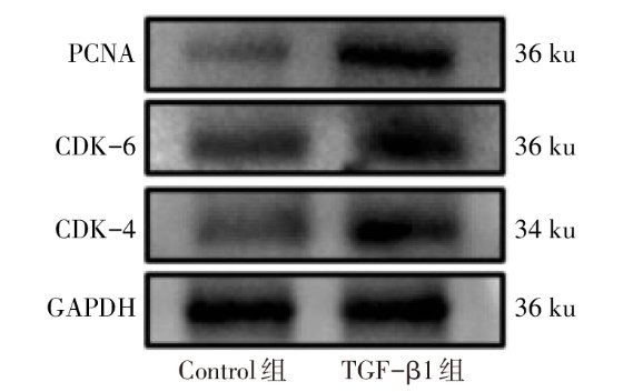
图1 2组系膜细胞中CDK-4、CDK-6及PCNA蛋白表达
Fig.1 The protein expression levels of CDK-4, CDK-6 and PCNA in mesangial cells of two groups
| 组别 | CDK-4 | CDK-6 | PCNA |
|---|---|---|---|
| Control组 | 0.66±0.05 | 0.69±0.05 | 0.49±0.03 |
| TGF-β1组 | 1.14±0.07 | 1.05±0.05 | 0.97±0.12 |
| t | 32.600** | 8.310* | 6.733* |
表2 2组系膜细胞中CDK-4、CDK-6及PCNA蛋白表达水平比较
Tab.2 Comparison of protein expression levels of CDK-4, CDK-6 and PCNA in mesangial cells between two groups (n=3,$\bar{x}±s$)
| 组别 | CDK-4 | CDK-6 | PCNA |
|---|---|---|---|
| Control组 | 0.66±0.05 | 0.69±0.05 | 0.49±0.03 |
| TGF-β1组 | 1.14±0.07 | 1.05±0.05 | 0.97±0.12 |
| t | 32.600** | 8.310* | 6.733* |
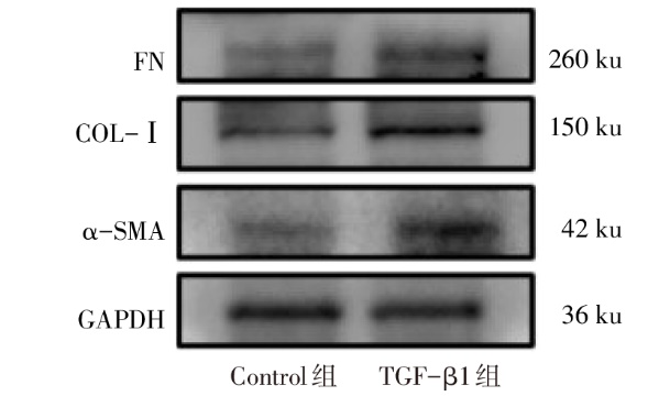
图2 2组系膜细胞中α-SMA、COL-Ⅰ及FN 蛋白表达
Fig.2 The protein expression levels of α-SMA, COL-Ⅰ and FN in mesangial cells of two groups
| 组别 | 蛋白 | mRNA | ||||
|---|---|---|---|---|---|---|
| α-SMA | COL-Ⅰ | FN | α-SMA | COL-Ⅰ | FN | |
| Control组 | 0.56±0.03 | 0.62±0.02 | 0.47±0.09 | 1.01±0.00 | 1.01±0.01 | 1.00±0.00 |
| TGF-β1组 | 1.05±0.09 | 1.03±0.04 | 0.99±0.05 | 4.36±0.44 | 4.21±0.35 | 4.64±0.74 |
| t | 14.930** | 21.230** | 7.386* | 13.340** | 15.870** | 8.520** |
表3 2组系膜细胞α-SMA、COL-Ⅰ及FN蛋白及mRNA表达水平比较
Tab.3 Comparison of protein and mRNA expression levels of α-SMA, COL-Ⅰ and FN in mesangial cells between two groups (n=3,$\bar{x}±s$)
| 组别 | 蛋白 | mRNA | ||||
|---|---|---|---|---|---|---|
| α-SMA | COL-Ⅰ | FN | α-SMA | COL-Ⅰ | FN | |
| Control组 | 0.56±0.03 | 0.62±0.02 | 0.47±0.09 | 1.01±0.00 | 1.01±0.01 | 1.00±0.00 |
| TGF-β1组 | 1.05±0.09 | 1.03±0.04 | 0.99±0.05 | 4.36±0.44 | 4.21±0.35 | 4.64±0.74 |
| t | 14.930** | 21.230** | 7.386* | 13.340** | 15.870** | 8.520** |
| 组别 | 0 h | 24 h | 48 h | 72 h | F |
|---|---|---|---|---|---|
| TGF-β1+0 ng/L AMPP2组 | 1.00±0.00 | 1.47±0.05A | 1.59±0.03AB | 1.58±0.06AB | 164.300** |
| TGF-β1+1 ng/L AMPP2组 | 1.00±0.00 | 1.46±0.11A | 1.48±0.04aA | 1.40±0.03aA | 174.300** |
| TGF-β1+10 ng/L AMPP2组 | 1.00±0.00 | 1.42±0.16A | 1.19±0.01aAB | 1.17±0.04aAB | 157.700** |
| TGF-β1+100 ng/L AMPP2组 | 1.00±0.00 | 1.32±0.02aA | 1.29±0.04aA | 1.27±0.04aAB | 96.870** |
| F | 20.810** | 112.400** | 45.840** |
表4 4组系膜细胞不同时间点增殖情况比较
Tab.4 Comparison of mesangial cell proliferation at different time points between four groups (n=3,$\bar{x}±s$)
| 组别 | 0 h | 24 h | 48 h | 72 h | F |
|---|---|---|---|---|---|
| TGF-β1+0 ng/L AMPP2组 | 1.00±0.00 | 1.47±0.05A | 1.59±0.03AB | 1.58±0.06AB | 164.300** |
| TGF-β1+1 ng/L AMPP2组 | 1.00±0.00 | 1.46±0.11A | 1.48±0.04aA | 1.40±0.03aA | 174.300** |
| TGF-β1+10 ng/L AMPP2组 | 1.00±0.00 | 1.42±0.16A | 1.19±0.01aAB | 1.17±0.04aAB | 157.700** |
| TGF-β1+100 ng/L AMPP2组 | 1.00±0.00 | 1.32±0.02aA | 1.29±0.04aA | 1.27±0.04aAB | 96.870** |
| F | 20.810** | 112.400** | 45.840** |
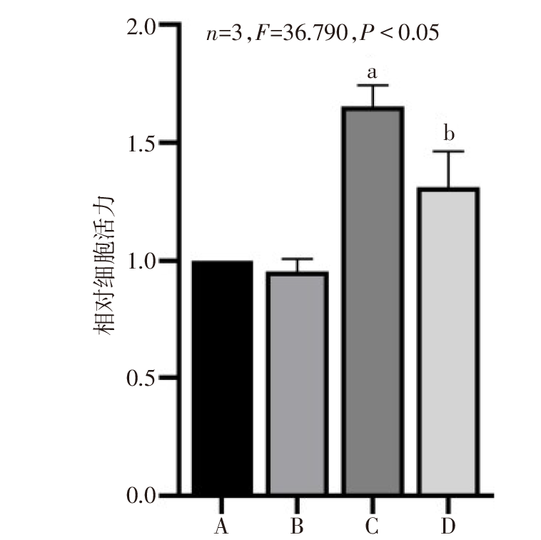
图3 AMPP2对细胞活力的影响 A:Control组;B:AMPP2组;C:TGF-β1组;D:TGF-β1+AMPP2组;a与Control组比较,b与TGF-β1组比较,P<0.05;图4—6同。
Fig.3 Effects of AMPP2 on mesangial cell proliferative activity
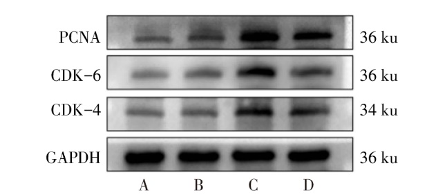
图4 4组系膜细胞中CDK-4、CDK-6及PCNA蛋白表达
Fig.4 The protein expression levels of CDK-4, CDK-6 and PCNA in mesangial cells in four groups
| 组别 | CDK-4 | CDK-6 | PCNA |
|---|---|---|---|
| Control组 | 0.85±0.05 | 1.09±0.13 | 0.79±0.15 |
| AMPP2组 | 0.81±0.11 | 1.09±0.22 | 0.78±0.26 |
| TGF-β1组 | 1.26±0.10a | 1.78±0.15a | 1.62±0.17a |
| TGF-β1+AMPP2组 | 0.86±0.14b | 1.30±0.15b | 0.96±0.22b |
| F | 16.420** | 11.480** | 11.390** |
表5 4组系膜细胞CDK-4、CDK-6及PCNA蛋白表达水平比较
Tab.5 Comparison of protein expression levels of CDK-4, CDK-6 and PCNA in mesangial cells between four groups (n=3,$\bar{x}±s$)
| 组别 | CDK-4 | CDK-6 | PCNA |
|---|---|---|---|
| Control组 | 0.85±0.05 | 1.09±0.13 | 0.79±0.15 |
| AMPP2组 | 0.81±0.11 | 1.09±0.22 | 0.78±0.26 |
| TGF-β1组 | 1.26±0.10a | 1.78±0.15a | 1.62±0.17a |
| TGF-β1+AMPP2组 | 0.86±0.14b | 1.30±0.15b | 0.96±0.22b |
| F | 16.420** | 11.480** | 11.390** |
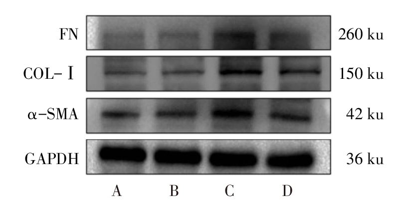
图5 4组系膜细胞中α-SMA、COL-Ⅰ及FN蛋白表达
Fig.5 The protein expression levels of α-SMA, COL-Ⅰ and FN in mesangial cells of four groups
| 组别 | α-SMA | COL-Ⅰ | FN |
|---|---|---|---|
| Control组 | 0.63±0.08 | 0.85±0.10 | 0.67±0.05 |
| AMPP2组 | 0.60±0.05 | 0.81±0.12 | 0.60±0.01 |
| TGF-β1组 | 1.20±0.11a | 1.23±0.08a | 1.24±0.19a |
| TGF-β1+AMPP2组 | 0.76±0.13b | 0.94±0.16b | 0.89±0.09b |
| F | 32.660** | 12.520** | 20.940** |
表6 4组系膜细胞α-SMA、COL-Ⅰ及FN蛋白表达水平比较
Tab.6 Comparison of mRNA expression levels of α-SMA, COL-Ⅰ and FN in mesangial cells between four groups (n=3,$\bar{x}±s$)
| 组别 | α-SMA | COL-Ⅰ | FN |
|---|---|---|---|
| Control组 | 0.63±0.08 | 0.85±0.10 | 0.67±0.05 |
| AMPP2组 | 0.60±0.05 | 0.81±0.12 | 0.60±0.01 |
| TGF-β1组 | 1.20±0.11a | 1.23±0.08a | 1.24±0.19a |
| TGF-β1+AMPP2组 | 0.76±0.13b | 0.94±0.16b | 0.89±0.09b |
| F | 32.660** | 12.520** | 20.940** |
| 组别 | α-SMA | COL-Ⅰ | FN |
|---|---|---|---|
| Control组 | 0.99±0.03 | 1.02±0.01 | 0.92±0.03 |
| AMPP2组 | 0.90±0.06 | 0.88±0.10 | 0.92±0.11 |
| TGF-β1组 | 4.03±0.45a | 4.37±0.25a | 4.12±0.60a |
| TGF-β1+AMPP2组 | 1.58±0.14b | 1.68±0.52b | 1.73±0.34b |
| F | 98.530** | 110.800** | 54.750** |
表7 4组系膜细胞α-SMA、COL-Ⅰ及FN mRNA表达水平比较
Tab.7 Comparison of mRNA expression levels of α-SMA, COL-Ⅰ and FN in mesangial cells between four groups (n=3,$\bar{x}±s$)
| 组别 | α-SMA | COL-Ⅰ | FN |
|---|---|---|---|
| Control组 | 0.99±0.03 | 1.02±0.01 | 0.92±0.03 |
| AMPP2组 | 0.90±0.06 | 0.88±0.10 | 0.92±0.11 |
| TGF-β1组 | 4.03±0.45a | 4.37±0.25a | 4.12±0.60a |
| TGF-β1+AMPP2组 | 1.58±0.14b | 1.68±0.52b | 1.73±0.34b |
| F | 98.530** | 110.800** | 54.750** |
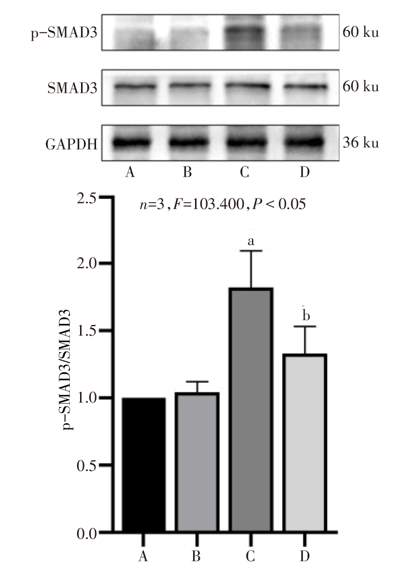
图6 4组系膜细胞中p-SMAD3/SMAD3蛋白表达
Fig.6 The protein expression levels of p-SMAD3/SMAD3 in mesangial cells in four groups
| [1] | PILLEBOUT E, SUNDERKOTTER C. IgA vasculitis[J]. Semin Immunopathol, 2021, 43(5):729-738. doi:10.1007/s00281-021-00874-9. |
| [2] | GARDNER-MEDWIN J M, DOLEZALOVA P, CUMMINS C, et al. Incidence of Henoch-Schonlein purpura,Kawasaki disease,and rare vasculitides in children of different ethnic origins[J]. Lancet, 2002, 360(9341):1197-1202. doi:10.1016/S0140-6736(02)11279-7. |
| [3] | YANG X, LI Q, HE Y, et al. Individualized medication based on pharmacogenomics and treatment progress in children with IgAV nephritis[J]. Front Pharmacol, 2022, 13:956397. doi:10.3389/fphar.2022.956397. |
| [4] | ZHANG H, DENG Z, WANG Y. Molecular insight in intrarenal inflammation affecting four main types of cells in nephrons in IgA nephropathy[J]. Front Med(Lausanne), 2023, 10:1128393. doi:10.3389/fmed.2023.1128393. |
| [5] | SUN G X, DING R, LI M, et al. Ghrelin attenuates renal fibrosis and inflammation of obstructive nephropathy[J]. J Urol, 2015, 193(6):2107-2115. doi:10.1016/j.juro.2014.11.098. |
| [6] | YUAN Q, REN Q, LI L, et al. A Klotho-derived peptide protects against kidney fibrosis by targeting TGF-β signaling[J]. Nat Commun, 2022, 13(1):438. doi:10.1038/s41467-022-28096-z. |
| [7] | LI Y K, MA D X, WANG Z M, et al. The glucagon-like peptide-1 (GLP-1)analog liraglutide attenuates renal fibrosis[J]. Pharmacol Res, 2018, 131:102-111. doi:10.1016/j.phrs.2018.03.004. |
| [8] | 施会敏, 鱼敏逸, 曲高婷, 等. 紫癜性肾炎患儿血清中多肽的差异表达研究[J]. 徐州医科大学学报, 2019, 39(4):239-244. |
| SHI H M, YU M Y, QU G T, et al. Differential expression of peptides in serum of children with purpura nephritis[J]. Acta Academiae Medicinae Xuzhou, 2019, 39(4):239-244. doi:10.3969/j.issn.2096-3882.2019.04.002. | |
| [9] | ZULIANI-ALVAREZ L, MIDWOOD K S. Fibrinogen-related proteins in tissue repair:how a unique domain with a common structure controls diverse aspects of wound healing[J]. Adv Wound Care(New Rochelle), 2015, 4(5):273-285. doi:10.1089/wound.2014.0599. |
| [10] | SCINDIA Y M, DESHMUKH U S, BAGAVANT H. Mesangial pathology in glomerular disease:targets for therapeutic intervention[J]. Adv Drug Deliv Rev, 2010, 62(14):1337-1343. doi:10.1016/j.addr.2010.08.011. |
| [11] | ONG C H, THAM C L, HARITH H H, et al. TGF-β-induced fibrosis:A review on the underlying mechanism and potential therapeutic strategies[J]. Eur J Pharmacol, 2021, 911:174510. doi:10.1016/j.ejphar.2021.174510. |
| [12] | YE X, LI J, LIU Z, et al. Peptide mediated therapy infibrosis:Mechanisms,advances and prospects[J]. Biomed Pharmacother, 2023, 157:113978. doi:10.1016/j.biopha.2022.113978. |
| [13] | LI D, NIE H, JIANG K, et al. Molecular characterization and expression analysis of fibrinogen related protein(FREP)genes of Manila clam(Ruditapes philippinarum)after lipopolysaccharides challenge[J]. Comp Biochem Physiol C Toxicol Pharmacol, 2020, 228:108672. doi:10.1016/j.cbpc.2019.108672. |
| [14] | SORIA J, MIRSHAHI S, MIRSHAHI S Q, et al. Fibrinogen alphaC domain:Its importance in physiopathology[J]. Res Pract Thromb Haemost, 2019, 3(2):173-183. doi:10.1002/rth2.12183. |
| [15] | TIAN Y, HONG M, JING S, et al. Clinical and prognostic effect of plasma fibrinogen in renal cell carcinoma:a meta-analysis[J]. Biomed Res Int, 2017, 2017:9591506. doi:10.1155/2017/9591506. |
| [16] | GAO J, WANG Y, DONG Z, et al. A novel differential diagnostic model based on multiple biological parameters for immunoglobulin A nephropathy[J]. BMC Med Inform Decis Mak, 2012, 12:58. doi:10.1186/1472-6947-12-58. |
| [17] | VILAR R, FISH R J, CASINI A, et al. Fibrin(ogen)in human disease:both friend and foe[J]. Haematologica, 2020, 105(2):284-296. doi:10.3324/haematol.2019.236901. |
| [18] | MIDGLEY A C, ROGERS M, HALLETT M B, et al. Transforming growth factor-beta1 (TGF-beta1)-stimulated fibroblast to myofibroblast differentiation is mediated by hyaluronan (HA)-facilitated epidermal growth factor receptor(EGFR)and CD44 co-localization in lipid rafts[J]. J Biol Chem, 2013, 288(21):14824-14838. doi:10.1074/jbc.M113.451336. |
| [19] | HU H H, CHEN D Q, WANG Y N, et al. New insights into TGF-β/Smad signaling in tissue fibrosis[J]. Chem Biol Interact, 2018, 292:76-83. doi:10.1016/j.cbi.2018.07.008. |
| [1] | 杨晓芳, 贾新燕, 丰文君. miR-181a-5p通过HMGB1/NF-κB信号通路调控狼疮性肾炎小鼠肾小球系膜细胞增殖和凋亡[J]. 天津医药, 2026, 54(3): 232-237. |
| [2] | 王喆, 邱林, 马贲. 番茄来源胞外囊泡样颗粒对口腔鳞状细胞癌的作用效果研究[J]. 天津医药, 2026, 54(2): 145-150. |
| [3] | 赵兰君, 李良惠, 马馨, 巩娇娇, 郑臣辉, 石琳. 穿心莲内酯调控STAT3/GPX4通路对骨髓瘤细胞增殖和凋亡的影响[J]. 天津医药, 2026, 54(1): 8-13. |
| [4] | 黄伟, 王健键, 黄英, 杨俊. 复方苦参注射液联合化疗及贝伐珠单抗对卵巢癌患者近期疗效的影响[J]. 天津医药, 2026, 54(1): 88-92. |
| [5] | 李硕, 张云鹏, 黄艳, 曹美然, 贾兰芳, 胡桂才, 黄兰, 段书众. 系统性免疫炎症指数与IgA肾病患者临床病理特征的关系[J]. 天津医药, 2025, 53(9): 932-936. |
| [6] | 刘虹, 张玥玥, 王一琳, 王彩丽, 王晓敏, 毛敏, 李燕. MicroRNA-34a通过调控Wnt途径影响慢性淋巴细胞白血病进展的机制探讨[J]. 天津医药, 2025, 53(8): 785-790. |
| [7] | 韩建存, 周谊. 川陈皮素调节FAK/AKT信号通路对喉鳞状细胞癌细胞增殖和凋亡的影响[J]. 天津医药, 2025, 53(6): 561-565. |
| [8] | 余朝霞, 马贲, 邱林, 高倩, 尼娜. 基于网络药理学和实验验证探究鲍式层孔菌多酚的抗头颈鳞癌机制[J]. 天津医药, 2025, 53(5): 456-461. |
| [9] | 李晨, 李占恩, 苏宏伟, 侯彩云, 董少文. KRT17调节Wnt/β-catenin信号通路对膀胱癌细胞增殖、凋亡及上皮间质转化的影响[J]. 天津医药, 2025, 53(5): 462-467. |
| [10] | 苏红见, 张春艳, 张卫东, 韩利, 乔亚红. 鸢尾素调控EBF3/ALOX15通路影响肺腺癌细胞增殖和迁移[J]. 天津医药, 2025, 53(4): 337-342. |
| [11] | 祁卫华, 黄广磊, 张媛媛, 班宏英, 毛诏旭. 连翘脂素调节cAMP/EPAC1/RAP1信号通路对肺癌细胞恶性进展的影响[J]. 天津医药, 2025, 53(4): 343-348. |
| [12] | 朱永红, 巴军凤. 血清总IgE、EOS、LDH和呼出气一氧化氮对过敏性鼻炎合并支气管哮喘的预测价值[J]. 天津医药, 2025, 53(4): 360-364. |
| [13] | 闫玲新, 李森, 郭改莉, 孟婉秋, 徐超. 异牡荆素通过miR-339-5p/HSPA8轴调节胰腺癌细胞的生物学行为[J]. 天津医药, 2025, 53(3): 230-235. |
| [14] | 胡烙, 章程, 胡志军. 血清TRPV1、TIMP4、TGF-β1水平对良性阵发性位置性眩晕复发的预测价值[J]. 天津医药, 2025, 53(3): 267-271. |
| [15] | 马莉莉, 李子沐, 王亮, 许彭, 李秀梅. 间充质干细胞外泌体对食管癌ECA109细胞生物学行为的影响[J]. 天津医药, 2025, 53(2): 113-117. |
| 阅读次数 | ||||||
|
全文 |
|
|||||
|
摘要 |
|
|||||