Tianjin Medical Journal ›› 2022, Vol. 50 ›› Issue (9): 907-911.doi: 10.11958/20220121
• Cell and Molecular Biology • Previous Articles Next Articles
PING Jing1( ), ZHOU Donghua1, ZHU Dajiang2, TIAN Jie1, CHEN Ying1, FAN Juhua1
Received:2022-01-21
Revised:2022-05-11
Published:2022-09-15
Online:2022-09-05
PING Jing, ZHOU Donghua, ZHU Dajiang, TIAN Jie, CHEN Ying, FAN Juhua. Expression and its clinical significance of Musashi2 in different molecular subtypes of breast invasive ductal carcinoma[J]. Tianjin Medical Journal, 2022, 50(9): 907-911.
CLC Number:
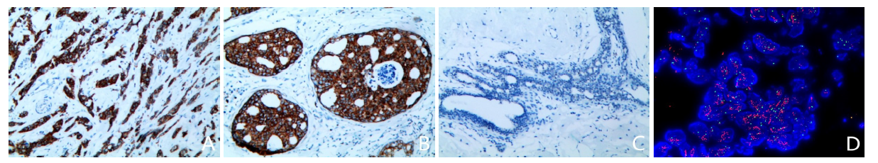
Fig.1 Immunohistochemistry staining and fluorescence in situ hybridization of different tissues in breast cancer
| 临床病理特征 | n | Musashi2阳性 | χ2 | |
|---|---|---|---|---|
| 年龄(岁) | ≤50 | 77 | 43(55.84) | 0.075 |
| >50 | 48 | 28(58.33) | ||
| 肿瘤直径(cm) | <2 | 50 | 32(64.00) | 1.858 |
| 2~5 | 66 | 34(51.52) | ||
| >5 | 9 | 5(55.56) | ||
| 脉管受累 | 否 | 84 | 46(54.76) | 0.434 |
| 是 | 41 | 25(60.98) | ||
| 淋巴结转移 | 阴性 | 53 | 33(62.26) | 1.120 |
| 阳性 | 72 | 38(52.78) | ||
| TNM分期 | 早期 | 85 | 54(63.53) | 4.902* |
| 晚期 | 40 | 17(42.50) | ||
| 组织学分级 | Ⅰ+Ⅱ | 101 | 61(60.40) | 2.772 |
| Ⅲ | 24 | 10(41.67) |
Tab.1 Comparison of positive expression rate of Musashi2 in invasive ductal carcinoma of breast between patients with different clinicopathological features
| 临床病理特征 | n | Musashi2阳性 | χ2 | |
|---|---|---|---|---|
| 年龄(岁) | ≤50 | 77 | 43(55.84) | 0.075 |
| >50 | 48 | 28(58.33) | ||
| 肿瘤直径(cm) | <2 | 50 | 32(64.00) | 1.858 |
| 2~5 | 66 | 34(51.52) | ||
| >5 | 9 | 5(55.56) | ||
| 脉管受累 | 否 | 84 | 46(54.76) | 0.434 |
| 是 | 41 | 25(60.98) | ||
| 淋巴结转移 | 阴性 | 53 | 33(62.26) | 1.120 |
| 阳性 | 72 | 38(52.78) | ||
| TNM分期 | 早期 | 85 | 54(63.53) | 4.902* |
| 晚期 | 40 | 17(42.50) | ||
| 组织学分级 | Ⅰ+Ⅱ | 101 | 61(60.40) | 2.772 |
| Ⅲ | 24 | 10(41.67) |
| 项目 | n | Musashi2阳性 | χ2 | r | |
|---|---|---|---|---|---|
| HER-2 | 阴性 | 78 | 46(58.97) | 0.400 | -0.057 |
| 阳性 | 47 | 25(53.19) | |||
| ER | 阴性 | 36 | 9(25.00) | 20.837** | 0.408* |
| 阳性 | 89 | 62(69.66) | |||
| PR | 阴性 | 42 | 12(28.57) | 20.541** | 0.405* |
| 阳性 | 83 | 59(71.08) | |||
| Ki67 | ≤14% | 29 | 20(68.97) | 2.278 | -0.135 |
| >14% | 96 | 51(53.13) | |||
Tab.2 The relationship between Musashi2 expression and molecular pathological features of breast invasive ductal carcinoma
| 项目 | n | Musashi2阳性 | χ2 | r | |
|---|---|---|---|---|---|
| HER-2 | 阴性 | 78 | 46(58.97) | 0.400 | -0.057 |
| 阳性 | 47 | 25(53.19) | |||
| ER | 阴性 | 36 | 9(25.00) | 20.837** | 0.408* |
| 阳性 | 89 | 62(69.66) | |||
| PR | 阴性 | 42 | 12(28.57) | 20.541** | 0.405* |
| 阳性 | 83 | 59(71.08) | |||
| Ki67 | ≤14% | 29 | 20(68.97) | 2.278 | -0.135 |
| >14% | 96 | 51(53.13) | |||
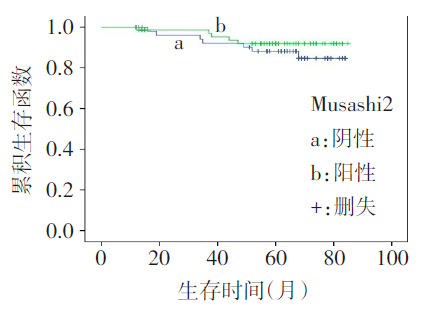
Fig.2 Survival curves of Musashi2 protein negative and positive expressions in patients with breast invasive ductal carcinoma
| [1] | OKANO H, IMAI T, OKABE M. Musashi:a translational regulator of cell fate[J]. J Cell Sci, 2002, 115(Pt7):1355-1359. doi: 10.1242/jcs.115.7.1355. |
| [2] | 李宇, 王鹏, 贾垚, 等. Msi2在上皮细胞肿瘤中的作用及临床价值[J]. 现代医学, 2017, 45(8):1167-1170. |
| LI Y, WANG P, JIA Y, et al. Clinical value and role of Msi2 in epithelial tumor[J]. Modern Medical Journal, 2017, 4(8):1167-1170. doi: 10.3969/j.issn.1671-7562.2017.08.026. | |
| [3] | 李捷, 刘紫蒙, 郑一琼, 等. 乳腺浸润性导管癌组织IRS1、PRSS3蛋白表达与磷酸化蛋白激酶B表达和预后的关系研究[J]. 现代生物医学进展, 2021, 21(24):4617-4622. |
| LI J, LIU Z M, ZHENG Y Q, et al. Relationship research between the IRS1,PRSS3 protein expression and phosphorylated protein kinase B expression and prognosis in breast invasive ductal carcinoma[J]. Progress in Modern Biomedicine, 2021, 21(24):4617-4622. doi: 10.13241/j.cnki.pmb.2021.24.003. | |
| [4] | 马建萍, 马芬兰. 不同分子分型乳腺癌的临床病理特征及预后的关系[J]. 实用癌症杂志, 2017, 32(12):2041-2044. |
| MA J P, MA F L. Clinicopathological features and prognosis of different molecular types of breast cancer[J]. The Practical Journal of Cancer, 2017, 32(12):2041-2044. doi: 10.3969/j.issn.1001-5930.2017.12.039. | |
| [5] | LAKHANI S R, ELLIS I O, SCHNITT S J, et al. WHO classification of tumours of the breast[M]. 4th ed. Lyon: International Agency for Research on Cancer (IARC) press, 2012:34-38. |
| [6] | GIULIANO A E, EDGE S B, HORTOBAGYI G N. Eighth edition of the AJCC cancer staging manual:breast cancer[J]. Ann Surg Oncol, 2018, 25(7):1783-1785. doi: 10.1245/s10434-018-6486-6. |
| [7] | 中国抗癌协会乳腺癌专业委员会. 中国抗癌协会乳腺癌诊治指南与规范(2015版)[J]. 中国癌症杂志, 2015, 25(9):692-754. |
| Chinese Anti-Cancer Association, Committee of Breast Cancer Society. Guidelines and norms for diagnosis and treatment of breast cancer of Chinese Anti-cancer Association (2015 edition)[J]. China Oncol, 2015, 25(9):692-754. doi: 10.3969/j.issn.1007-3969.2015.09.010. | |
| [8] | 《乳腺癌HER2检测指南2014版》编写组. 乳腺癌HER2检测指南(2014版)[J]. 中华病理学杂志, 2014, 43(4):262-267. |
| “Breast cancer HER2 detection gudelines (2014 edition)” Compilling Group. Breast cancer HER2 detection guidelines (2014 edition)[J]. Chin J Pathol, 2014, 43(4):262-267. doi: 10.3760/cma.j.issn.0529-5807.2014.04.012. | |
| [9] | KUDINOV A E, KARANICOLAS J, GOLEMIS E A, et al. Musashi RNA-binding proteins as cancer drivers and novel therapeutic targets[J]. Clin Cancer Res, 2017, 23(9):2143-2153. doi: 10.1158/1078-0432.CCR-16-2728. |
| [10] | 王小刚, 刘静, 郭庚, 等. MSI2生物学特性的研究进展[J]. 国际遗传学杂志, 2017, 40(4):228-232. |
| WANG X G, LIU J, GUO G, et al. Research progress of the biological characteristics of MSI2[J]. International Journal of Genetics, 2017, 40(4):228-232.doi: 10.3760/cma.j.issn.1673-4386.2017.04.008. | |
| [11] | DAS CHAGAS P F, BARONI M, BRASSESCO M S, et al. Interplay between the RNA binding-protein Musashi and developmental signaling pathways[J]. J Gene Med, 2020, 22(1):e3136. doi: 10.1002/jgm.3136. |
| [12] | LIU Y, FAN Y, WANG X, et al. Musashi-2 is a prognostic marker for the survival of patients with cervical cancer[J]. Oncol Lett, 2018, 15(4):5425-5432. doi: 10.3892/ol.2018.8077. |
| [13] | ZHAO J, ZHANG Y, LIU X S, et al. RNA-binding protein Musashi2 stabilizing androgen receptor drives prostate cancer progression[J]. Cancer Sci, 2020, 111(2):369-382. doi: 10.1111/cas.14280. |
| [14] | YANG Z, LI J, SHI Y, et al. Increased musashi 2 expression indicates a poor prognosis and promotes malignant phenotypes in gastric cancer[J]. Oncol Lett, 2019, 17(3):2599-2606. doi: 10.3892/ol.2019.9889. |
| [15] | SHENG W, DONG M, CHEN C, et al. Musashi2 promotes the development and progression of pancreatic cancer by down-regulating Numb protein[J]. Oncotarget, 2017, 8(9):14359-14373. doi: 10.18632/oncotarget.8736. |
| [16] | 张苏杰, 殷华奇, 叶雄俊, 等. Musashi-2在恶性肿瘤发生发展及诊疗靶点中的作用[J]. 协和医学杂志, 2017, 8(4):289-293. |
| ZHANG S J, YIN H Q, YE X J, et al. Role of Musashi-2 in the occurrence and development and as the diagnostic and therapeutic target of malignant tumours[J]. Medical Journal of Peking Union Medical College Hospital, 2017, 8(4):289-293. doi: 10.3969/j.issn.1674-9081.2017.05.018. | |
| [17] | EMADI-BAYGI M, NIKPOUR P, MOHAMMAD-HASHEM F, et al. MSI2 expression is decreased in grade II of gastric carcinoma[J]. Pathol Res Pract, 2013, 209(11):689-691. doi: 10.1016/j.prp.2013.07.008. |
| [18] | 王琳, 沈佳丽, 熊世双, 等. JAG1和Notch3在乳腺癌组织中的表达及预后价值[J]. 临床肿瘤学杂志, 2020, 25(1):13-19. |
| WANG L, SHEN J L, XIONG S S, et al. Expression of JAG1 and Notch3 in breast cancer tissues and their prognostic value[J]. Chinese Clinical Oncology, 2020, 25(1):13-19. doi: 10.3969/j.issn.1009-0460.2020.01.003. | |
| [19] | SORLIE T, TIBSHIRANI R, PARKER J, et al. Repeated observation of breast tumor subtypes in independent gene expression data sets[J]. Proc Natl Acad Sci U S A, 2003, 100(14):8418-8423. doi: 10.1073/pnas.0932692100. |
| [20] | KATZ Y, LI F, LAMBERT N J, et al. Musashi proteins are post-transcriptional regulators of the epithelial-luminal cell state[J]. Elife, 2014, 3:e03915. doi: 10.7554/eLife.03915. |
| [21] | 李菲菲. Musashi在乳腺发育和肿瘤发生中的功能与分子作用机制[D]. 北京: 中国农业大学, 2016. |
| LI F F. The function and molecular mechanism of Musashi on mammary gland development and tumorigenesis[D]. Beijing: China Agricultural University, 2016. | |
| [22] | 张莹, 任占平, 张芫. Ki-67表达与乳腺癌分子分型及临床病理特征的关系[J]. 临床与实验病理学杂志, 2014, 30(11):1220-1223. |
| ZHANG Y, REN Z P, ZHANG Y. Relationship between the expression of Ki-67 and molecular classification and clinical pathological features in breast cancerr[J]. J Clin Exp Pathol, 2014, 30(11):1220-1223. doi: 10.13315/j.cnki.cjcep.2014.11.005. | |
| [23] | KANG M H, JEONG K J, KIM W Y, et al. Musashi RNA-binding protein 2 regulates estrogen receptor 1 function in breast cancer[J]. Oncogene, 2017, 36(12):1745-1752. doi: 10.1038/onc.2016.327. |
| [1] | HUANG Huiqi, WU Qiuyuan, ZHANG Kun, LI Peixian, XIONG Yaming, YE Guolin, ZHOU Dan. Research on the anti-tumor mechanism of toosendanin combined with olaparib in triple negative breast cancer [J]. Tianjin Medical Journal, 2025, 53(9): 897-902. |
| [2] | WANG Chao, ZHANG Junmei, ZHANG Peng, LIU Li, WANG Xiaochun. Comparison of efficacy of recombinant human interleukin-11 and herombopag olamine tablets on chemotherapy-induced thrombocytopenia in breast cancer [J]. Tianjin Medical Journal, 2025, 53(4): 365-369. |
| [3] | LI Na, HE Ying, TENG Fei, HE Wenshu, GUO Caifeng, ZHONG Na, WU Qiong, LI Jun. The application value of ultrasound BI-RADS classification combined with serum FGFR1 and GDF3 in the differential diagnosis of benign and malignant breast masses [J]. Tianjin Medical Journal, 2025, 53(3): 247-251. |
| [4] | WANG Wei, XIA Haishui, MA Shang. Efficacy of trastuzumab combined with neratinib in the treatment of HER-2 positive metastatic breast cancer [J]. Tianjin Medical Journal, 2025, 53(3): 321-325. |
| [5] | WANG Linna, WANG Di, DONG Yanhong. Clinical effect of aribulin combined with NP regimen in the treatment of patients with advanced breast cancer [J]. Tianjin Medical Journal, 2025, 53(3): 326-330. |
| [6] | HUANG Xiaoqing, LIU Yuanyuan, YAN Liang, YI Shuping. Dosimetric comparison of two irradiation modes after radical mastectomy for breast cancer [J]. Tianjin Medical Journal, 2025, 53(11): 1204-1207. |
| [7] | LIU Yuanyuan, HUANG Xiaoqing, YAN Liang, YI Shuping. Preventive efficacy of rhGM-CSF combined with Kangfuxin liquid on radiation dermatitis of breast cancer [J]. Tianjin Medical Journal, 2025, 53(10): 1076-1080. |
| [8] | MAN Yi, XU Ya, HE Xiancheng, SONG Shaofeng, LIU Aiguo. Relationship between expression levels of EGFR, Ki-67, P53 and CTC and the prognosis of triple negative breast cancer [J]. Tianjin Medical Journal, 2024, 52(8): 862-867. |
| [9] | ZHU Gangming, DONG Yongde, ZHU Ruiting, TAN Yuanman, TAO Juan, LIU Xiao, CHEN Decheng, YANG Gai. The value of magnetic resonance relaxation time quantitative imaging in predicting molecular subtypes of invasive ductal carcinoma [J]. Tianjin Medical Journal, 2024, 52(7): 770-774. |
| [10] | CHEN Zhiyan, WU Qiuyuan, DENG Yuhua, ZHOU Dan. Status and application of organoid technology in breast cancer research [J]. Tianjin Medical Journal, 2024, 52(6): 668-672. |
| [11] | LIU Danyang, LI Yongtao, ZHANG Haiyan, LI Lin, LIU Yang, SHEN Lei. Effect of breast cancer cell conditioned medium on biological behavior of bone marrow mesenchymal stem cells [J]. Tianjin Medical Journal, 2024, 52(5): 454-458. |
| [12] | LU Xinyi, DU Weipo, LI Jinggang, GUO Fangfang, ZHANG Xiaolei, LIU Jing. Correlation between serum miR-193a-3p, ATF5 levels and chemotherapy efficacy in patients with triple negative breast cancer [J]. Tianjin Medical Journal, 2024, 52(12): 1313-1316. |
| [13] | XIE Haoran, LI Yihao, LIU Cheng, XIA Yuting, QIU Shenglei, XIONG Bin, FENG Qizhen. Research of predictive factors of axillary lymph node metastasis in breast cancer under the context of DIP payment of medical insurance [J]. Tianjin Medical Journal, 2024, 52(11): 1193-1196. |
| [14] | WU Haixiao, MA Wenjuan, LI Zhijun, ZHANG Chao. Relationship between pectoralis muscle index and prognosis of breast cancer patients with bone metastases based on radiomics [J]. Tianjin Medical Journal, 2023, 51(9): 1007-1010. |
| [15] | YANG Pingping, ZHANG Jing, CHEN Hongyu, ZHANG Miao, ZHOU Dingan, FANG Wen. Tumor suppressor gene SASH1 binds Vimentin and negatively regulates the expression of Vimentin in breast cancer [J]. Tianjin Medical Journal, 2023, 51(9): 904-908. |
| Viewed | ||||||
|
Full text |
|
|||||
|
Abstract |
|
|||||