Tianjin Medical Journal ›› 2023, Vol. 51 ›› Issue (6): 607-612.doi: 10.11958/20221430
• Experimental Research • Previous Articles Next Articles
SUN Liyan1( ), LIU Zeru2, SU Yongsheng3, AI Hongliang1
Received:2022-09-06
Revised:2022-12-22
Published:2023-06-15
Online:2023-06-20
SUN Liyan, LIU Zeru, SU Yongsheng, AI Hongliang. Effect of asperuloside on pyroptosis of lung tissue in septic rats by regulating NLRP3/Caspase-1/GSDMD signaling pathway[J]. Tianjin Medical Journal, 2023, 51(6): 607-612.
CLC Number:
| 组别 | MV(mL) | Ri(kPa-1·L-1·s-1) | p(O2)(mmHg) |
|---|---|---|---|
| C组 | 7.69±0.86 | 24.97±4.65 | 98.82±10.61 |
| M组 | 2.71±0.42a | 59.68±6.80a | 54.75±5.15a |
| AL组 | 3.91±0.48b | 50.98±6.12b | 65.12±6.24b |
| AM组 | 4.80±0.51bc | 42.24±5.16bc | 75.82±7.43bc |
| AH组 | 7.13±0.73bcd | 26.43±4.52bcd | 95.93±9.52bcd |
| AH+N组 | 2.93±0.38e | 55.32±6.03e | 58.12±5.36e |
| F | 129.083** | 69.525** | 61.205** |
Tab.1 Comparison of lung function indexes and p(O2) of rats between the six groups
| 组别 | MV(mL) | Ri(kPa-1·L-1·s-1) | p(O2)(mmHg) |
|---|---|---|---|
| C组 | 7.69±0.86 | 24.97±4.65 | 98.82±10.61 |
| M组 | 2.71±0.42a | 59.68±6.80a | 54.75±5.15a |
| AL组 | 3.91±0.48b | 50.98±6.12b | 65.12±6.24b |
| AM组 | 4.80±0.51bc | 42.24±5.16bc | 75.82±7.43bc |
| AH组 | 7.13±0.73bcd | 26.43±4.52bcd | 95.93±9.52bcd |
| AH+N组 | 2.93±0.38e | 55.32±6.03e | 58.12±5.36e |
| F | 129.083** | 69.525** | 61.205** |
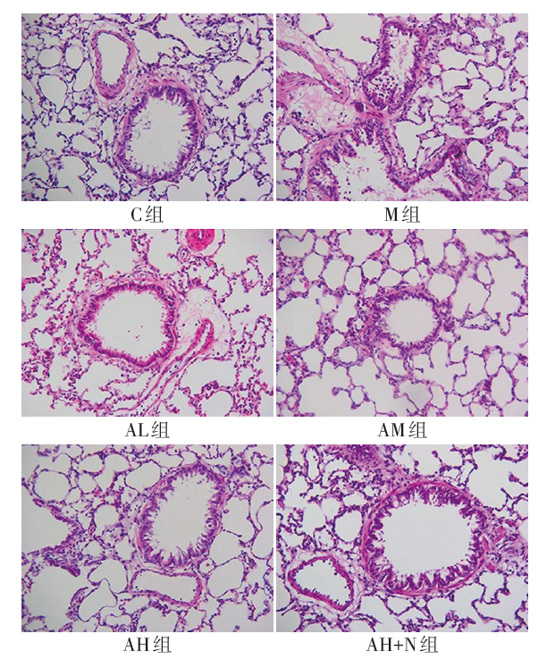
Fig.1 Pathological morphology of rat lung tissue detected by HE staining (×200)
| 组别 | 中性粒细胞 | 巨噬细胞 | 淋巴细胞 |
|---|---|---|---|
| C组 | 2.68±0.65 | 22.76±2.43 | 5.09±0.67 |
| M组 | 13.10±1.12a | 54.20±6.51a | 81.94±17.62a |
| AL组 | 10.68±0.95b | 46.59±5.27b | 64.75±13.41b |
| AM组 | 8.19±0.70bc | 39.84±4.25bc | 45.01±9.46bc |
| AH组 | 3.15±0.57bcd | 25.90±2.56bcd | 6.13±0.81bcd |
| AH+N组 | 12.38±1.31e | 53.32±6.03e | 80.61±15.73e |
| F | 242.906** | 80.336** | 88.312** |
Tab.2 Comparison of the number of inflammatory cells in BALF between the six groups
| 组别 | 中性粒细胞 | 巨噬细胞 | 淋巴细胞 |
|---|---|---|---|
| C组 | 2.68±0.65 | 22.76±2.43 | 5.09±0.67 |
| M组 | 13.10±1.12a | 54.20±6.51a | 81.94±17.62a |
| AL组 | 10.68±0.95b | 46.59±5.27b | 64.75±13.41b |
| AM组 | 8.19±0.70bc | 39.84±4.25bc | 45.01±9.46bc |
| AH组 | 3.15±0.57bcd | 25.90±2.56bcd | 6.13±0.81bcd |
| AH+N组 | 12.38±1.31e | 53.32±6.03e | 80.61±15.73e |
| F | 242.906** | 80.336** | 88.312** |
| 组别 | TNF-α | IL-8 | IL-1β |
|---|---|---|---|
| C组 | 27.35±5.02 | 204.94±32.25 | 16.87±3.03 |
| M组 | 183.46±21.53a | 968.40±92.30a | 242.64±28.12a |
| AL组 | 136.59±17.84b | 761.25±77.62b | 193.45±20.51b |
| AM组 | 107.86±13.46bc | 589.32±54.92bc | 134.45±12.51bc |
| AH组 | 31.12±6.32bcd | 229.86±30.74bcd | 21.13±3.54bcd |
| AH+N组 | 164.63±20.14e | 960.11±90.58e | 230.36±25.92e |
| F | 185.239** | 251.860** | 296.171** |
Tab.3 Comparison of serum levels of IL-6, IL-8 and IL-1β between the six groups
| 组别 | TNF-α | IL-8 | IL-1β |
|---|---|---|---|
| C组 | 27.35±5.02 | 204.94±32.25 | 16.87±3.03 |
| M组 | 183.46±21.53a | 968.40±92.30a | 242.64±28.12a |
| AL组 | 136.59±17.84b | 761.25±77.62b | 193.45±20.51b |
| AM组 | 107.86±13.46bc | 589.32±54.92bc | 134.45±12.51bc |
| AH组 | 31.12±6.32bcd | 229.86±30.74bcd | 21.13±3.54bcd |
| AH+N组 | 164.63±20.14e | 960.11±90.58e | 230.36±25.92e |
| F | 185.239** | 251.860** | 296.171** |
| 组别 | n | NLRP3 | GSDMD |
|---|---|---|---|
| C组 | 10 | 1.00±0.00 | 1.00±0.00 |
| M组 | 10 | 2.89±0.36a | 2.57±0.32a |
| AL组 | 10 | 2.36±0.29b | 2.19±0.26b |
| AM组 | 10 | 2.02±0.25bc | 1.84±0.29bc |
| AH组 | 10 | 1.16±0.20bcd | 1.13±0.17bcd |
| AH+N组 | 10 | 2.64±0.34e | 2.31±0.33e |
| F | 84.386** | 63.873** |
Tab.4 Comparison of relative fluorescence intensity of NLRP3 and GSDMD in lung tissue of rats between the six groups
| 组别 | n | NLRP3 | GSDMD |
|---|---|---|---|
| C组 | 10 | 1.00±0.00 | 1.00±0.00 |
| M组 | 10 | 2.89±0.36a | 2.57±0.32a |
| AL组 | 10 | 2.36±0.29b | 2.19±0.26b |
| AM组 | 10 | 2.02±0.25bc | 1.84±0.29bc |
| AH组 | 10 | 1.16±0.20bcd | 1.13±0.17bcd |
| AH+N组 | 10 | 2.64±0.34e | 2.31±0.33e |
| F | 84.386** | 63.873** |

Fig.2 Co-expression of NLRP3 and GSDMD in rat lung tissue detected by immunofluorescence staining (×200)
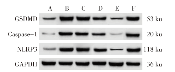
Fig.3 The expression of NLRP3/Caspase-1/GSDMD signaling pathway related proteins in lung tissue of each group detected by Western blot assay
| 组别 | n | NLRP3 | Caspase-1 | GSDMD |
|---|---|---|---|---|
| C组 | 10 | 0.19±0.04 | 0.10±0.02 | 0.21±0.04 |
| M组 | 10 | 1.20±0.16a | 1.08±0.13a | 1.29±0.15a |
| AL组 | 10 | 0.98±0.13b | 0.81±0.11b | 0.95±0.12b |
| AM组 | 10 | 0.71±0.10bc | 0.59±0.07bc | 0.78±0.09bc |
| AH组 | 10 | 0.22±0.05bcd | 0.13±0.03bcd | 0.25±0.06bcd |
| AH+N组 | 10 | 1.16±0.11e | 1.04±0.14e | 1.26±0.16e |
| F | 178.049** | 204.821** | 178.037** |
Tab.5 Comparison of relative expression levels of NLRP3/Caspase-1/GSDMD pathway related proteins in lung tissue of rats between the six groups
| 组别 | n | NLRP3 | Caspase-1 | GSDMD |
|---|---|---|---|---|
| C组 | 10 | 0.19±0.04 | 0.10±0.02 | 0.21±0.04 |
| M组 | 10 | 1.20±0.16a | 1.08±0.13a | 1.29±0.15a |
| AL组 | 10 | 0.98±0.13b | 0.81±0.11b | 0.95±0.12b |
| AM组 | 10 | 0.71±0.10bc | 0.59±0.07bc | 0.78±0.09bc |
| AH组 | 10 | 0.22±0.05bcd | 0.13±0.03bcd | 0.25±0.06bcd |
| AH+N组 | 10 | 1.16±0.11e | 1.04±0.14e | 1.26±0.16e |
| F | 178.049** | 204.821** | 178.037** |
| [1] | EVANS L, RHODES A, ALHAZZANI W, et al. Surviving sepsis campaign:international guidelines for management of sepsis and septic shock 2021[J]. Intensive Care Med, 2021, 47(11):1181-1247. doi:10.1007/s00134-021-06506-y. |
| [2] | MA J, XU L Y, SUN Q H, et al. Inhibition of miR-1298-5p attenuates sepsis lung injury by targeting SOCS6[J]. Mol Cell Biochem, 2021, 476(10):3745-3756. doi:10.1007/s11010-021-04170-w. |
| [3] | JIAO Y, ZHANG T, ZHANG C, et al. Exosomal miR-30d-5p of neutrophils induces M1 macrophage polarization and primes macrophage pyroptosis in sepsis-related acute lung injury[J]. Crit Care, 2021, 25(1):356-370. doi:10.1186/s13054-021-03775-3. |
| [4] | ZHANG Z T, ZHANG D Y, XIE K, et al. Luteolin activates Tregs to promote IL-10 expression and alleviating caspase-11-dependent pyroptosis in sepsis-induced lung injury[J]. Int Immunopharmacol, 2021, 99(5):107914-107922. doi:10.1016/j.intimp.2021.107914. |
| [5] | YAO F, JIN Z, ZHENG Z, et al. HDAC11 promotes both NLRP3/caspase-1/GSDMD and caspase-3/GSDME pathways causing pyroptosis via ERG in vascular endothelial cells[J]. Cell Death Discov, 2022, 8(1):112-122. doi:10.1038/s41420-022-00906-9. |
| [6] | LIU B, WANG Z, HE R, et al. Buformin alleviates sepsis-induced acute lung injury via inhibiting NLRP3-mediated pyroptosis through an AMPK-dependent pathway[J]. Clin Sci (Lond), 2022, 136(4):273-289. doi:10.1042/CS20211156. |
| [7] | 洪庆, 徐曼丽, 汤建. 车叶草苷的研究进展[J]. 中国野生植物资源, 2018, 37(4):43-45,69. |
| HONG Q, XU M L, TANG J. Advances of study on the iridoid asperuloside[J]. Chin Wild Plant Res, 2018, 37(4):43-45,69. doi:10.3969/j.issn.1006-9690.2018.04.009. | |
| [8] | 屠万倩, 李宁, 张留记, 等. 不同加工方式生产的杜仲叶中8种化学成分的含量及抗氧化活性研究[J]. 中国新药杂志, 2020, 29(16):1863-1867. |
| TU W Q, LI N, ZHANG L J, et al. Comparison of contents and antioxidant activity of eight components in Eucommia ulmoides leaves processed by different methods[J]. Chin J New Drugs, 2020, 29(16):1863-1867. doi:10.3969/j.issn.1003-3734.2020.16.013. | |
| [9] | 王海燕, 高令心, 莫美娜, 等. 补肾益肺法联合家庭氧疗治疗慢性阻塞性肺疾病稳定期40例[J]. 中医研究, 2019, 32(3):26-29. |
| WANG H Y, GAO L X, MO M N, et al. Treatment of 40 patients with chronic obstructive pulmonary disease by Tonifying kidney and nourishing lung combined with home oxygen therapy[J]. Trad Chin Med Res, 2019, 32(3):26-29. doi:10.3969/j.issn.1001-6910.2019.03.11. | |
| [10] | 陈剑明, 彭美哲, 陈奕杉, 等. 参附黄制剂联合亚低温对脓毒症大鼠下丘脑AMPK-POMC相关靶点的影响[J]. 中华中医药杂志, 2022, 37(5):2475-2480. |
| CHEN J M, PENG M Z, CHEN Y S, et al. Effects of Shenfuhuang Preparation combined with subhypothermia on AMPK-POMC-related targets in the hypothalamic brain of septic rats[J]. Chin J Tradit Chin Med Pharm, 2022, 37(5):2475-2480. | |
| [11] | 初巍巍, 袁菲阳, 丁国臣, 等. 车叶草苷对小鼠Lewis肺癌的抑瘤、抗炎作用及其机制[J]. 武警医学, 2020, 31(5):401-404. |
| CHU W W, YUAN F Y, DING G C, et al. Anti-tumor and anti-inflammatory effect of asperuloside on mice with Lewis lung cancer[J]. Med J Chin PAPF, 2020, 31(5):401-404. doi:10.3969/j.issn.1004-3594.2020.05.009. | |
| [12] | SHAN W, LIAO X, TANG Y, et al. Dexmedetomidine alleviates inflammation in neuropathic pain by suppressing NLRP3 via Nrf2 activation[J]. Exp Ther Med, 2021, 22(4):1046-1054. doi:10.3892/etm.2021.10479. |
| [13] | LV X, ZHANG X Y, ZHANG Q, et al. lncRNA NEAT1 aggravates sepsis-induced lung injury by regulating the miR-27a/PTEN axis[J]. Lab Invest, 2021, 101(10):1371-1381. doi:10.1038/s41374-021-00620-7. |
| [14] | ACKERMAN M H, AHRENS T, KELLY J, et al. Sepsis[J]. Crit Care Nurs Clin North Am, 2021, 33(4):407-418. doi:10.1016/j.cnc.2021.08.003. |
| [15] | ZHUO Y, YANG L, LI D, et al. Syringaresinol resisted sepsis-induced acute lung injury by suppressing pyroptosis via the oestrogen receptor-β signalling pathway[J]. Inflammation, 2022, 45(2):824-837. doi:10.1007/s10753-021-01587-9. |
| [16] | DING H, YANG J, CHEN L, et al. Memantine alleviates acute lung injury via inhibiting macrophage pyroptosis[J]. Shock, 2021, 56(6):1040-1048. doi:10.1097/SHK.0000000000001790. |
| [17] | WANG L X, REN C, YAO R Q, et al. Sestrin2 protects against lethal sepsis by suppressing the pyroptosis of dendritic cells[J]. Cell Mol Life Sci, 2021, 78(24):8209-8227. doi:10.1007/s00018-021-03970-z. |
| [18] | LI S, SUN Y, SONG M, et al. NLRP3/caspase-1/GSDMD-mediated pyroptosis exerts a crucial role in astrocyte pathological injury in mouse model of depression[J]. JCI Insight, 2021, 6(23):e146852-e146865. doi:10.1172/jci.insight.146852. |
| [19] | WANG Y C, LIU Q X, ZHENG Q, et al. Dihydromyricetin alleviates sepsis-induced acute lung injury through inhibiting NLRP3 inflammasome-dependent pyroptosis in mice model[J]. Inflammation, 2019, 42(4):1301-1310. doi:10.1007/s10753-019-00990-7. |
| Viewed | ||||||
|
Full text |
|
|||||
|
Abstract |
|
|||||