Tianjin Medical Journal ›› 2024, Vol. 52 ›› Issue (2): 129-135.doi: 10.11958/20230437
• Experimental Research • Previous Articles Next Articles
ZHONG Jiashuai( ), FENG Yumei△()
Received:2023-03-27
Revised:2023-05-08
Published:2024-02-15
Online:2024-01-26
Contact:
△ E-mail:ZHONG Jiashuai, FENG Yumei. Comparative study on the directed differentiation ability of mouse bone marrow and adipose-derived mesenchymal stem cells[J]. Tianjin Medical Journal, 2024, 52(2): 129-135.
CLC Number:
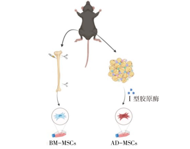
Fig.1 Isolation and culture of BM-MSCs and AD-MSCs
| 基因名称 | 引物序列(5′→3′) | 产物大小/bp |
|---|---|---|
| Gapdh | 上游:ATTGTCAGCAATGCATCCTG | 102 |
| 下游:ATGGACTGTGGTCATGAGCC | ||
| CD29 | 上游:CCTGTAACTCCGACGCCTTT | 137 |
| 下游:AAGGTCCCCACTCAGCAATG | ||
| CD34 | 上游:TTTCCTGATGAACCGTCGCA | 165 |
| 下游:GCAGGGTTGTGAGGTACTGT | ||
| CD44 | 上游:ACCTTGGCCACCACTCCTAAT | 118 |
| 下游:TCACATGGGAGTCTTCACTTGG | ||
| CD45 | 上游:CTTTGCTTATGTGGCGTGTGT | 139 |
| 下游:TTATCCCCTTCTGATGCGCC | ||
| CD90 | 上游:TCCAAGTCGGAACTCTTGGC | 133 |
| 下游:TCCAGGCGAAGGTTTTGGTT | ||
| Runx2 | 上游:CTCTGCACCAAGTCCTTTTAATC | 104 |
| 下游:AGGAGGGGTAAGACTGGTCATAG | ||
| Sp7 | 上游:CTGAGAGAGGAGCAGATCCC | 135 |
| 下游:GTGAGCTTCTTCCTGGGTAGG | ||
| Sox9 | 上游:AGGAAGTCGGTGAAGAACGG | 167 |
| 下游:GGACCCTGAGATTGCCCAGA | ||
| Col2a1 | 上游:ATGAGGGAGCGGTAGAGACC | 190 |
| 下游:GCCCTAATTTTCGGGCATCC | ||
| Pparg | 上游:GAGCACTTCACAAGAAATTACC | 192 |
| 下游:GAACTCCATAGTGGAAGCCT | ||
| Cebpa | 上游:CAAGAACAGCAACGAGTACCG | 120 |
| 下游:AGGCGGTCATTGTCACTGGT |
Tab.1 Primer sequences
| 基因名称 | 引物序列(5′→3′) | 产物大小/bp |
|---|---|---|
| Gapdh | 上游:ATTGTCAGCAATGCATCCTG | 102 |
| 下游:ATGGACTGTGGTCATGAGCC | ||
| CD29 | 上游:CCTGTAACTCCGACGCCTTT | 137 |
| 下游:AAGGTCCCCACTCAGCAATG | ||
| CD34 | 上游:TTTCCTGATGAACCGTCGCA | 165 |
| 下游:GCAGGGTTGTGAGGTACTGT | ||
| CD44 | 上游:ACCTTGGCCACCACTCCTAAT | 118 |
| 下游:TCACATGGGAGTCTTCACTTGG | ||
| CD45 | 上游:CTTTGCTTATGTGGCGTGTGT | 139 |
| 下游:TTATCCCCTTCTGATGCGCC | ||
| CD90 | 上游:TCCAAGTCGGAACTCTTGGC | 133 |
| 下游:TCCAGGCGAAGGTTTTGGTT | ||
| Runx2 | 上游:CTCTGCACCAAGTCCTTTTAATC | 104 |
| 下游:AGGAGGGGTAAGACTGGTCATAG | ||
| Sp7 | 上游:CTGAGAGAGGAGCAGATCCC | 135 |
| 下游:GTGAGCTTCTTCCTGGGTAGG | ||
| Sox9 | 上游:AGGAAGTCGGTGAAGAACGG | 167 |
| 下游:GGACCCTGAGATTGCCCAGA | ||
| Col2a1 | 上游:ATGAGGGAGCGGTAGAGACC | 190 |
| 下游:GCCCTAATTTTCGGGCATCC | ||
| Pparg | 上游:GAGCACTTCACAAGAAATTACC | 192 |
| 下游:GAACTCCATAGTGGAAGCCT | ||
| Cebpa | 上游:CAAGAACAGCAACGAGTACCG | 120 |
| 下游:AGGCGGTCATTGTCACTGGT |
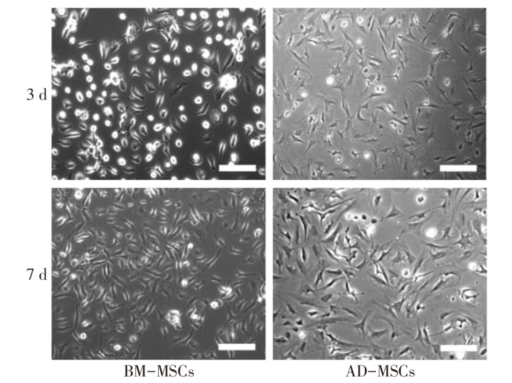
Fig.2 Comparison of cell morphology of MSCs
| 细胞 | CD29/×10-3 | CD44/×10-3 | CD90/×10-3 | CD34 | CD45 |
|---|---|---|---|---|---|
| BM-MSCs | 1.861±0.288 | 0.562±0.120 | 0.456±0.017 | 0 | 0 |
| AD-MSCs | 0.805±0.208 | 0.433±0.158 | 0.851±0.261 | 0 | 0 |
Tab.2 The gene expression levels of two types of MSCs
| 细胞 | CD29/×10-3 | CD44/×10-3 | CD90/×10-3 | CD34 | CD45 |
|---|---|---|---|---|---|
| BM-MSCs | 1.861±0.288 | 0.562±0.120 | 0.456±0.017 | 0 | 0 |
| AD-MSCs | 0.805±0.208 | 0.433±0.158 | 0.851±0.261 | 0 | 0 |
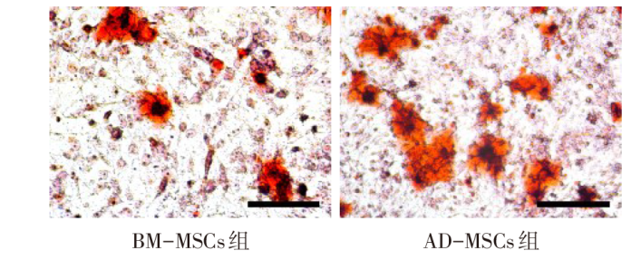
Fig.3 Osteogenic differentiation degree of MSCs in two groups (Alizarin red staining, ×200)
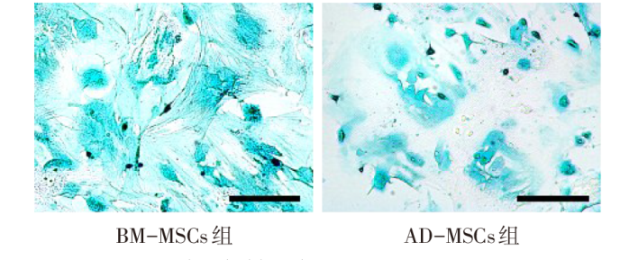
Fig.4 Degree of chondroblast differentiation of MSCs in two groups (Alcian Blue staining, ×200)
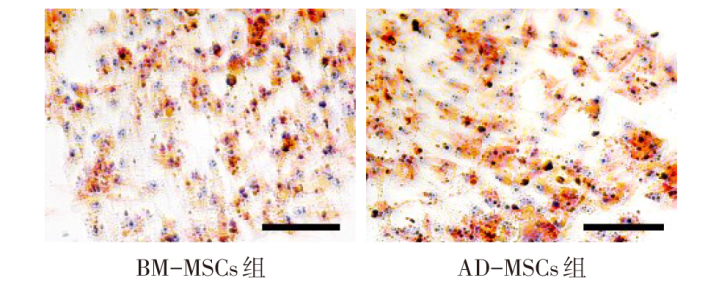
Fig.5 Degree of adipogenic differentiation of MSCs in two groups (Oil Red O staining, ×200)
| 细胞 | 诱导分化前 | 诱导分化后 | ||
|---|---|---|---|---|
| Runx2/×10-5 | Sp7/×10-6 | Runx2/×10-4 | Sp7/×10-5 | |
| BM-MSCs | 4.85±0.99 | 6.11±1.17 | 2.42±0.44 | 4.66±3.73 |
| AD-MSCs | 4.36±1.13 | 2.01±1.22 | 4.61±0.92 | 4.17±3.54 |
| t | 0.510 | 3.024* | 3.923* | 1.171 |
Tab.3 Comparison of expression levels of MSCs marker genes before and after osteogenic differentiation between the two groups
| 细胞 | 诱导分化前 | 诱导分化后 | ||
|---|---|---|---|---|
| Runx2/×10-5 | Sp7/×10-6 | Runx2/×10-4 | Sp7/×10-5 | |
| BM-MSCs | 4.85±0.99 | 6.11±1.17 | 2.42±0.44 | 4.66±3.73 |
| AD-MSCs | 4.36±1.13 | 2.01±1.22 | 4.61±0.92 | 4.17±3.54 |
| t | 0.510 | 3.024* | 3.923* | 1.171 |
| 细胞 | 诱导分化前 | 诱导分化后 | ||
|---|---|---|---|---|
| Sox9/×10-5 | Col2a1/×10-6 | Sox9/×10-3 | Col2a1/×10-5 | |
| BM-MSCs | 9.22±1.67 | 2.12±1.88 | 1.71±0.08 | 2.35±1.48 |
| AD-MSCs | 4.24±0.99 | 1.58±1.16 | 0.98±0.15 | 1.80±1.38 |
| t | 4.282* | 0.326 | 4.786* | 0.365 |
Tab.4 Comparison of expression levels of MSCs marker genes before and after induction of chondroblast differentiation between two groups
| 细胞 | 诱导分化前 | 诱导分化后 | ||
|---|---|---|---|---|
| Sox9/×10-5 | Col2a1/×10-6 | Sox9/×10-3 | Col2a1/×10-5 | |
| BM-MSCs | 9.22±1.67 | 2.12±1.88 | 1.71±0.08 | 2.35±1.48 |
| AD-MSCs | 4.24±0.99 | 1.58±1.16 | 0.98±0.15 | 1.80±1.38 |
| t | 4.282* | 0.326 | 4.786* | 0.365 |
| 细胞 | 诱导分化前 | 诱导分化后 | ||
|---|---|---|---|---|
| Pparg/×10-5 | Cebpa/×10-4 | Pparg/×10-3 | Cebpa/×10-2 | |
| BM-MSCs | 4.92±1.48 | 6.91±0.95 | 1.70±0.87 | 0.72±0.05 |
| AD-MSCs | 4.86±2.54 | 7.47±1.96 | 10.50±6.52 | 2.00±0.35 |
| t | 0.032 | 0.406 | 2.123* | 5.593* |
Tab.5 Comparison of expression levels of MSCs marker genes before and after induction of adipogenic differentiation between the two groups
| 细胞 | 诱导分化前 | 诱导分化后 | ||
|---|---|---|---|---|
| Pparg/×10-5 | Cebpa/×10-4 | Pparg/×10-3 | Cebpa/×10-2 | |
| BM-MSCs | 4.92±1.48 | 6.91±0.95 | 1.70±0.87 | 0.72±0.05 |
| AD-MSCs | 4.86±2.54 | 7.47±1.96 | 10.50±6.52 | 2.00±0.35 |
| t | 0.032 | 0.406 | 2.123* | 5.593* |
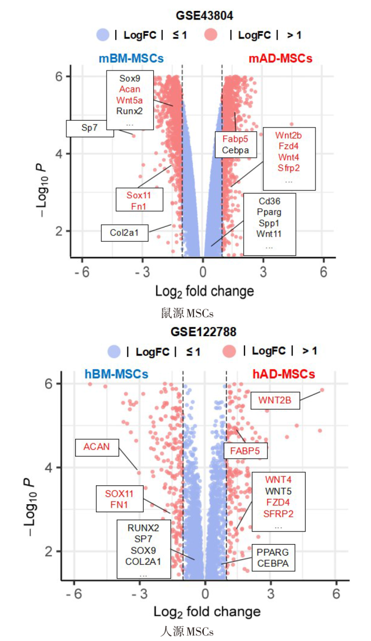
Fig.6 Volcanic map of significant difference genes between BM-MSCs and AD-MSCs
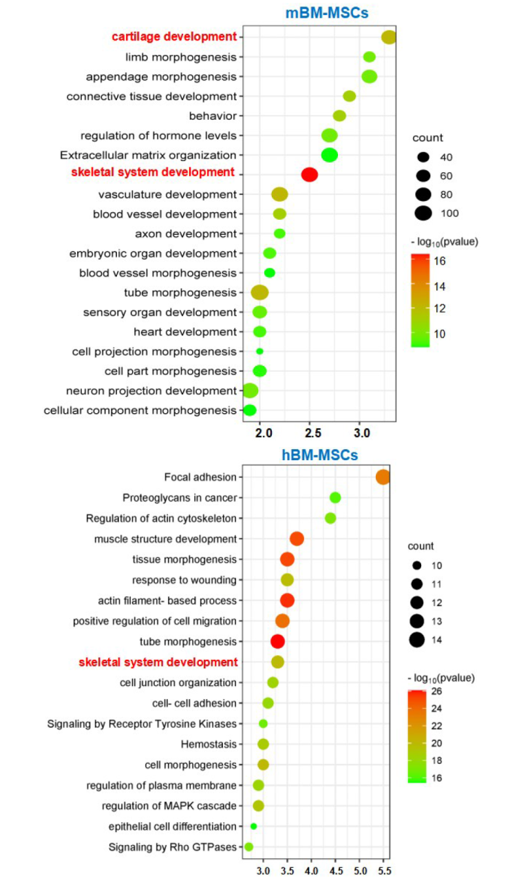
Fig.7 GO enrichment analysis of high expression gene sets in mBM-MSCs and hBM-MSCs
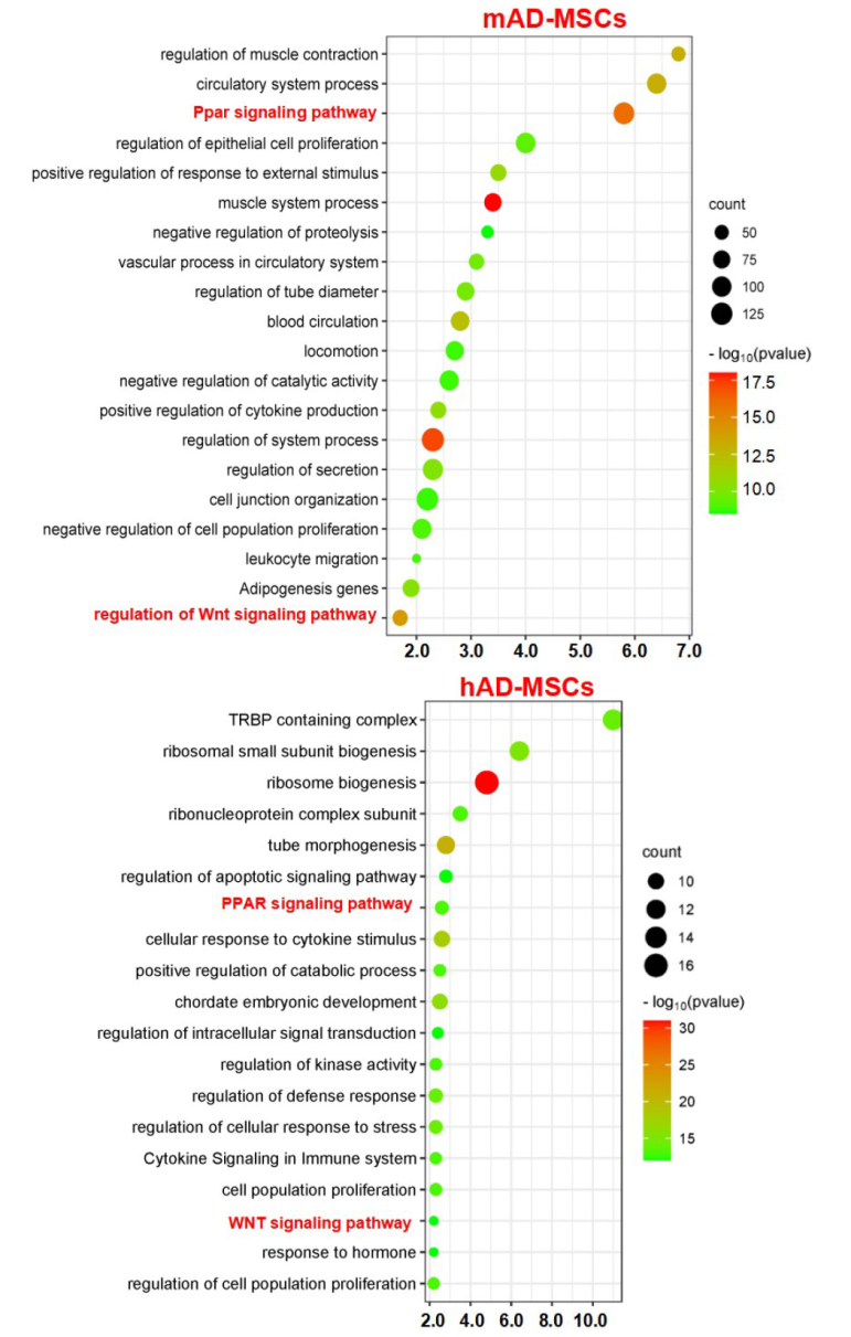
Fig.8 GO enrichment analysis of high-expression gene sets in mAD-MSCs and hAD-MSCs
| [1] | ZHANG L, MA X J, FEI Y Y, et al. Stem cell therapy in liver regeneration:Focus on mesenchymal stem cells and induced pluripotent stem cells[J]. Pharmacol Ther, 2022, 232:108004. doi:10.1016/j.pharmthera.2021.108004. |
| [2] | HOANG D M, PHAM P T, BACH T Q, et al. Stem cell-based therapy for human diseases[J]. Signal Transduct Target Ther, 2022, 7(1):272. doi:10.1038/s41392-022-01134-4. |
| [3] | MOHAMED-AHMED S, YASSIN M A, RASHAD A, et al. Comparison of bone regenerative capacity of donor-matched human adipose-derived and bone marrow mesenchymal stem cells[J]. Cell Tissue Res, 2021, 383(3):1061-1075. doi:10.1007/s00441-020-03315-5. |
| [4] | ZHU X, YAN T, CHENG C, et al. Mesenchymal stem cells(MSCs)in targeted drug delivery:Literature review and exploratory data on migrating and differentiation capacities of bone MSCs into hepatic progenitor cells[J]. Curr Top Med Chem, 2021, 21(14):1251-1267. doi:10.2174/1568026621666210708092728. |
| [5] | GUO Z, SUN C, YANG H, et al. Regulation of neural differentiation of ADMSCs using graphene-mediated wireless-localized electrical signals driven by electromagnetic induction[J]. Adv Sci(Weinh), 2022, 9(14):e2104424. doi:10.1002/advs.202104424. |
| [6] | XU Y, LIU X, LI Y, et al. SPION-MSCs enhance therapeutic efficacy in sepsis by regulating MSC-expressed TRAF1-dependent macrophage polarization[J]. Stem Cell Res Ther, 2021, 12(1):531. doi:10.1186/s13287-021-02593-2. |
| [7] | CHEN R, XIE Y, ZHONG X, et al. MSCs derived from amniotic fluid and umbilical cord require different administration schemes and exert different curative effects on different tissues in rats with CLP-induced sepsis[J]. Stem Cell Res Ther, 2021, 12(1):164. doi:10.1186/s13287-021-02218-8. |
| [8] | RUSCH R M, OGAWA Y, SATO S, et al. MSCs become collagen-type I producing cells with different phenotype in allogeneic and syngeneic bone marrow transplantation[J]. Int J Mol Sci, 2021, 22(9):4895. doi:10.3390/ijms22094895. |
| [9] | HUANG C P, HSU K C, WU C P, et al. Osteogenic differentiation from mouse adipose-derived stem cells and bone marrow stem cells[J]. Chin J Physiol, 2022, 65(1):21-29. doi:10.4103/cjp.cjp_64_21. |
| [10] | STUKEL SHAH J M, LUNDQUIST B, MACAITIS J, et al. Comparative evaluation of mesenchymal stromal cell growth and osteogenic differentiation on a shape memory polymer scaffold[J]. J Biomed Mater Res B Appl Biomater, 2022, 110(9):2063-2074. doi:10.1002/jbm.b.35061. |
| [11] | GROTTKAU B E, YANG X, ZHANG L, et al. Comparison of effects of mechanical stretching on osteogenic potential of ASCs and BMSCs[J]. Bone Res, 2013, 1(3):282-290. doi:10.4248/BR201303006. |
| [12] | HIWATASHI N, HIRANO S, SUZUKI R, et al. Comparison of ASCs and BMSCs combined with atelocollagen for vocal fold scar regeneration[J]. Laryngoscope, 2016, 126(5):1143-1150. doi:10.1002/lary.25667. |
| [13] | MOHAMED-AHMED S, FRISTAD I, LIE S A, et al. Adipose-derived and bone marrow mesenchymal stem cells:a donor-matched comparison[J]. Stem Cell Res Ther, 2018, 9(1):168. doi:10.1186/s13287-018-0914-1. |
| [14] | HAYASHI O, KATSUBE Y, HIROSE M, et al. Comparison of osteogenic ability of rat mesenchymal stem cells from bone marrow,periosteum,and adipose tissue[J]. Calcif Tissue Int, 2008, 82(3):238-247. doi:10.1007/s00223-008-9112-y. |
| [15] | ALMALKI S G, AGRAWAL D K. Key transcription factors in the differentiation of mesenchymal stem cells[J]. Differentiation, 2016, 92(1/2):41-51. doi:10.1016/j.diff.2016.02.005. |
| [16] | ZHAO Z, ZHAO M, XIAO G, et al. Gene transfer of the Runx2 transcription factor enhances osteogenic activity of bone marrow stromal cells in vitro and in vivo[J]. Mol Ther, 2005, 12(2):247-253. doi:10.1016/j.ymthe.2005.03.009. |
| [17] | SU X, LIAO L, SHUAI Y, et al. MiR-26a functions oppositely in osteogenic differentiation of BMSCs and ADSCs depending on distinct activation and roles of Wnt and BMP signaling pathway[J]. Cell Death Dis, 2015, 6(8): e1851. doi:10.1038/cddis.2015.221. |
| [18] | IAQUINTA M R, LANZILLOTTI C, MAZZIOTTA C, et al. The role of microRNAs in the osteogenic and chondrogenic differentiation of mesenchymal stem cells and bone pathologies[J]. Theranostics, 2021, 11(13):6573-6591. doi:10.7150/thno.55664. |
| [19] | HOANG D M, PHAM P T, BACH T Q, et al. Stem cell-based therapy for human diseases[J]. Signal Transduct Target Ther, 2022, 7(1):272. doi:10.1038/s41392-022-01134-4. |
| [1] | LIU Hong, ZHANG Yueyue, WANG Yilin, WANG Caili, WANG Xiaomin, MAO Min, LI Yan. The research on the mechanism of microRNA-34a influencing the progression of chronic lymphocytic leukemia by regulating the Wnt pathway [J]. Tianjin Medical Journal, 2025, 53(8): 785-790. |
| [2] | YU Xiaomeng, SUO Rui, DU Xintao, SUO Ying, ASIHAER Ayala, HAO Tianxu, ZHAO Xiaoyun. Effects of human umbilical cord-derived mesenchymal stem cells on chronic intermittent hypoxia in mice [J]. Tianjin Medical Journal, 2025, 53(8): 814-819. |
| [3] | LI Linsen, FENG Yumei. Study on the characteristics of bone remodeling in ovariectomized mice [J]. Tianjin Medical Journal, 2025, 53(6): 566-570. |
| [4] | FAN Huijuan, LIU Taotao, LI Nan, XIA Shihai. Mechanism and research progress of mesenchymal stem cell therapy for acute pancreatitis [J]. Tianjin Medical Journal, 2025, 53(3): 331-336. |
| [5] | MA Lili, LI Zimu, WANG Liang, XU Peng, LI Xiumei. Effects of mesenchymal stem cell exosomes on biological behavior of esophageal carcinoma ECA109 cells [J]. Tianjin Medical Journal, 2025, 53(2): 113-117. |
| [6] | SU Yi, ZHOU Jinliang, DING Qiang. The mechanism of total flavonoids of Rhizoma drynariae improving lipid metabolism disorders in steroid-induced avascular necrosis of femoral head in rabbits [J]. Tianjin Medical Journal, 2025, 53(11): 1138-1144. |
| [7] | YANG Jian, LI Min, LI Yueyang, TIAN Chen. T-ALL derived bone marrow stromal stem cells promote T-ALL proliferation through the FGF2-FGFR2 pathway [J]. Tianjin Medical Journal, 2025, 53(1): 29-34. |
| [8] | ZHOU Liyun, WANG Yan, DONG Benchao, YANG Peichuan, SHEN Jiahui, MA Jianxiong, MA Xinlong. Advances in the use of stem cell mechanical sensitivity against osteoporosis [J]. Tianjin Medical Journal, 2024, 52(8): 877-881. |
| [9] | LIU Danyang, LI Yongtao, ZHANG Haiyan, LI Lin, LIU Yang, SHEN Lei. Effect of breast cancer cell conditioned medium on biological behavior of bone marrow mesenchymal stem cells [J]. Tianjin Medical Journal, 2024, 52(5): 454-458. |
| [10] | WANG Funing, DAI Huibo, SHAN Yun, YU Manshu, SHENG Meixiao. Study on the effect and mechanism of bone marrow mesenchymal stem cells on apoptosis of peritoneal mesothelial cells [J]. Tianjin Medical Journal, 2024, 52(2): 113-118. |
| [11] | WANG Xinyao, YANG Hui, LI Bingbing. Research progress of mesenchymal stem cells in endometriosis [J]. Tianjin Medical Journal, 2024, 52(2): 215-219. |
| [12] | HOU Yongbo, YU Junma, ZHU Haijuan. New research progress of exosomes in the treatment of myocardial infarction [J]. Tianjin Medical Journal, 2023, 51(9): 1016-1019. |
| [13] | WANG Juan, ZHAO Yuxia, RU Yawei, WANG Zihan, FU Ting, LI Guang. Study of effects of metformin on osteogenic differentiation of mesenchymal stem cells and bone tissue regeneration in type I osteogenesis imperfecta mice [J]. Tianjin Medical Journal, 2023, 51(8): 830-833. |
| [14] | NING Yinkuan, LIU Linzhi, CHEN Xianping. Experimental study on the mineralization of rabbit bone marrow mesenchymal stem cells after transfection with BMP2 recombinant lentivirus [J]. Tianjin Medical Journal, 2023, 51(5): 454-459. |
| [15] | YANG Yutao, WANG Yuan, XIE Jiaxin, FU Xiafei. Isolation, identification and ovarian uptake of exosomes derived from human bone marrow mesenchymal stem cells [J]. Tianjin Medical Journal, 2023, 51(5): 468-472. |
| Viewed | ||||||
|
Full text |
|
|||||
|
Abstract |
|
|||||