Tianjin Medical Journal ›› 2023, Vol. 51 ›› Issue (5): 454-459.doi: 10.11958/20221496
• Cell and Molecular Biology • Previous Articles Next Articles
NING Yinkuan( ), LIU Linzhi△(), CHEN Xianping
Received:2022-09-16
Revised:2022-11-18
Published:2023-05-15
Online:2023-05-05
Contact:
△E-mail:liulinzhi1010@163.com
NING Yinkuan, LIU Linzhi, CHEN Xianping. Experimental study on the mineralization of rabbit bone marrow mesenchymal stem cells after transfection with BMP2 recombinant lentivirus[J]. Tianjin Medical Journal, 2023, 51(5): 454-459.
CLC Number:
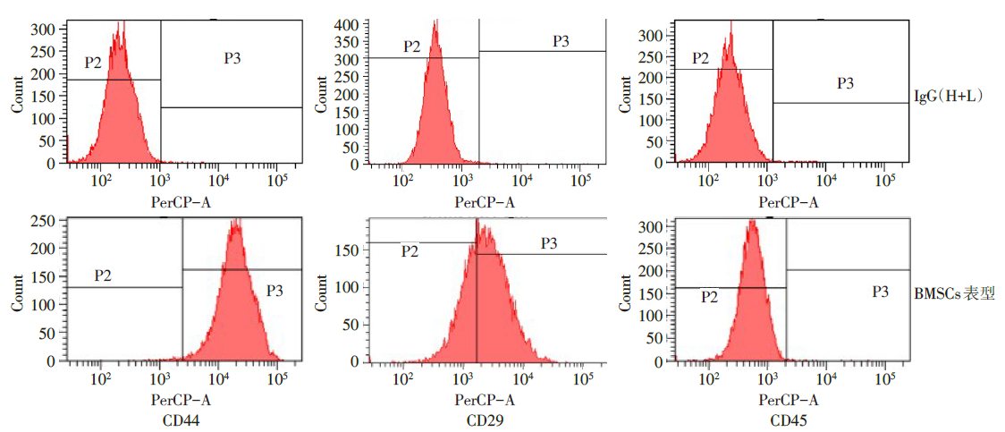
Fig.1 The characteristics of cell surface markers in BMSCs

Fig.2 The cell morphology of BMSCs under fluorescence microscope (×100)
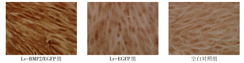
Fig.3 The SP immunochemical staining of BMP2 expression (DAB stanining, ×200)
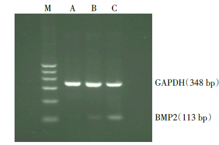
Fig.4 The BMP2 gene expression in BMSCs measured by RT-PCR
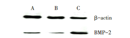
Fig.5 The protein expression of BMP2 measured by Western blot assay
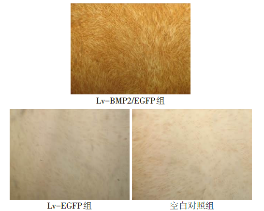
Fig.6 The I collagen immunohistochemical staining of BMSCs (×100)
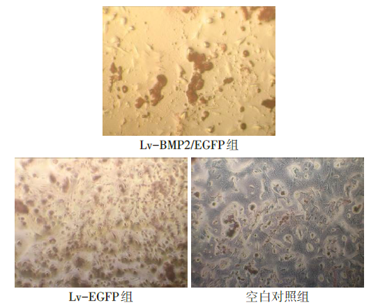
Fig.7 The alizarin red S staining of BMSCs (×50)
| 组别 | 第7天 | 第14天 | 第21天 |
|---|---|---|---|
| 空白对照组 | 2.88±0.57 | 4.99±0.47 | 5.76±0.50 |
| Lv-EGFP组 | 2.76±0.50 | 4.76±0.50 | 5.71±0.44 |
| Lv-BMP2/EGFP组 | 6.76±0.91ab | 16.42±2.22ab | 24.39±2.54ab |
| F | 98.659** | 222.143** | 452.715** |
Tab.1 Comparison of ALP levels at different time points between the three groups
| 组别 | 第7天 | 第14天 | 第21天 |
|---|---|---|---|
| 空白对照组 | 2.88±0.57 | 4.99±0.47 | 5.76±0.50 |
| Lv-EGFP组 | 2.76±0.50 | 4.76±0.50 | 5.71±0.44 |
| Lv-BMP2/EGFP组 | 6.76±0.91ab | 16.42±2.22ab | 24.39±2.54ab |
| F | 98.659** | 222.143** | 452.715** |
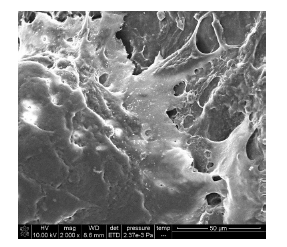
Fig.8 The microscopic morphology of the mineralized nodule surface observed by SEM
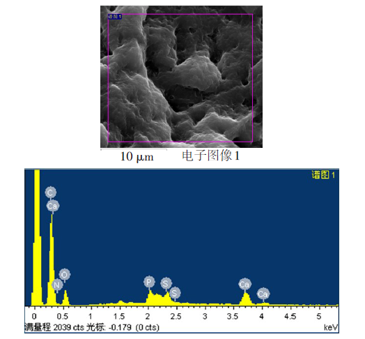
Fig.9 The energy spectrum analysis of the scanned area on the surface of the mineralized nodule
| 元素 | 原子质量百分比(%) | 原子个数百分比(%) |
|---|---|---|
| C K | 0.41 | 13.08 |
| N K | 0.95 | 25.79 |
| O K | 2.19 | 51.93 |
| P K | 0.23 | 2.78 |
| S K | 0.18 | 2.16 |
| Ca K | 0.45 | 4.27 |
| 总量 | 4.42 | - |
| Ca/P | 1.52 | - |
Tab.2 The elemental analysis of the scanned area on the surface of the mineralized nodule
| 元素 | 原子质量百分比(%) | 原子个数百分比(%) |
|---|---|---|
| C K | 0.41 | 13.08 |
| N K | 0.95 | 25.79 |
| O K | 2.19 | 51.93 |
| P K | 0.23 | 2.78 |
| S K | 0.18 | 2.16 |
| Ca K | 0.45 | 4.27 |
| 总量 | 4.42 | - |
| Ca/P | 1.52 | - |
| [1] | CHEN M, XU Y, ZHANG T, et al. Mesenchymal stem cell sheets:A new cell-based strategy for bone repair and regeneration[J]. Biotechnol Lett, 2019, 41(3):305-318. doi:10.1007/s10529-019-02649-7. |
| [2] | HU T, NAIDU M, YANG Z, et al. Bone regeneration by controlled release of bone morphogenetic protein-2:A rabbit spinal fusion chamber molecular study[J]. Tissue Eng Part A, 2019, 25(19/20):1356-1368. doi:10.1089/ten.TEA.2018.0281. |
| [3] | HALLORAN D, DURBANO H W, NOHE A. Bone morphogenetic protein-2 in development and bone homeostasis[J]. J Dev Biol, 2020, 8(3):19. doi:10.3390/jdb8030019. |
| [4] | HONG M H, LEE J H, JUNG H S, et al. Biomineralization of bone tissue:calcium phosphate-based inorganics in collagen fibrillar organic matrices[J]. Biomater Res, 2022, 26(1):42. doi:10.1186/s40824-022-00288-0. |
| [5] | WANG J, LIU Q, GUO Z, et al. Progress on biomimetic mineralization and materials for hard tissue regeneration[J]. ACS Biomater Sci Eng, 2023, 9(4):1757-1773. doi: 10.1021/acsbiomaterials.1c01070. |
| [6] | SHANG F, YU Y, LIU S, et al. Advancing application of mesenchymal stem cell-based bone tissue regeneration[J]. Bioact Mater, 2021, 6(3):666-683. doi:10.1016/j.bioactmat.2020.08.014. |
| [7] | DE LA VEGA R E, ATASOY-ZEYBEK A, PANOS J A, et al. Gene therapy for bone healing:Lessons learned and new approaches[J]. Transl Res, 2021, 236:1-16. doi:10.1016/j.trsl.2021.04.009. |
| [8] | 谭靓, 李泰明. 腺相关病毒载体在基因治疗领域中的应用和挑战[J]. 科技与创新, 2020,(13):157-159. |
| TAN L, LI T M. Application and challenges of adeno-associated viral vectors in gene therapy[J]. Science and Technology & Innovation, 2020,(13):157-159. doi:10.15913/j.cnki.kjycx.2020.13.066. | |
| [9] | MILONE M C, O'DOHERTY U. Clinical use of lentiviral vectors[J]. Leukemia, 2018, 32(7):1529-1541. doi:10.1038/s41375-018-0106-0. |
| [10] | PERRY C, RAYAT A. Lentiviral vector bioprocessing[J]. Viruses, 2021, 13(2):268. doi:10.3390/v13020268. |
| [11] | 李诗鹏, 李强, 石正松, 等. 基因重组腺病毒载体与慢病毒载体转染兔骨髓间充质干细胞的对比[J]. 中国组织工程研究, 2017, 21(9):1340-1345. |
| LI S P, LI Q, SHI Z S, et al. Comparison of lentiviral vector and adenoviral vector mediated gene transfer into rabbit bone marrow mesenchymal stem cells[J]. Chinese Journal of Tissue Engineering Research, 2017, 21(9):1340-1345. doi:10.3969/j.issn.2095-4344.2017.09.006. | |
| [12] | KONG J, WANG Y, QI W, et al. Green fluorescent protein inspired fluorophores[J]. Adv Colloid Interface Sci, 2020, 285:102286. doi:10.1016/j.cis.2020.102286. |
| [13] | 赵怡心. 骨细胞BMP信号调控成骨分化作用与机制研究[D]. 重庆: 重庆医科大学, 2021. |
| ZHAO Y X. The effect and mechanism of BMP signaling on osteogenic differentiation of osteocyte[D]. Chongqing: Chongqing Medical University, 2021. | |
| [14] | YUAN Z, LI Q, LUO S, et al. PPARγ and wnt signaling in adipogenic and osteogenic differentiation of mesenchymal stem cells[J]. Curr Stem Cell Res Ther, 2016, 11(3):216-225. doi:10.2174/1574888x10666150519093429. |
| [15] | LI L, KHONG M L, LUI E, et al. Long-chain polyphosphate in osteoblast matrix vesicles: Enrichment and inhibition of mineralization[J]. Biochim Biophys Acta Gen Subj, 2019, 1863(1):199-209. doi:10.1016/j.bbagen.2018.10.003. |
| [16] | MURSHED M. Mechanism of bone mineralization[J]. Cold Spring Harb Perspect Med, 2018, 8(12):a031229. doi:10.1101/cshperspect.a031229. |
| [17] | KITAURA H, MARAHLEH A, OHORI F, et al. Osteocyte-related cytokines regulate osteoclast formation and bone resorption[J]. Int J Mol Sci, 2020, 21(14):5169. doi:10.3390/ijms21145169. |
| [18] | HASEGAWA T. Ultrastructure and biological function of matrix vesicles in bone mineralization[J]. Histochem Cell Biol, 2018, 149(4):289-304. doi:10.1007/s00418-018-1646-0. |
| Viewed | ||||||
|
Full text |
|
|||||
|
Abstract |
|
|||||