Tianjin Medical Journal ›› 2025, Vol. 53 ›› Issue (3): 301-306.doi: 10.11958/20241833
• Applied Research • Previous Articles Next Articles
LI Xiaoshuo1( ), HAN Zixu2, LI Yang2, LI Mengning3,△()
Received:2024-11-13
Revised:2024-12-30
Published:2025-03-15
Online:2025-03-31
Contact:
E-mail:LI Xiaoshuo, HAN Zixu, LI Yang, LI Mengning. Predictive value of vertebral CT parameters for early fusion sink after TLIF[J]. Tianjin Medical Journal, 2025, 53(3): 301-306.
CLC Number:
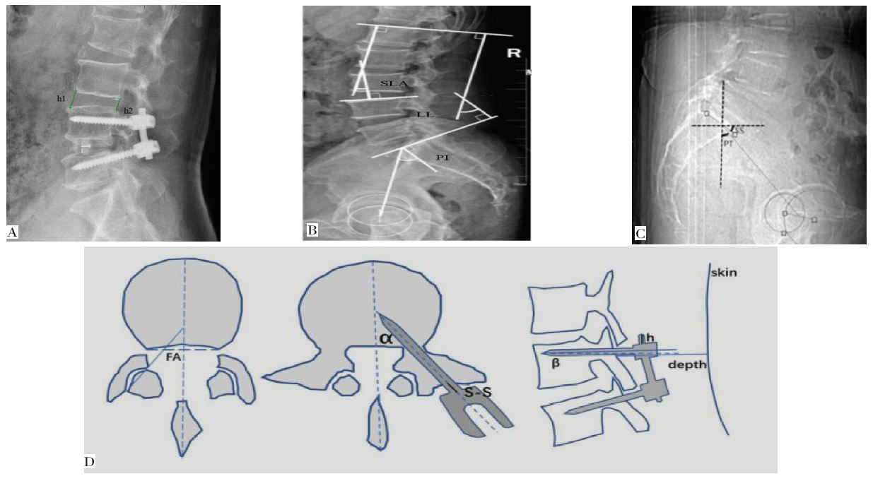
Fig.1 Imaging parameter measurement
| 组别 | n | 年龄 | 性别 | BMI/(kg/m2) | 病程/月 | ||||||||||||||||||
|---|---|---|---|---|---|---|---|---|---|---|---|---|---|---|---|---|---|---|---|---|---|---|---|
| <60岁 | ≥60岁 | 男 | 女 | ||||||||||||||||||||
| 无沉降组 | 121 | 65(53.72) | 56(46.28) | 60(49.59) | 61(50.41) | 23.87±2.13 | 45.76±8.34 | ||||||||||||||||
| 沉降组 | 57 | 20(35.09) | 37(64.91) | 23(40.35) | 34(59.65) | 24.01±2.08 | 46.84±8.14 | ||||||||||||||||
| χ2或t | 5.391* | 1.328 | 0.412 | 0.814 | |||||||||||||||||||
| 组别 | 合并症 | 疾病类型 | |||||||||||||||||||||
| 高血压 | 糖尿病 | 高脂血症 | 骨质疏松症 | 腰椎间盘突出症 | 腰椎滑脱 | 腰椎管狭窄 | |||||||||||||||||
| 无沉降组 | 29(23.97) | 27(22.31) | 24(19.83) | 30(24.79) | 34(28.10) | 41(33.88) | 46(38.02) | ||||||||||||||||
| 沉降组 | 15(26.32) | 14(24.56) | 13(22.81) | 24(42.11) | 23(40.35) | 15(26.32) | 19(33.33) | ||||||||||||||||
| χ2 | 0.115 | 0.110 | 0.208 | 5.495* | 2.754 | ||||||||||||||||||
| 组别 | 手术节段 | 融合器长度/mm | 融合器高度/mm | 融合器接触面积/% | 手术时间/min | 术中出血量/mL | |||||||||||||||||
| L2—3 | L3—4 | L4—5 | L5—S1 | ||||||||||||||||||||
| 无沉降组 | 26(21.49) | 25(20.66) | 20(16.53) | 50(41.32) | 8.99±1.21 | 10.64±2.12 | 34.5±2.3 | 175.34±34.38 | 121.35±35.85 | ||||||||||||||
| 沉降组 | 2(3.51) | 4(7.02) | 16(28.07) | 35(61.40) | 9.35±1.18 | 11.21±2.24 | 35.6±2.4 | 181.58±34.30 | 131.23±45.34 | ||||||||||||||
| χ2或t | 18.213** | 1.871 | 1.670 | 2.787** | 1.131 | 1.572 | |||||||||||||||||
Tab.1 Comparison of general data between the sedimentation group and the non-ssedimentation group
| 组别 | n | 年龄 | 性别 | BMI/(kg/m2) | 病程/月 | ||||||||||||||||||
|---|---|---|---|---|---|---|---|---|---|---|---|---|---|---|---|---|---|---|---|---|---|---|---|
| <60岁 | ≥60岁 | 男 | 女 | ||||||||||||||||||||
| 无沉降组 | 121 | 65(53.72) | 56(46.28) | 60(49.59) | 61(50.41) | 23.87±2.13 | 45.76±8.34 | ||||||||||||||||
| 沉降组 | 57 | 20(35.09) | 37(64.91) | 23(40.35) | 34(59.65) | 24.01±2.08 | 46.84±8.14 | ||||||||||||||||
| χ2或t | 5.391* | 1.328 | 0.412 | 0.814 | |||||||||||||||||||
| 组别 | 合并症 | 疾病类型 | |||||||||||||||||||||
| 高血压 | 糖尿病 | 高脂血症 | 骨质疏松症 | 腰椎间盘突出症 | 腰椎滑脱 | 腰椎管狭窄 | |||||||||||||||||
| 无沉降组 | 29(23.97) | 27(22.31) | 24(19.83) | 30(24.79) | 34(28.10) | 41(33.88) | 46(38.02) | ||||||||||||||||
| 沉降组 | 15(26.32) | 14(24.56) | 13(22.81) | 24(42.11) | 23(40.35) | 15(26.32) | 19(33.33) | ||||||||||||||||
| χ2 | 0.115 | 0.110 | 0.208 | 5.495* | 2.754 | ||||||||||||||||||
| 组别 | 手术节段 | 融合器长度/mm | 融合器高度/mm | 融合器接触面积/% | 手术时间/min | 术中出血量/mL | |||||||||||||||||
| L2—3 | L3—4 | L4—5 | L5—S1 | ||||||||||||||||||||
| 无沉降组 | 26(21.49) | 25(20.66) | 20(16.53) | 50(41.32) | 8.99±1.21 | 10.64±2.12 | 34.5±2.3 | 175.34±34.38 | 121.35±35.85 | ||||||||||||||
| 沉降组 | 2(3.51) | 4(7.02) | 16(28.07) | 35(61.40) | 9.35±1.18 | 11.21±2.24 | 35.6±2.4 | 181.58±34.30 | 131.23±45.34 | ||||||||||||||
| χ2或t | 18.213** | 1.871 | 1.670 | 2.787** | 1.131 | 1.572 | |||||||||||||||||
| 组别 | n | 腰椎CT 值/HU | 椎间隙 高度/mm | 节段性 前凸角/° | 腰椎 前凸角/° | 骨盆 入射角/° | 骨盆 倾斜角/° | 螺钉 内倾角/° | 螺钉 尾倾角/° |
|---|---|---|---|---|---|---|---|---|---|
| 无沉降组 | 121 | 131.03±11.19 | 12.30±1.24 | 17.99±1.87 | 45.28±13.59 | 49.14±9.32 | 32.85±4.99 | 13.49±2.87 | 11.85±3.67 |
| 沉降组 | 57 | 110.80±10.92 | 13.57±1.15 | 20.45±1.95 | 47.25±12.24 | 48.70±8.24 | 31.68±5.26 | 13.12±2.98 | 11.25±3.87 |
| t | 11.337** | 6.562** | 8.070** | 0.936 | 0.298 | 1.423 | 0.771 | 0.982 |
Tab.2 Comparison of CT image parameters after TLIF between the sedimentation group and the non-sedimentation group
| 组别 | n | 腰椎CT 值/HU | 椎间隙 高度/mm | 节段性 前凸角/° | 腰椎 前凸角/° | 骨盆 入射角/° | 骨盆 倾斜角/° | 螺钉 内倾角/° | 螺钉 尾倾角/° |
|---|---|---|---|---|---|---|---|---|---|
| 无沉降组 | 121 | 131.03±11.19 | 12.30±1.24 | 17.99±1.87 | 45.28±13.59 | 49.14±9.32 | 32.85±4.99 | 13.49±2.87 | 11.85±3.67 |
| 沉降组 | 57 | 110.80±10.92 | 13.57±1.15 | 20.45±1.95 | 47.25±12.24 | 48.70±8.24 | 31.68±5.26 | 13.12±2.98 | 11.25±3.87 |
| t | 11.337** | 6.562** | 8.070** | 0.936 | 0.298 | 1.423 | 0.771 | 0.982 |
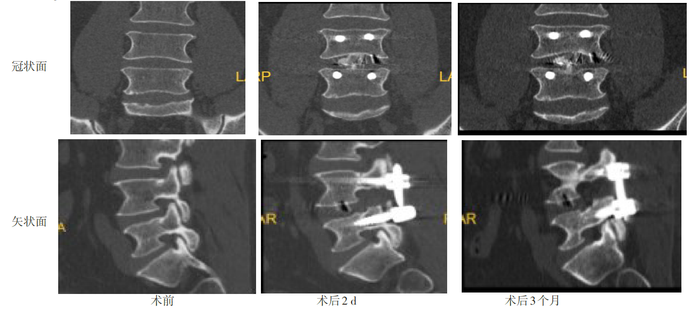
Fig.2 Preoperative and postoperative CT images of a patient with fusion sink after TLIF
| 变量 | β | SE | Wald χ2 | P | OR | 95%CI |
|---|---|---|---|---|---|---|
| 年龄 | 0.536 | 0.197 | 7.403 | 0.026 | 1.709 | 1.162~2.515 |
| 手术节段 | 0.641 | 0.215 | 8.889 | 0.012 | 1.898 | 1.246~2.893 |
| 椎间隙高度 | 0.685 | 0.203 | 11.386 | <0.001 | 1.984 | 1.333~2.953 |
| 节段性前凸角 | 0.988 | 0.312 | 10.028 | <0.001 | 2.686 | 1.457~4.951 |
| 腰椎CT值 | -0.789 | 0.192 | 16.887 | <0.001 | 0.454 | 0.312~0.662 |
| 常数项 | -0.421 | 0.184 | 5.235 | 0.045 | - | - |
Tab.3 Logistic regression analysis of multiple factors affecting the early fusion settlement after TLIF
| 变量 | β | SE | Wald χ2 | P | OR | 95%CI |
|---|---|---|---|---|---|---|
| 年龄 | 0.536 | 0.197 | 7.403 | 0.026 | 1.709 | 1.162~2.515 |
| 手术节段 | 0.641 | 0.215 | 8.889 | 0.012 | 1.898 | 1.246~2.893 |
| 椎间隙高度 | 0.685 | 0.203 | 11.386 | <0.001 | 1.984 | 1.333~2.953 |
| 节段性前凸角 | 0.988 | 0.312 | 10.028 | <0.001 | 2.686 | 1.457~4.951 |
| 腰椎CT值 | -0.789 | 0.192 | 16.887 | <0.001 | 0.454 | 0.312~0.662 |
| 常数项 | -0.421 | 0.184 | 5.235 | 0.045 | - | - |
| 因素 | AUC | 95%CI | 截断值 | 敏感 度/% | 特异 度/% | 约登 指数 |
|---|---|---|---|---|---|---|
| 临床因素 | ||||||
| 年龄≥60岁 | 0.593 | 0.517~0.666 | - | 64.9 | 53.7 | 0.186 |
| 手术节段 L5—S1 | 0.600 | 0.524~0.673 | - | 61.4 | 58.7 | 0.201 |
| 椎体CT参数 | ||||||
| 腰椎CT值 | 0.872 | 0.814~0.917 | 126.20 HU | 89.5 | 72.7 | 0.622 |
| 椎间隙高度 | 0.810 | 0.745~0.865 | 12.61 mm | 82.5 | 69.4 | 0.519 |
| 节段性前凸角 | 0.735 | 0.663~0.798 | 18.93° | 71.9 | 71.0 | 0.430 |
| 椎体CT参数 联合 | 0.960 | 0.920~0.984 | - | 93.0 | 88.4 | 0.814 |
Tab.4 The predictive value of clinical factors and vertebral CT parameters for early fusion subsidence after TLIF
| 因素 | AUC | 95%CI | 截断值 | 敏感 度/% | 特异 度/% | 约登 指数 |
|---|---|---|---|---|---|---|
| 临床因素 | ||||||
| 年龄≥60岁 | 0.593 | 0.517~0.666 | - | 64.9 | 53.7 | 0.186 |
| 手术节段 L5—S1 | 0.600 | 0.524~0.673 | - | 61.4 | 58.7 | 0.201 |
| 椎体CT参数 | ||||||
| 腰椎CT值 | 0.872 | 0.814~0.917 | 126.20 HU | 89.5 | 72.7 | 0.622 |
| 椎间隙高度 | 0.810 | 0.745~0.865 | 12.61 mm | 82.5 | 69.4 | 0.519 |
| 节段性前凸角 | 0.735 | 0.663~0.798 | 18.93° | 71.9 | 71.0 | 0.430 |
| 椎体CT参数 联合 | 0.960 | 0.920~0.984 | - | 93.0 | 88.4 | 0.814 |
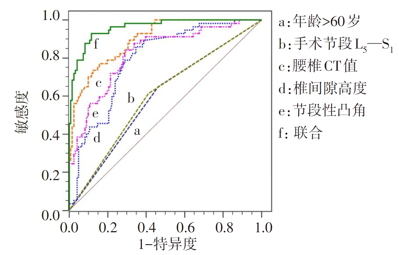
Fig.3 ROC curve of clinical factors and vertebral CT parameters predicting early fusion vessel subsidence after TLIF
| [1] | HEO D H, KIM J Y, PARK J Y, et al. Clinical experiences of 3-dimensional biportal endoscopic spine surgery for lumbar degenerative disease[J]. Oper Neurosurg(Hagerstown), 2022, 22(4):231-238. doi:10.1227/ONS.0000000000000090. |
| [2] | MACKI M, HAMILTON T, HADDAD Y W, et al. Expandable cage technology-transforaminal,anterior,and lateral lumbar interbody fusion[J]. Oper Neurosurg(Hagerstown), 2021, 21(Suppl 1):S69-S80. doi:10.1093/ons/opaa342. |
| [3] | GODOLIAS P, TATARYN Z L, PLÜMER J, et al. Cage subsidence-a multifactorial matter![J]. Orthopadie (Heidelb), 2023, 52(8):662-669. doi:10.1007/s00132-023-04363-9. |
| [4] | ROBERTSON T S, PIJLS B G, MUNN Z, et al. Change in CT-measured acetabular bone density following total hip arthroplasty:a systematic review and meta-analysis[J]. Acta Orthop, 2023,94:191-199. doi:10.2340/17453674.2023.11635. |
| [5] | PU X, WANG D, GU S. Advances in Hounsfield units value for predicting cage subsidence on spinal interbody fusion surgery[J]. Eur Spine J, 2023, 32(9):3149-3157. doi:10.1007/s00586-023-07805-2. |
| [6] | 范宇澄, 蔡东岭, 郭伟俊, 等. 腰椎终板形态对经椎间孔腰椎椎间融合术后融合器沉降的影响[J]. 实用骨科杂志, 2023, 29(3):193-197,213. |
| FAN Y C, CAI D L, GUO W J, et al. Effect of lumbar endplate morphology on cage subsidence after transforaminal lumbar interbody fusion[J]. Journal of Practical Orthopedics, 2023, 29(3):193-197,213. | |
| [7] | DHAR U K, MENZER E L, LIN M, et al. Factors influencing cage subsidence in anterior cervical corpectomy and discectomy:a systematic review[J]. Eur Spine J, 2023, 32(3):957-968. doi:10.1007/s00586-023-07530-w. |
| [8] | MARCHI L, ABDALA N, OLIVEIRA L, et al. Radiographic and clinical evaluation of cage subsidence after stand-alone lateral interbody fusion[J]. J Neurosurg Spine, 2013, 19(1):110-118. doi:10.3171/2013.4.SPINE12319. |
| [9] | WANG L, LI H, ZHAO Y, et al. Ligamentum-preserved/temporary preserved minimally invasive transforaminal lumbar interbody fusion for lumbar spondylolisthesis:technical note and 2-year follow-up[J]. Spine(Phila Pa 1976), 2022, 47(8):E328-E336. doi:10.1097/BRS.0000000000004136. |
| [10] | SEBAALY A, KREICHATI G, TARCHICHI J, et al. Transforaminal lumbar interbody fusion using banana-shaped and straight cages:meta-analysis of clinical and radiological outcomes[J]. Eur Spine J, 2023, 32(9):3158-3166. doi:10.1007/s00586-023-07797-z. |
| [11] | 李根, 李鑫, 杨硕, 等. 经椎间孔腰椎椎间融合术术后融合器沉降预测模型的建立与验证[J]. 医学研究杂志, 2023, 52(5):55-61. |
| LI G, LI X, YANG S, et al. Development and validation of a predictive model for cage subsidence after transforaminal lumbar interbody fusion[J]. Journal of Medical Research, 2023, 52(5):55-61. doi:10.11969/j.issn.1673-548X.2023.05.013. | |
| [12] | SUN W X, LIU H N, CHEN M T, et al. Meta-analysis of the clinical efficacy and safety of oblique lateral interbody fusion and transforaminal interbody fusion in the treatment of degenerative lumbar spondylolisthesis[J]. EFORT Open Rev, 2022, 7(9):663-670. doi:10.1530/EOR-22-0042. |
| [13] | 金锋, 王焕, 唐世技, 等. TLIF术后融合器下沉的危险因素调查[J]. 颈腰痛杂志, 2023, 44(4):663-665. |
| JIN F, WANG H, TANG S J, et al. Investigation on the risk factors of fusion apparatus sinking after TLIF operation[J]. The Journal of Cervicodynia and Lumbodynia, 2023, 44(4):663-665. doi:10.3969/j.issn.1005-7234.2023.04.046. | |
| [14] | AMORIM-BARBOSA T, PEREIRA C, CATELAS D, et al. Risk factors for cage subsidence and clinical outcomes after transforaminal and posterior lumbar interbody fusion[J]. Eur J Orthop Surg Traumatol, 2022, 32(7):1291-1299. doi:10.1007/s00590-021-03103-z. |
| [15] | TOUBAN B M, SAYEGH M J, GALINA J, et al. Computed tomography measured psoas cross sectional area is associated with bone mineral density measured by dual energy X-ray absorptiometry[J]. J Clin Densitom, 2022, 25(4):592-598. doi:10.1016/j.jocd.2022.04.001. |
| [16] | 谭波, 胡豇, 卢冰, 等. 骨质疏松症患者血清H2S的变化及临床意义[J]. 天津医药, 2022, 50(8):832-835. |
| TAN B, HU J, LU B, et al. Changes of serum H2S in patients with osteoporosis and its clinical significance[J]. Tianjin Med J, 2022, 50(8):832-835. doi:10.11958/20212020. | |
| [17] | 田爱现, 马剑雄, 马信龙, 等. 极外侧路径下椎体间融合术与传统开放后路椎体间融合术治疗椎间盘突出的效果比较[J]. 中国中西医结合外科杂志, 2023, 29(3):317-321. |
| TIAN A X, MA J X, MA X L et al. Perioperative efficacy of extreme lateral interbody fusion versus traditional open posterior interbody fusion for disc herniation[J]. Chinese Journal of Surgery of Integrated Traditional and Western Medicine, 2023, 29(3):317-321. doi:10.3969/j.issn.1007-6948.2023.03.006. | |
| [18] | JONES C, OKANO I, SALZMANN S N, et al. Endplate volumetric bone mineral density is a predictor for cage subsidence following lateral lumbar interbody fusion:a risk factor analysis[J]. Spine J, 2021, 21(10):1729-1737. doi:10.1016/j.spinee.2021.02.021. |
| [19] | HUANG W, GONG Z, ZHENG C, et al. Preoperative assessment of bone density using MRI-based vertebral bone quality score modified for patients undergoing cervical spine surgery[J]. Global Spine J, 2024, 14(4):1238-1247. doi:10.1177/21925682221138261. |
| [20] | 傅赛琼, 常磊, 张明彦, 等. 老年腰椎退行性疾病患者MIS-TLIF术后融合器后移的影响因素[J]. 中国老年学杂志, 2022, 42(7):1637-1640. |
| FU S Q, CHANG L, ZHANG M Y, et al. Influence factors of fusion apparatus retraction in elderly patients with lumbar degenerative diseases after MIS-TLIF surgery[J]. Chinese Journal of Gerontology, 2022, 42(7):1637-1640. doi:10.3969/j.issn.1005-9202.2022.07.031. | |
| [21] | YAO Y C, CHAO H, KAO K Y, et al. CT Hounsfield unit is a reliable parameter for screws loosening or cages subsidence in minimally invasive transforaminal lumbar interbody fusion[J]. Sci Rep, 2023, 13(1):1620. doi:10.1038/s41598-023-28555-7. |
| [22] | WU H, SHAN Z, ZHAO F, et al. Poor bone quality,multilevel surgery,and narrow and tall cages are associated with intraoperative endplate injuries and late-onset cage subsidence in lateral lumbar interbody fusion:a systematic review[J]. Clin Orthop Relat Res, 2022, 480(1):163-188. doi:10.1097/CORR.0000000000001915. |
| [23] | 袁照, 梁体洪, 孙红, 等. 采用椎间融合器实施PLIF的LDD患者术后椎间融合器下沉影响因素研究进展[J]. 山东医药, 2023, 63(6):96-100. |
| YUAN Z, LIANG T H, SUN H, et al. Research progress on the influencing factors of post-operative fusion organ sinking in LDD patients undergoing PLIF with interbody fusion organ[J]. Shandong Medicine Journal, 2023, 63(6):96-100. doi:10.3969/j.issn.1002-266X.2023.06.024. |
| [1] | YU Fang, ZHAO Xiaoyong, REN Yuan, LIU Hongyang. Application effect of percutaneous vertebroplasty combined with 3D-printed personalized vertebral body stents in the repair of thoracolumbar vertebral fractures [J]. Tianjin Medical Journal, 2025, 53(7): 730-735. |
| [2] | JIANG Zehua, CUI Haojun, ZHANG Boyu, REN Zhishuai, MA Junfeng, ZHANG Hongjie, ZHU Rusen. Research progress on the correlation between sagittal spinopelvic parameters and the prognosis of lumbar fusion surgery [J]. Tianjin Medical Journal, 2025, 53(1): 103-107. |
| [3] | YUE Zong-jin, LIU Ru-yin△, YU Lu, WANG Xin-li, FENG Zhong-kai, WANG Xi-bin. Puerarin saponins induce the differentiation of BMSCs into nucleus pulposus-like cells via NAMPT-Sirt1 axis#br# [J]. Tianjin Medical Journal, 2021, 49(9): 897-903. |
| [4] | LIU Yue, JIANG Hong-feng, HUANG Hong-chao, LI Ning, XU Bao-shan. Controversial issues related to the treatment of degenerative lumbar spondylolisthesis [J]. Tianjin Medical Journal, 2021, 49(8): 883-886. |
| [5] | XU Bao-shan, LIU Yang, JIANG Hong-feng, LIU Yue, WANG Tao, LI Ning, XU Hai-wei, HUANG Hong-chao, JI Ning. The design and application of self-anchored lateral lumbar interbody fusion [J]. Tianjin Med J, 2019, 47(9): 937-942. |
| [6] | LI Shuang1, SUN Xiao-lei1, MA Xin-long1△, ZHANG Yang2, DENG Shu-cai1, HAO Yong-hong1. The response of cyclic tensile strain on the BMSCs co-cultured human degenerative anulus fibrosus cells [J]. Tianjin Med J, 2017, 45(6): 571-576. |
| [7] | XU Baoshan, MA Xinlong, YANG Qiang, LIU Yue, JIANG Hongfeng, XU Haiwei, JI Ning. The design and clinical application of MED-TLIF with mobile microendoscopic discectomy technique. [J]. Tianjin Med J, 2016, 44(7): 910-913. |
| Viewed | ||||||
|
Full text |
|
|||||
|
Abstract |
|
|||||