
Tianjin Medical Journal ›› 2026, Vol. 54 ›› Issue (6): 637-642.doi: 10.11958/20253436
• Applied Research • Previous Articles Next Articles
KANG Xiaona( ), LIU Yaping, FENG Yun, LUO Yumei, JIA Ai△()
Received:2025-11-27
Revised:2026-02-25
Published:2026-06-15
Online:2026-06-15
Contact:
△E-mail:JiaAi2663@163.com
KANG Xiaona, LIU Yaping, FENG Yun, LUO Yumei, JIA Ai. Development of a preoperative assessment model for the malignant potential in gastrointestinal stromal tumor based on endoscopic ultrasound and contrast-enhanced computed tomography[J]. Tianjin Medical Journal, 2026, 54(6): 637-642.
CLC Number:
| 组别 | n | 性别 | 年龄/岁 | 发病部位 | ||||||||||||
|---|---|---|---|---|---|---|---|---|---|---|---|---|---|---|---|---|
| 男 | 女 | 食管 | 胃 | 十二指肠 | 小肠 | 直肠 | ||||||||||
| 低恶性潜能组 | 145 | 71(48.97) | 74(51.03) | 55.82±10.79 | 3(2.07) | 123(84.83) | 10(6.90) | 7(4.83) | 2(1.38) | |||||||
| 高恶性潜能组 | 46 | 26(56.53) | 20(43.48) | 56.91±11.10 | 1(2.17) | 36(78.26) | 3(6.52) | 4(8.70) | 2(4.35) | |||||||
| t或χ2 | 0.798 | 0.594 | 2.576 | |||||||||||||
| 组别 | 临床表现 | |||||||||||||||
| 腹痛、腹胀 | 恶心、呕吐 | 反酸、烧心、嗳气 | 胃肠道出血 | 肠梗阻 | 黑便 | 乏力、纳差 | ||||||||||
| 低恶性潜能组 | 126(86.90) | 32(22.07) | 43(29.66) | 27(18.62) | 7(4.83) | 22(15.17) | 7(4.83) | |||||||||
| 高恶性潜能组 | 43(93.48) | 16(34.78) | 19(41.30) | 14(30.43) | 5(10.87) | 11(23.91) | 5(10.87) | |||||||||
| χ2 | 1.484 | 3.000 | 2.162 | 2.891 | 2.165 | 1.867 | 2.165 | |||||||||
Tab.1 Comparison of general data between the two groups
| 组别 | n | 性别 | 年龄/岁 | 发病部位 | ||||||||||||
|---|---|---|---|---|---|---|---|---|---|---|---|---|---|---|---|---|
| 男 | 女 | 食管 | 胃 | 十二指肠 | 小肠 | 直肠 | ||||||||||
| 低恶性潜能组 | 145 | 71(48.97) | 74(51.03) | 55.82±10.79 | 3(2.07) | 123(84.83) | 10(6.90) | 7(4.83) | 2(1.38) | |||||||
| 高恶性潜能组 | 46 | 26(56.53) | 20(43.48) | 56.91±11.10 | 1(2.17) | 36(78.26) | 3(6.52) | 4(8.70) | 2(4.35) | |||||||
| t或χ2 | 0.798 | 0.594 | 2.576 | |||||||||||||
| 组别 | 临床表现 | |||||||||||||||
| 腹痛、腹胀 | 恶心、呕吐 | 反酸、烧心、嗳气 | 胃肠道出血 | 肠梗阻 | 黑便 | 乏力、纳差 | ||||||||||
| 低恶性潜能组 | 126(86.90) | 32(22.07) | 43(29.66) | 27(18.62) | 7(4.83) | 22(15.17) | 7(4.83) | |||||||||
| 高恶性潜能组 | 43(93.48) | 16(34.78) | 19(41.30) | 14(30.43) | 5(10.87) | 11(23.91) | 5(10.87) | |||||||||
| χ2 | 1.484 | 3.000 | 2.162 | 2.891 | 2.165 | 1.867 | 2.165 | |||||||||
| 组别 | n | 肿瘤直径/mm | 外观不光滑 | 形状不规则 | 边界不清晰 | ||||||||||
|---|---|---|---|---|---|---|---|---|---|---|---|---|---|---|---|
| 低恶性潜能组 | 145 | 43.91±13.61 | 56(38.62) | 54(37.24) | 15(10.34) | ||||||||||
| 高恶性潜能组 | 46 | 49.60±17.29 | 24(52.17) | 24(52.17) | 14(30.43) | ||||||||||
| t或χ2 | 2.303* | 2.635 | 3.223 | 10.945** | |||||||||||
| 组别 | 病灶起源层 | 回声强度 | |||||||||||||
| 固有肌层 | 非固有肌层 | 低 | 高 | ||||||||||||
| 低恶性潜能组 | 119(82.07) | 26(17.93) | 136(93.79) | 9(6.21) | |||||||||||
| 高恶性潜能组 | 38(82.61) | 8(17.39) | 46(100.00) | 0 | |||||||||||
| χ2 | 0.007 | 1.773 | |||||||||||||
| 组别 | 回声不均匀 | 钙化 | 囊性病变 | 局部侵犯 | |||||||||||
| 低恶性潜能组 | 58(40.00) | 28(19.31) | 33(22.76) | 31(21.38) | |||||||||||
| 高恶性潜能组 | 25(54.35) | 15(32.61) | 16(34.78) | 16(34.78) | |||||||||||
| χ2 | 2.926 | 3.540 | 2.647 | 3.382 | |||||||||||
| 组别 | 血流信号 | ||||||||||||||
| 未见信号 | 信号不丰富 | 信号丰富 | |||||||||||||
| 低恶性潜能组 | 19(13.10) | 91(62.76) | 35(24.14) | ||||||||||||
| 高恶性潜能组 | 7(15.22) | 21(45.65) | 18(39.13) | ||||||||||||
| χ2 | 4.686 | ||||||||||||||
Tab.2 Comparison of EUS image features bwtween the two groups
| 组别 | n | 肿瘤直径/mm | 外观不光滑 | 形状不规则 | 边界不清晰 | ||||||||||
|---|---|---|---|---|---|---|---|---|---|---|---|---|---|---|---|
| 低恶性潜能组 | 145 | 43.91±13.61 | 56(38.62) | 54(37.24) | 15(10.34) | ||||||||||
| 高恶性潜能组 | 46 | 49.60±17.29 | 24(52.17) | 24(52.17) | 14(30.43) | ||||||||||
| t或χ2 | 2.303* | 2.635 | 3.223 | 10.945** | |||||||||||
| 组别 | 病灶起源层 | 回声强度 | |||||||||||||
| 固有肌层 | 非固有肌层 | 低 | 高 | ||||||||||||
| 低恶性潜能组 | 119(82.07) | 26(17.93) | 136(93.79) | 9(6.21) | |||||||||||
| 高恶性潜能组 | 38(82.61) | 8(17.39) | 46(100.00) | 0 | |||||||||||
| χ2 | 0.007 | 1.773 | |||||||||||||
| 组别 | 回声不均匀 | 钙化 | 囊性病变 | 局部侵犯 | |||||||||||
| 低恶性潜能组 | 58(40.00) | 28(19.31) | 33(22.76) | 31(21.38) | |||||||||||
| 高恶性潜能组 | 25(54.35) | 15(32.61) | 16(34.78) | 16(34.78) | |||||||||||
| χ2 | 2.926 | 3.540 | 2.647 | 3.382 | |||||||||||
| 组别 | 血流信号 | ||||||||||||||
| 未见信号 | 信号不丰富 | 信号丰富 | |||||||||||||
| 低恶性潜能组 | 19(13.10) | 91(62.76) | 35(24.14) | ||||||||||||
| 高恶性潜能组 | 7(15.22) | 21(45.65) | 18(39.13) | ||||||||||||
| χ2 | 4.686 | ||||||||||||||
| 组别 | n | 密度不均匀 | 形态不规则 | 边界不清晰 | |||||||
|---|---|---|---|---|---|---|---|---|---|---|---|
| 低恶性潜能组 | 145 | 35(24.14) | 38(26.21) | 31(21.38) | |||||||
| 高恶性潜能组 | 46 | 17(36.96) | 26(56.52) | 16(34.78) | |||||||
| χ2 | 2.896 | 14.404** | 3.382 | ||||||||
| 组别 | 生长方式 | ||||||||||
| 腔内型 | 腔外型 | 混合型 | |||||||||
| 低恶性潜能组 | 67(46.21) | 39(26.90) | 39(26.90) | ||||||||
| 高恶性潜能组 | 17(36.96) | 19(41.30) | 10(21.74) | ||||||||
| χ2 | 3.429 | ||||||||||
| 组别 | 溃疡 | 肿瘤血管影 | 不均匀强化 | 钙化 | |||||||
| 低恶性潜能组 | 46(31.72) | 41(28.28) | 67(46.21) | 10(6.90) | |||||||
| 高恶性潜能组 | 21(45.65) | 18(39.13) | 39(84.78) | 6(13.04) | |||||||
| χ2 | 2.975 | 1.927 | 21.041** | 1.719 | |||||||
Tab.3 Comparison of enhanced CT imaging features between the two groups
| 组别 | n | 密度不均匀 | 形态不规则 | 边界不清晰 | |||||||
|---|---|---|---|---|---|---|---|---|---|---|---|
| 低恶性潜能组 | 145 | 35(24.14) | 38(26.21) | 31(21.38) | |||||||
| 高恶性潜能组 | 46 | 17(36.96) | 26(56.52) | 16(34.78) | |||||||
| χ2 | 2.896 | 14.404** | 3.382 | ||||||||
| 组别 | 生长方式 | ||||||||||
| 腔内型 | 腔外型 | 混合型 | |||||||||
| 低恶性潜能组 | 67(46.21) | 39(26.90) | 39(26.90) | ||||||||
| 高恶性潜能组 | 17(36.96) | 19(41.30) | 10(21.74) | ||||||||
| χ2 | 3.429 | ||||||||||
| 组别 | 溃疡 | 肿瘤血管影 | 不均匀强化 | 钙化 | |||||||
| 低恶性潜能组 | 46(31.72) | 41(28.28) | 67(46.21) | 10(6.90) | |||||||
| 高恶性潜能组 | 21(45.65) | 18(39.13) | 39(84.78) | 6(13.04) | |||||||
| χ2 | 2.975 | 1.927 | 21.041** | 1.719 | |||||||
| 变量 | β | SE | Wald χ2 | P | OR | OR 95%CI |
|---|---|---|---|---|---|---|
| 肿瘤直径 | 0.033 | 0.013 | 6.036 | 0.014 | 1.033 | 1.007~1.061 |
| 肿瘤边界 | 1.177 | 0.479 | 6.047 | 0.014 | 3.244 | 1.270~8.287 |
| 肿瘤形态 | 1.392 | 0.400 | 12.137 | <0.001 | 4.022 | 1.838~8.801 |
| 强化方式 | 1.879 | 0.471 | 15.907 | <0.001 | 6.546 | 2.600~16.479 |
| 常数项 | -4.756 | 0.855 | 30.925 | <0.001 | 0.009 |
Tab.4 Multivariate Logistic regression analysis of the influencing factors of high malignant potential in GIST patients
| 变量 | β | SE | Wald χ2 | P | OR | OR 95%CI |
|---|---|---|---|---|---|---|
| 肿瘤直径 | 0.033 | 0.013 | 6.036 | 0.014 | 1.033 | 1.007~1.061 |
| 肿瘤边界 | 1.177 | 0.479 | 6.047 | 0.014 | 3.244 | 1.270~8.287 |
| 肿瘤形态 | 1.392 | 0.400 | 12.137 | <0.001 | 4.022 | 1.838~8.801 |
| 强化方式 | 1.879 | 0.471 | 15.907 | <0.001 | 6.546 | 2.600~16.479 |
| 常数项 | -4.756 | 0.855 | 30.925 | <0.001 | 0.009 |
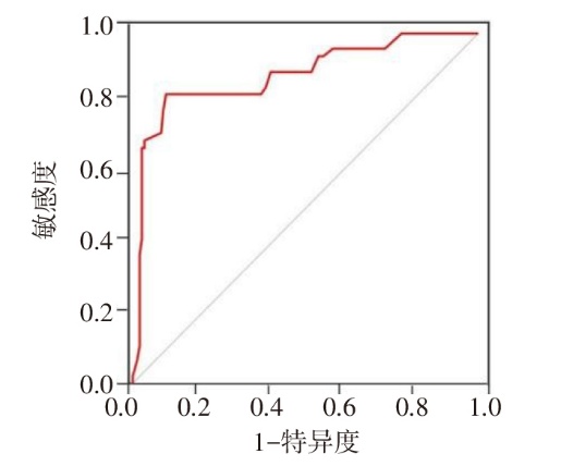
Fig.1 ROC curve diagram for the risk assessment model of high malignant potential in GIST patients
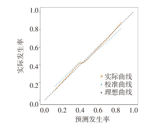
Fig.2 The calibration curve of the model for assessing the high malignant potential of GIST patients
| [1] | SHARMA A K, KIM T S, BAUER S, et al. Gastrointestinal stromal tumor:new insights for a multimodal approach[J]. Surg Oncol Clin N Am, 2022, 31(3):431-446. doi:10.1016/j.soc.2022.03.007. |
| [2] | CICALA C M, BAUER S, HEINRICH M C, et al. Gastrointestinal stromal tumor:current approaches and future directions in the treatment of advanced disease[J]. Hematol Oncol Clin North Am, 2025, 39(4):773-784. doi:10.1016/j.hoc.2025.04.006. |
| [3] | 杨易, 冯峰. 影像组学在宫颈癌异质性分析中的研究进展[J]. 放射学实践, 2020, 35(4):564-568. |
| YANG Y, FENG F. Research progress of radiomics in heterogeneity analysis of cervical cancer[J]. Radiologic Practice, 2020, 35(4):564-568. doi:10.13609/j.cnki.1000-0313.2020.04.032. | |
| [4] | ASLANIAN H R, MUNIRAJ T, NAGAR A, et al. Endoscopic ultrasound in cancer staging[J]. Gastrointest Endosc Clin N Am, 2024, 34(1):37-49. doi:10.1016/j.giec.2023.09.009. |
| [5] | XIA F, GUO F, LIU Z, et al. Enhanced CT combined with texture analysis for differential diagnosis of pleomorphic adenoma and adenolymphoma[J]. BMC Med Imaging, 2023, 23(1):169. doi:10.1186/s12880-023-01129-9. |
| [6] | 《胃肠间质瘤病理诊断临床实践指南(2022版)》编写专家委员会(2022版). 胃肠间质瘤病理诊断临床实践指南(2022版)[J]. 中华病理学杂志, 2022, 51(10):959-969. |
| Expert Committee for Clinical Practice Guidelines for the Pathological Diagnosis of Gastrointestinal Stromal Tumors(2022 version). Clinical practice guideline for the pathological diagnosis of gastrointestinal stromal tumor(2022 version)[J]. Chin J Pathol, 2022, 51(10):959-969. doi:10.3760/cma.j.cn112151-20220512-00405. | |
| [7] | JANG S H, KWON J E, KIM J H, et al. Prediction of tumor recurrence in patients with non-gastric gastrointestinal stromal tumors following resection according to the Modified National Institutes of Health Criteria[J]. Intest Res, 2014, 12(3):229-235. doi:10.5217/ir.2014.12.3.229. |
| [8] | STRAUSS G, GEORGE S. Gastrointestinal stromal tumors[J]. Curr Oncol Rep, 2025, 27(3):312-321. doi:10.1007/s11912-025-01636-8. |
| [9] | ZHU H, YANG G, MA Y, et al. Update of epidemiology,survival and initial treatment in patients with gastrointestinal stromal tumour in the USA:a retrospective study based on SEER database[J]. BMJ Open, 2023, 13(7):e072945. doi:10.1136/bmjopen-2023-072945. |
| [10] | SUN X, SUN Y, XIONG R, et al. Hidden risk factors for recurrence of jejunoileal gastrointestinal stromal tumors with low NIH classification: implications for surveillance and adjuvant therapy[J]. J Transl Med, 2025, 23(1):1156. doi:10.1186/s12967-025-07163-1. |
| [11] | 刘东鑫, 余荣国, 徐超. 超声内镜对胃肠道间质瘤诊断价值的荟萃分析[J]. 内科理论与实践, 2022, 17(1):63-69. |
| LIU D X, YU R G, XU C. Diagnostic value of endoscopic ultrasonography in assessment of gastrointestinal stromal tumors: a meta-analysis[J]. J Intern Med Concepts Pract, 2022, 17(1):63-69. doi:10.16138/j.1673-6087.2022.01.012. | |
| [12] | 蒋颖, 闫骏. 胃肠道间质瘤术前及术后诊断的研究进展[J]. 南通大学学报(医学版), 2020, 40(3):272-275. |
| JIANG Y, YAN J. Research progress of preoperative and postoperative diagnosis of gastrointestinal stromal tumor[J]. Journal of Nantong University(Medical Sciences), 2020, 40(3):272-275. doi:10.16424/j.cnki.cn32-1807/r.2020.03.020. | |
| [13] | ZHANG Y, HUANG Z X, SONG B. Role of imaging in evaluating the response after neoadjuvant treatment for pancreatic ductal adenocarcinoma[J]. World J Gastroenterol, 2021, 27(22):3037-3049. doi:10.3748/wjg.v27.i22.3037. |
| [14] | SATO R, HARADA R, HASHIMOTO K, et al. Gastrointestinal stromal tumors in the duodenum show increased contrast enhancement compared with those in the stomach on computed tomography[J]. Mol Clin Oncol, 2022, 17(4):144. doi:10.3892/mco.2022.2577. |
| [15] | 罗泰钊, 王盛集, 吴惠凤, 等. 胃肠道间质瘤MSCT影像特征与危险度分级的相关性分析[J]. 影像研究与医学应用, 2024, 8(8):60-62. |
| LUO T Z, WANG S J, WU H F, et al. Correlation analysis of MSCT imaging features of gastrointestinal stromal tumors and risk grading[J]. Journal of Imaging Research and Medical Applications, 2024, 8(8):60-62. doi:10.3969/j.issn.2096-3807.2024.08.018. | |
| [16] | 马冲菲. 基于增强CT图像的不同影像组学模型在预测胃间质瘤的危险度分级的应用[D]. 石家庄: 河北医科大学, 2021. |
| MA C F. The application of different radiomics models based on enhanced CT images in predicting the risk grade of gastric stromal tumors[D]. Shijiazhuang: Hebei Medical University, 2021. | |
| [17] | DOITA S, WATANABE M, OGAWA T, et al. Utility of follow-up computed tomography (CT)and fluorodeoxyglucose-positron emission tomography/CT(FDG-PET/CT)in diagnosing gastrointestinal stromal tumor(GIST)presenting as spontaneous hemoperitoneum: a case report[J]. Cureus, 2024, 16(9):e69799. doi:10.7759/cureus.69799. |
| [18] | 宋时东. 胃肠道间质瘤的CT影像学特征及其诊断价值分析[J]. 影像研究与医学应用, 2024, 8(19):119-121,124. |
| SONG S D. Analysis of CT imaging characteristics and diagnostic value of gastrointestinal stromal tumor[J]. Journal of Imaging Research and Medical Applications, 2024, 8(19):119-121,124. doi:10.3969/j.issn.2096-3807. | |
| [19] | 王燕. 增强CT及超声内镜对胃肠道间质瘤术前危险度评估及诊断价值分析[D]. 兰州: 兰州大学, 2021. |
| WANG Y. Analysis of the diagnostic value of enhanced CT and endoscopic ultrasound in the preoperative risk assessment of gastrointestinal stromal tumors[D]. Lanzhou: Lanzhou University, 2021. | |
| [20] | 吴琼, 张瑾, 粱田, 等. 基于CT特征的列线图模型对GIST核分裂象数的预测价值[J]. 临床放射学杂志, 2024, 43(4):610-614. |
| WU Q, ZHANG J, LIANG T, et al. The value of the nomogram based on CT feature for predicting the mitotic index of gastrointestinal stromal tumorss[J]. Journal of Clinical Radiology, 2024, 43(4):610-614. doi:10.13437/j.cnki.jcr.2024.04.037. | |
| [21] | 陈长波, 马俊, 兰小惠. 胃肠道间质瘤CT诊断及与病理肿瘤恶性潜能相关性分析[J]. 中国CT和MRI杂志, 2022, 20(6):156-158. |
| CHEN C B, MA J, LAN X H. CT diagnosis of gastrointestinal stromal tumors(GISTs)and its correlation with pathological malignant potential of tumor[J]. Chinese Journal of CT and MRI, 2022, 20(6):156-158. doi:10.3969/j.issn.1672-5131.2022.06.051. | |
| [22] | WANG T T, LIU W W, LIU X H, et al. Relationship between multi-slice computed tomography features and pathological risk stratification assessment in gastric gastrointestinal stromal tumors[J]. World J Gastrointest Oncol, 2023, 15(6):1073-1085. doi:10.4251/wjgo.v15.i6.1073. |
| Viewed | ||||||
|
Full text |
|
|||||
|
Abstract |
|
|||||
