Tianjin Medical Journal ›› 2022, Vol. 50 ›› Issue (9): 948-952.doi: 10.11958/20220168
• Clinical Research • Previous Articles Next Articles
ZHU Yunlin1,2( ), YIN Guangli3,△(), DAI Pingtao3
Received:2022-01-28
Revised:2022-03-26
Published:2022-09-15
Online:2022-09-05
Contact:
YIN Guangli
E-mail:scuky2011@126.com;443539894@qq.com
ZHU Yunlin, YIN Guangli, DAI Pingtao. Evaluation value of carotid ultrasound combined with serum HbA1c and FIB in acute anterior circulation cerebral infarction[J]. Tianjin Medical Journal, 2022, 50(9): 948-952.
CLC Number:
| 组别 | n | PSV (cm/s) | EDV (cm/s) | IMT (mm) | HbA1c (%) | FIB (g/L) |
|---|---|---|---|---|---|---|
| 对照组 | 50 | 69.1±8.3 | 18.59±4.37 | 0.94±0.10 | 7.83±1.54 | 2.83±0.51 |
| 脑梗死组 | 93 | 77.8±8.9 | 23.05±3.02 | 1.12±0.14 | 9.33±1.03 | 4.01±1.08 |
| t | 5.689** | 7.160** | 8.189** | 6.937** | 7.294** |
Tab.1 Comparison of carotid artery ultrasound examination indexes and serum HbA1c and FIB levels between the two groups
| 组别 | n | PSV (cm/s) | EDV (cm/s) | IMT (mm) | HbA1c (%) | FIB (g/L) |
|---|---|---|---|---|---|---|
| 对照组 | 50 | 69.1±8.3 | 18.59±4.37 | 0.94±0.10 | 7.83±1.54 | 2.83±0.51 |
| 脑梗死组 | 93 | 77.8±8.9 | 23.05±3.02 | 1.12±0.14 | 9.33±1.03 | 4.01±1.08 |
| t | 5.689** | 7.160** | 8.189** | 6.937** | 7.294** |
| 指标 | β | SE | Wald χ2 | P | OR | OR 95%CI |
|---|---|---|---|---|---|---|
| PSV | 0.076 | 0.044 | 2.939 | 0.086 | 1.079 | 0.989~1.176 |
| EDV | 0.364 | 0.108 | 11.363 | 0.001 | 1.438 | 1.164~1.777 |
| IMT | 10.716 | 3.068 | 12.198 | <0.001 | 45 092.968 | 110.241~1.844×10⁷ |
| HbA1c | 1.243 | 0.332 | 14.053 | <0.001 | 3.466 | 1.810~6.639 |
| FIB | 1.285 | 0.641 | 4.014 | 0.045 | 3.615 | 1.028~12.710 |
| 常量 | -37.976 | 7.457 | 25.934 | <0.001 | <0.001 | - |
Tab.2 Analysis of influencing factors of cerebral infarction
| 指标 | β | SE | Wald χ2 | P | OR | OR 95%CI |
|---|---|---|---|---|---|---|
| PSV | 0.076 | 0.044 | 2.939 | 0.086 | 1.079 | 0.989~1.176 |
| EDV | 0.364 | 0.108 | 11.363 | 0.001 | 1.438 | 1.164~1.777 |
| IMT | 10.716 | 3.068 | 12.198 | <0.001 | 45 092.968 | 110.241~1.844×10⁷ |
| HbA1c | 1.243 | 0.332 | 14.053 | <0.001 | 3.466 | 1.810~6.639 |
| FIB | 1.285 | 0.641 | 4.014 | 0.045 | 3.615 | 1.028~12.710 |
| 常量 | -37.976 | 7.457 | 25.934 | <0.001 | <0.001 | - |
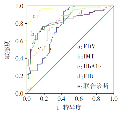
Fig.1 ROC analysis of EDV, IMT, HbA1c and FIB in the diagnosis of cerebral infarction
| 指标 | AUC及95%CI | 截断值 | 敏感度 (%) | 特异度 (%) | 约登 指数 |
|---|---|---|---|---|---|
| EDV | 0.770(0.683~0.857) | 19.6 cm/s | 93.50 | 58.00 | 0.515 |
| IMT | 0.852(0.788~0.917) | 1.02 mm | 73.10 | 84.00 | 0.571 |
| HbA1c | 0.799(0.717~0.880) | 8.42% | 82.80 | 70.00 | 0.528 |
| FIB | 0.829(0.762~0.896) | 3.04 g/L | 76.30 | 82.00 | 0.583 |
| 联合诊断 | 0.970(0.947~0.992) | - | 83.90 | 96.00 | 0.799 |
Tab.3 Analysis of values of EDV, IMT, HbA1c and FIB in the diagnosis of cerebral infarction
| 指标 | AUC及95%CI | 截断值 | 敏感度 (%) | 特异度 (%) | 约登 指数 |
|---|---|---|---|---|---|
| EDV | 0.770(0.683~0.857) | 19.6 cm/s | 93.50 | 58.00 | 0.515 |
| IMT | 0.852(0.788~0.917) | 1.02 mm | 73.10 | 84.00 | 0.571 |
| HbA1c | 0.799(0.717~0.880) | 8.42% | 82.80 | 70.00 | 0.528 |
| FIB | 0.829(0.762~0.896) | 3.04 g/L | 76.30 | 82.00 | 0.583 |
| 联合诊断 | 0.970(0.947~0.992) | - | 83.90 | 96.00 | 0.799 |
| 组别 | n | PSV(cm/s) | EDV(cm/s) | IMT(mm) | HbA1c(%) | FIB(g/L) |
|---|---|---|---|---|---|---|
| 轻度组 | 37 | 75.9±7.2 | 22.35±2.11 | 1.02±0.10 | 9.15±0.96 | 3.35±0.91 |
| 中度组 | 34 | 77.9±9.1 | 23.79±3.74 | 1.17±0.13a | 9.01±1.17 | 4.03±1.02a |
| 重度组 | 22 | 80.9±10.7 | 23.08±2.92 | 1.22±0.10a | 10.10±0.16ab | 5.06±0.46ab |
| F | 2.259 | 2.083 | 25.089** | 10.134** | 26.177** |
Tab.4 Comparison of carotid artery ultrasound examination indexes and serum HbA1c and FIB levels between the three groups of patients with different conditions of cerebral infarction
| 组别 | n | PSV(cm/s) | EDV(cm/s) | IMT(mm) | HbA1c(%) | FIB(g/L) |
|---|---|---|---|---|---|---|
| 轻度组 | 37 | 75.9±7.2 | 22.35±2.11 | 1.02±0.10 | 9.15±0.96 | 3.35±0.91 |
| 中度组 | 34 | 77.9±9.1 | 23.79±3.74 | 1.17±0.13a | 9.01±1.17 | 4.03±1.02a |
| 重度组 | 22 | 80.9±10.7 | 23.08±2.92 | 1.22±0.10a | 10.10±0.16ab | 5.06±0.46ab |
| F | 2.259 | 2.083 | 25.089** | 10.134** | 26.177** |
| 指标 | β | SE | Wald χ2 | P | OR | OR 95%CI |
|---|---|---|---|---|---|---|
| PSV | 0.043 | 0.026 | 2.765 | 0.096 | 1.044 | 0.992~1.098 |
| EDV | 0.138 | 0.077 | 3.193 | 0.074 | 1.148 | 0.987~1.336 |
| IMT | 13.676 | 2.850 | 23.022 | <0.001 | 8.694×10⁵ | 3.259×10³~2.319×10⁸ |
| HbA1c | 0.284 | 0.211 | 1.808 | 0.179 | 1.328 | 0.878~2.008 |
| FIB | 1.107 | 0.257 | 18.503 | <0.001 | 3.025 | 1.827~5.010 |
Tab.5 Univariate Logistic regression analysis of moderate and severe cerebral infarction
| 指标 | β | SE | Wald χ2 | P | OR | OR 95%CI |
|---|---|---|---|---|---|---|
| PSV | 0.043 | 0.026 | 2.765 | 0.096 | 1.044 | 0.992~1.098 |
| EDV | 0.138 | 0.077 | 3.193 | 0.074 | 1.148 | 0.987~1.336 |
| IMT | 13.676 | 2.850 | 23.022 | <0.001 | 8.694×10⁵ | 3.259×10³~2.319×10⁸ |
| HbA1c | 0.284 | 0.211 | 1.808 | 0.179 | 1.328 | 0.878~2.008 |
| FIB | 1.107 | 0.257 | 18.503 | <0.001 | 3.025 | 1.827~5.010 |
| 指标 | β | SE | Wald χ2 | P | OR | OR 95%CI |
|---|---|---|---|---|---|---|
| IMT | 11.630 | 2.949 | 15.554 | <0.001 | 1.124×10⁵ | 347.276~3.638×10⁷ |
| FIB | 0.759 | 0.294 | 6.660 | 0.010 | 2.136 | 1.200~3.802 |
| 常量 | -15.305 | 3.292 | 21.609 | <0.001 | - | - |
Tab.6 Multivariate Logistic regression analysis of moderate and severe cerebral infarction
| 指标 | β | SE | Wald χ2 | P | OR | OR 95%CI |
|---|---|---|---|---|---|---|
| IMT | 11.630 | 2.949 | 15.554 | <0.001 | 1.124×10⁵ | 347.276~3.638×10⁷ |
| FIB | 0.759 | 0.294 | 6.660 | 0.010 | 2.136 | 1.200~3.802 |
| 常量 | -15.305 | 3.292 | 21.609 | <0.001 | - | - |
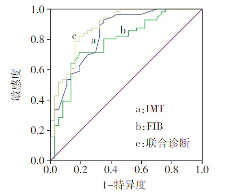
Fig.2 Analysis of values of IMT, FIB and their combined evaluation of moderate and severe cerebral infarction
| 指标 | AUC及95%CI | 截断值 | 敏感度 (%) | 特异度 (%) | 约登 指数 |
|---|---|---|---|---|---|
| IMT | 0.840(0.758~0.923) | 1.03 mm | 92.90 | 64.90 | 0.578 |
| FIB | 0.784(0.687~0.880) | 3.90 g/L | 71.40 | 81.10 | 0.525 |
| 联合诊断 | 0.882(0.809~0.954) | - | 82.10 | 81.10 | 0.632 |
Tab.7 Analysis of values of IMT, FIB and their combination in the evaluation of moderate and severe cerebral infarction
| 指标 | AUC及95%CI | 截断值 | 敏感度 (%) | 特异度 (%) | 约登 指数 |
|---|---|---|---|---|---|
| IMT | 0.840(0.758~0.923) | 1.03 mm | 92.90 | 64.90 | 0.578 |
| FIB | 0.784(0.687~0.880) | 3.90 g/L | 71.40 | 81.10 | 0.525 |
| 联合诊断 | 0.882(0.809~0.954) | - | 82.10 | 81.10 | 0.632 |
| [1] | LONG H, ZENG X, LIU Q, et al. Burden of osteoarthritis in China,1990-2017:Findings from the global burden of disease study 2017[J]. Lancet Rheumatol, 2020, 2(3):e164-e172. doi: 10.1186/s12916-021-01905-w. |
| [2] | 国家卫生健康委员会. 2019年中国卫生健康统计提要[M]. 北京: 中国协和医科大学出版社, 2019:10. |
| National Health Commission. China Health Statistical Digest[M]. Beijing: China Union Medical College Press, 2019:10. | |
| [3] | KNIGSTEIN K, SCHENCK U V, BÜSCHGES JC, et al. Carotid IMT and stiffness in the KiGGS 2 national survey:Third-generation measurement, quality algorithms and determinants of completeness[J]. Ultrasound Med Biol, 2021, 47(2):296-308. doi: 10.1016/j.ultrasmedbio.2020.10.015. |
| [4] | 梁蓉. 颈动脉超声联合血清五聚素3、脂蛋白相关磷脂酶A2检测对动脉粥样硬化脑梗死的诊断价值[J]. 中国动脉硬化杂志, 2019, 10(9):791-795. |
| LIANG R. Diagnostic value of carotid ultrasonography combined with serum pentraxin 3 and lipoprotein-associated phospholipase A2 in atherosclerotic cerebral infarction[J]. Chin J Arterioscls, 2019, 10(9):791-795. doi: 10.3969/j.issn.1007-3949.2019.09.012. | |
| [5] | POPNICOLAE O. Study of the relationship between glycosylated hemoglobin,Homa IR and serum glucose in acute ischemic stroke in the diabetic patient[J]. Int Med, 2020, 17(2):17-21. doi: 10.2478/inmed-2020-0106. |
| [6] | ROSSELLO X, RAPOSEIRAS-ROUBIN S, OLIVA B, et al. Glycated hemoglobin and subclinical atherosclerosis in people without diabetes[J]. J Am Coll Cardiol, 2021, 77(22):2777-2791. doi: 10.1016/j.jacc.2021.03.335. |
| [7] | MANERS J, GILL D, PANKRATZ N, et al. A Mendelian randomization of γ′ and total fibrinogen levels in relation to venous thromboembolism and ischemic stroke[J]. Blood, 2020, 136(26):445-447. doi: 10.1182/blood.2019004781. |
| [8] | 王怡萌, 栾波, 郭鹏, 等. 血脂沉积指数与冠心病动脉粥样硬化的相关性研究[J]. 中国动脉硬化杂志, 2020, 10(7):552-554. |
| WANG Y M, LUAN B, GUO P, et al. Study on the correlation between blood lipid deposition index and coronary heart disease atherosclerosis[J]. Chin J Arterioscl, 2020, 10(7):552-554. doi: 10.3969/j.issn.1007-3949.2020.07.007. | |
| [9] | GOLDSTEIN L B, SAMSA G P. Reliability of the National Institutes of Health Stroke Scale. Extension to non-neurologists in the context of a clinical trial[J]. Stroke, 1997, 28(2):307-310. doi: 10.1161/01.STR.28.2.307. |
| [10] | NORBY F L, ALONSO A, ROONEY M R, et al. Association of ventricular arrhythmias with dementia:the atherosclerosis risk in communities (ARIC) study[J]. Neurology, 2020, 96(6):665-668. doi: 10.1212/WNL.0000000000011122. |
| [11] | CATALANO O, BENDOTTI G, MORI A, et al. Evolving determinants of carotid atherosclerosis vulnerability in asymptomatic patients from the MAGNETIC observational study[J]. Sci Rep, 2021, 11(1):2327. doi: 10.1038/s41598-021-81247-y. |
| [12] | 周利, 徐莉, 邵汝升, 等. 颈动脉超声定量参数联合血清miR-128b,miR-146a对急性缺血性脑卒中的诊断价值及对预后的评价意义[J]. 影像科学与光化学, 2020, 39(3):6-8. |
| ZHOU L, XU L, SHAO R S, et al. The diagnostic value of carotid ultrasound quantitative parameters combined with serum miR-128b and miR-146a in acute ischemic stroke and the significance of prognostic evaluation[J]. Imag Sci Photochem, 2020, 39(3):6-8. doi: 10.3969/j.issn.1673-5110.2017.07.004. | |
| [13] | RICE C J, CHO S M, STROHM T, et al. Ultrasound criteria for assessment of vertebral artery origins[J]. J Neur, 2019, 30(1):222-225. doi: 10.1016/j.jacc.2011.02.064. |
| [14] | TIGKIROPOULOS K, KARAMANOS D, STAVRIDIS K, et al. Endovascular stent-graft repair of combined renal artery aneurysm and arteriovenous fistula[J]. Ann Vascul Surg, 2018, 55(2):325-329. doi: 10.1016/j.avsg.2018.07.054. |
| [15] | WANG M Y, WANG S Y, WANG X W, et al. Carotid intima-media thickness,genetic risk,and ischemic stroke:A family-based study in rural China[J]. Int J Environm Res Publ Health, 2020, 18(1):119. doi: 10.3390/ijerph18010119. |
| [16] | ZHOU P Y, SHEN Y, WANG L Y, et al. Association between carotid intima media thickness and small dense low-density lipoprotein cholesterol in acute ischaemic stroke[J]. Lipids Health Dis, 2020, 19(1):441-447. doi: 10.1186/s12944-020-01353-0. |
| [17] | MESA A, COFÁN M, ESMATJES E, et al. Biomarkers of fatty acid intake are independently associated with preclinical atherosclerosis in individuals with type 1 diabetes[J]. Eur J Nutr, 2021, 60(8):4595-4605. doi: 10.1007/s00394-021-02611-2. |
| [18] | 潘勇, 吴非, 骆文静, 等. 糖尿病与颅内动脉多支狭窄的相关性研究[J]. 临床神经病学杂志, 2019, 32(3):181-184. |
| PAN Y, WU F, LUO W J, et al. Correlation between diabetes mellitus and intracranial multi-arterial stenosis[J]. J Clin Neurol, 2019, 32(3):181-184. doi: 10.3969/j.issn.1004-1648.2019.03.008. | |
| [19] | REN J, DONG X, NAO J. Serum cystatin C is associated with carotid atherosclerosis in patients with acute ischemic stroke[J]. Neurol Sci, 2020, 41(5):445. doi: 10.1007/s10072-020-04383-9. |
| [20] | 胡玉海, 田佩孚. 血清CK-MB和FIB水平与急性心肌梗死患者的冠状动脉狭窄程度关联[J]. 基因组学与应用生物学, 2019, 38(4):1864-1868. |
| HU Y H, TIAN P F. Serum levels of CK-MB and FIB associated with the degree of coronary artery stenosis in patients with acute myocardial infarction[J]. Genom Appl Biol, 2019, 38(4):1864-1868. doi: 10.13417/j.gab.038.001864. | |
| [21] | 谭万江, 刘书红, 高伟, 等. 阿托伐他汀钙片对冠心病患者小而密低密度脂蛋白胆固醇水平的影响[J]. 中国临床药理学杂志, 2020, 36(2):103-105,117. |
| TAN W J, LIU S H, GAO W, et al. Effect of atorvastatin calcium on small dense low density lipoprotein cholesterol in patients with coronary heart disease[J]. Chin J Clin Pharmacol, 2020, 36(2):103-105,117. doi: 10.13699/j.cnki.1001-6821.2020.02.002. | |
| [22] | LIU Y, ZHU J, DENG X, et al. Serum level of lipoprotein-associated phospholipase A2 is a potential biomarker of vertebrobasilar dolichoectasia and its progression to cerebral infarction[J]. Neurol Sci, 2020, 4(1):552-557. doi: 10.1007/s10072-020-04563-7. |
| [23] | 许莉, 李慧英. 脑梗死急性期患者糖化血红蛋白和纤维蛋白原水平与认知功能障碍的关系[J]. 中华高血压杂志, 2019, 27(1):100. |
| XU L, LI H Y. The relationship between glycosylated hemoglobin and fibrinogen levels and cognitive dysfunction in patients with acute cerebral infarction[J]. Chin J Hypert, 2019, 27(1):100. doi: 10.16439/j.cnki.1673-7245.2019.01.032. | |
| [24] | 栗静, 田婷, 石正洪, 等. 纤维蛋白原、C反应蛋白及同型半胱氨酸与大动脉粥样硬化型卒中患者颈动脉易损性斑块的相关性分析[J]. 解放军医学杂志, 2017, 42(1):41-46. |
| LI J, TIAN T, SHI Z H, et al. Correlation analysis of fibrinogen, C-reactive protein and homocysteine with vulnerable carotid plaque in patients with large atherosclerotic stroke[J]. PLA Med J, 2017, 42(1):41-46. doi: 10.11855/j.issn.0577-7402.2017.01.08. |
| Viewed | ||||||
|
Full text |
|
|||||
|
Abstract |
|
|||||