Tianjin Medical Journal ›› 2024, Vol. 52 ›› Issue (10): 1100-1105.doi: 10.11958/20231935
• Applied Research • Previous Articles Next Articles
WANG Zhong( ), ZHAO Jingwen, WANG Tianchi, TANG Ying△()
Received:2023-12-18
Revised:2024-06-28
Published:2024-10-15
Online:2024-10-14
Contact:
△ E-mail:WANG Zhong, ZHAO Jingwen, WANG Tianchi, TANG Ying. The application value of ultrasound radiomics in the histological classification of nephritis[J]. Tianjin Medical Journal, 2024, 52(10): 1100-1105.
CLC Number:
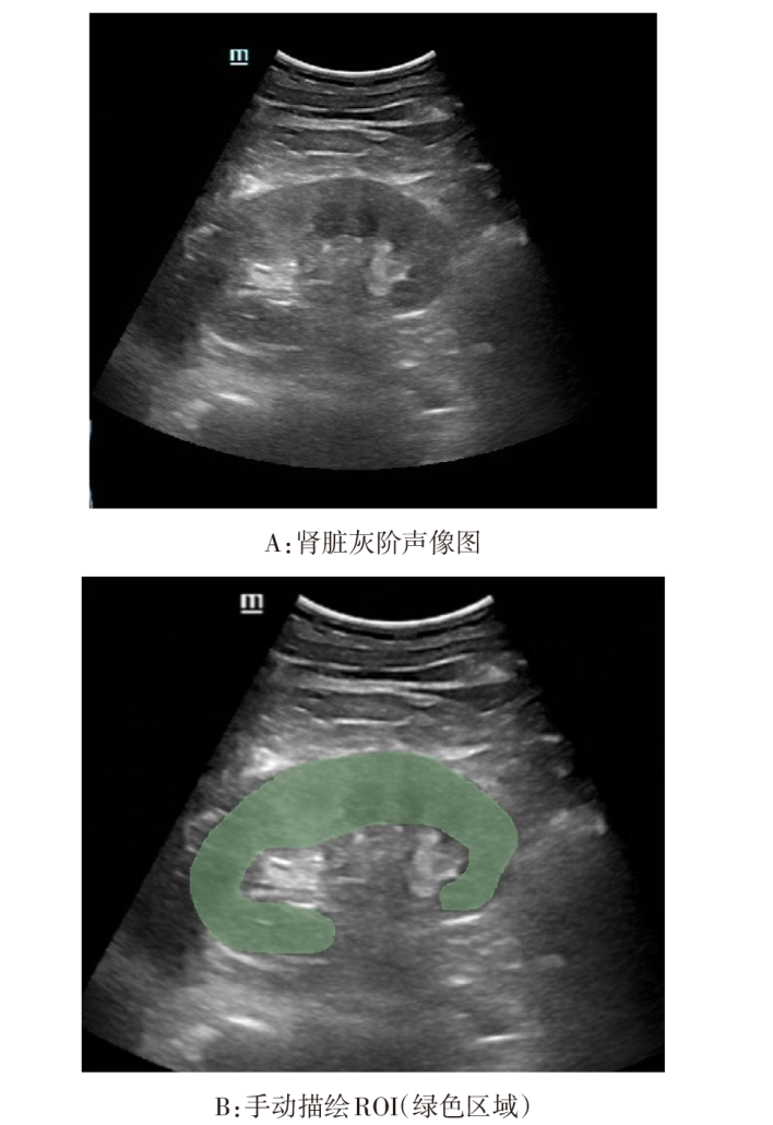
Fig.1 Selection of standard 2D grayscale images and delineation of ROI
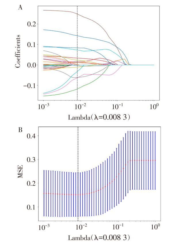
Fig.2 Omics feature screening
| 模型 | 数据集 | AUC(95%CI) | 敏感度 | 特异度 |
|---|---|---|---|---|
| LR | 训练集 | 0.956(0.940~0.973) | 0.872 | 0.944 |
| 验证集 | 0.944(0.916~0.973) | 0.878 | 0.867 | |
| D | 0.247 | |||
| KNN | 训练集 | 0.968(0.956~0.980) | 0.842 | 0.944 |
| 验证集 | 0.908(0.870~0.946) | 0.569 | 0.989 | |
| D | 1.582 | |||
| SVM | 训练集 | 0.976(0.961~0.990) | 0.936 | 0.929 |
| 验证集 | 0.943(0.914~0.972) | 0.886 | 0.856 | |
| D | 1.017 | |||
| RF | 训练集 | 0.948(0.929~0.968) | 0.909 | 0.888 |
| 验证集 | 0.914(0.875~0.953) | 0.821 | 0.878 | |
| D | 0.963 |
Tab.1 Comparison of the prediction performance of the four models
| 模型 | 数据集 | AUC(95%CI) | 敏感度 | 特异度 |
|---|---|---|---|---|
| LR | 训练集 | 0.956(0.940~0.973) | 0.872 | 0.944 |
| 验证集 | 0.944(0.916~0.973) | 0.878 | 0.867 | |
| D | 0.247 | |||
| KNN | 训练集 | 0.968(0.956~0.980) | 0.842 | 0.944 |
| 验证集 | 0.908(0.870~0.946) | 0.569 | 0.989 | |
| D | 1.582 | |||
| SVM | 训练集 | 0.976(0.961~0.990) | 0.936 | 0.929 |
| 验证集 | 0.943(0.914~0.972) | 0.886 | 0.856 | |
| D | 1.017 | |||
| RF | 训练集 | 0.948(0.929~0.968) | 0.909 | 0.888 |
| 验证集 | 0.914(0.875~0.953) | 0.821 | 0.878 | |
| D | 0.963 |
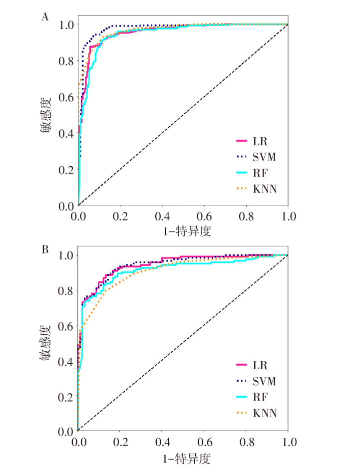
Fig.3 ROC curves for four predictive models

Fig.4 GiViTI calibration curves predicted by LR and SVM
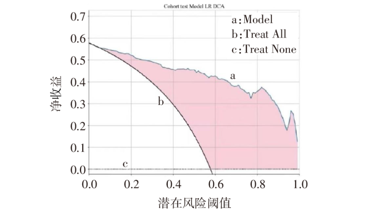
Fig.5 DCA curves for LR predictive models
| [1] | WU J, DUAN S W, YANG H T, et al. Efficacy and safety of Shenyankangfu Tablet,a Chinese patent medicine,for primary glomerulonephritis:A multicenter randomized controlled trial[J]. J Integr Med, 2021, 19(2):111-119. doi:10.1016/j.joim.2021.01.009. |
| [2] | HU R, QUAN S, WANG Y, et al. Spectrum of biopsy proven renal disease in Central China:a 10-year retrospective study based on 34,630 cases[J]. Sci Rep, 2020, 10(1):10994. doi:10.1038/s41598-020-67910-w. |
| [3] | LAMBIN P, RIOSVELAZQUEZ E, LEIJENAAR R, et al. Radiomics:Extracting more information from medical images using advanced feature analysis[J]. EJC, 2012, 48(4):441-446. doi:10.1016/j.ejca.2011.11.036. |
| [4] | 杨丽勤, 王希明. MRI影像组学用于前列腺癌包膜外侵犯的研究进展[J]. 国际医学放射学杂志, 2023, 46(2):202-206. |
| YANG L Q, WANG X M. Research progress of MRI radiomics in assessing extracapsular extension of prostate cancer[J]. Int J Med Radiol, 2023, 46(2):202-206. doi:10.19300/j.2023.Z20177. | |
| [5] | 王天驰, 王众, 牛宁宁, 等. 超声影像组学对移植肾实质性病变鉴别诊断的价值[J]. 天津医药, 2023, 51(6):653-657. |
| WANG T C, WANG Z, NIU N N, et al. The value of ultrasonography in the differential diagnosis of parenchymal lesions of transplanted kidney[J]. Tianjin Med J, 2023, 51(6):653-657. doi:10.11958/20230025. | |
| [6] | 周瑾, 常才, 周世崇. 多模态超声组学在乳腺癌术前诊断中的研究进展[J]. 肿瘤影像学, 2021, 30(5):327-331. |
| ZHOU J, CHANG C, ZHOU S C. The progress of multimodal ultrasound-radiomics in the preoperative diagnosis of breast cancer[J]. Oncoradiology, 2021, 30(5):327-331. doi:10.19732/j.cnki.2096-6210.2021.05.002. | |
| [7] | 淡一波, 陶虹月, 王一达, 等. 基于预建模的影像组学特征选择方法[J]. 信息技术, 2022(4):1-6,12. |
| DAN Y B, TAO H Y, WANG Y D, et al. Feature selection method of radiomics based on scout models[J]. Information Technology, 2022(4):1-6, 12. doi:10.13274/j.cnki.hdzy.2022.04.001. | |
| [8] | 吴昕怡, 包呼和. 纹理分析在超声医学中的应用研究现状[J]. 影像研究与医学应用, 2020, 4(20):5-7. |
| WU X Y, BAO H H. The status quo of the application of texture analysis in ultrasound medicine[J]. Journal of Imaging Research and Medical Applications, 2020, 4(20):5-7. | |
| [9] | 杨熠, 钱旭升, 周志勇, 等. 采用影像组学的肾肿瘤组织学亚型分类[J]. 浙江大学学报(工学版), 2019, 53(12):2381-2388. |
| YANG Y, QIAN X S, ZHOU Z Y, et al. Classification of renal tumor histology subtypes using radiomics[J]. Journal of Zhejiang University(Engineering Science), 2019, 53(12):2381-2388. doi:10.3785/j.issn.1008-973X.2019.12.016. | |
| [10] | ZHANG L, CHEN Z, FENG L, et al. Preliminary study on the application of renal ultrasonography radiomics in the classification of glomerulopathy[J]. BMC Med Imaging, 2021, 21(1):115. doi:10.1186/s12880-021-00647-8. |
| [11] | 陈剑霖, 付睿, 陈青, 等. 基于逻辑回归算法的移植肾功能延迟恢复发生风险因素分析及预测模型的建立[J]. 实用器官移植电子杂志, 2023, 11(5):457-463. |
| CHEN J L, FU R, CHEN Q, et al. Analysis of risk factors and prediction model of delayed recovery of transplanted kidney function based on logistic regression algorithm[J]. Prac J Organ Transplant(Electronic Version), 2023, 11(5):457-463. doi:10.3969/j.issn.2095-5332.2023.05.013. | |
| [12] | 王芳, 夏雨薇, 柴象飞, 等. 影像组学分析流程及临床应用的研究进展[J]. 中华解剖与临床杂志, 2021, 26(2):236-241. |
| WANG F, XIA Y W, CHAI X F, et al. Analysis process and clinical application of radiomics[J]. Chin J Anat Clin, 2021, 26(2):236-241. doi:10.3760/cma.j.cn101202-20200701-00200. | |
| [13] | SUAREZ-IBARROlA R, BASULTO-MARTINEZ M, HEINZE A, et al. Radiomics applications in renal tumor assessment:A comprehensive review of the literature[J]. Cancers(Basel), 2020, 12(6):1387. doi:10.3390/cancers12061387. |
| [14] | LIM E J, YEN J, FONG K Y, et al. Radiomics in Kidney Transplantation:A scoping review of current applications,limitations,and future directions[J]. Transplantation, 2024, 108(3):643-653. doi:10.1097/TP.0000000000004711. |
| [15] | LI C, QIAO G, LI J, et al. An ultrasonic-based radiomics nomogram for distinguishing between benign and malignant solid renal masses[J]. Front Oncol, 2022, 12:847805. doi:10.3389/fonc.2022.847805. |
| [16] | YANG Y, CHEN F, LIANG H, et al. CNN-based automatic segmentations and radiomics feature reliability on contrast-enhanced ultrasound images for renal tumors[J]. Front Oncol, 2023, 13:1166988. doi:10.3389/fonc.2023.1166988. |
| [17] | CHEN Z, YING M T C, WANG Y L, et al. Ultrasound‑based radiomics analysis in the assessment of renal fibrosis in patients with chronic kidney disease[J]. Abdom Radiol(NY), 2023, 48(8):2649-2657. doi:10.1007/s00261-023-03965-3. |
| [18] | 刘清华, 刘恺怡, 侯淳容. 梅州地区近10年肾穿病理活检结果分析[J]. 广东药科大学学报, 2021, 37(4):122-126. |
| LIU Q H, LIU K Y, HOU C R. Analysis of renal biopsy results in Meizhou area in recent 10 years[J]. Journal of Guangdong Pharmaceutical University, 2021, 37(4):122-126. doi:10.16809/j.cnki.2096-3653.2021041903. |
| Viewed | ||||||
|
Full text |
|
|||||
|
Abstract |
|
|||||