
Tianjin Medical Journal ›› 2026, Vol. 54 ›› Issue (6): 570-576.doi: 10.11958/20251716
• Cell and Molecular Biology • Previous Articles Next Articles
DENG Xinyi1( ), YANG Chen1,2, CAO Jianlin1,3, YANG Lei4, TAN Jian1, JIA Qiang1, MENG Zhaowei1,5,△()
Received:2025-08-07
Revised:2025-12-20
Published:2026-06-15
Online:2026-06-15
Contact:
△E-mail:zmeng@tmu.edu.cn
DENG Xinyi, YANG Chen, CAO Jianlin, YANG Lei, TAN Jian, JIA Qiang, MENG Zhaowei. Auranofin inhibits glucose uptake and induces ferroptosis in anaplastic thyroid cancer[J]. Tianjin Medical Journal, 2026, 54(6): 570-576.
CLC Number:
| 基因名称 | 引物序列(5'→3') | 产物大小/bp |
|---|---|---|
| GLUT1 | 上游:GGCCAAGAGTGTGCTAAAGAA 下游:ACAGCGTTGATGCCAGACAG | 201 |
| ACSL4 | 上游:CATCCCTGGAGCAGATACTCT 下游:TCACTTAGGATTTCCCTGGTCC | 96 |
| GPX4 | 上游:GAGGCAAGACCGAAGTAAACTAC 下游:CCGAACTGGTTACACGGGAA | 100 |
| GAPDH | 上游:GGAGCGAGATCCCTCCAAAAT 下游:GGCTGTTGTCATACTTCTCATGG | 197 |
Tab.1 Primer sequence
| 基因名称 | 引物序列(5'→3') | 产物大小/bp |
|---|---|---|
| GLUT1 | 上游:GGCCAAGAGTGTGCTAAAGAA 下游:ACAGCGTTGATGCCAGACAG | 201 |
| ACSL4 | 上游:CATCCCTGGAGCAGATACTCT 下游:TCACTTAGGATTTCCCTGGTCC | 96 |
| GPX4 | 上游:GAGGCAAGACCGAAGTAAACTAC 下游:CCGAACTGGTTACACGGGAA | 100 |
| GAPDH | 上游:GGAGCGAGATCCCTCCAAAAT 下游:GGCTGTTGTCATACTTCTCATGG | 197 |
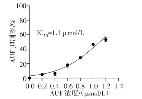
Fig.1 The inhibitory rate of AUF on CAL-62 cells
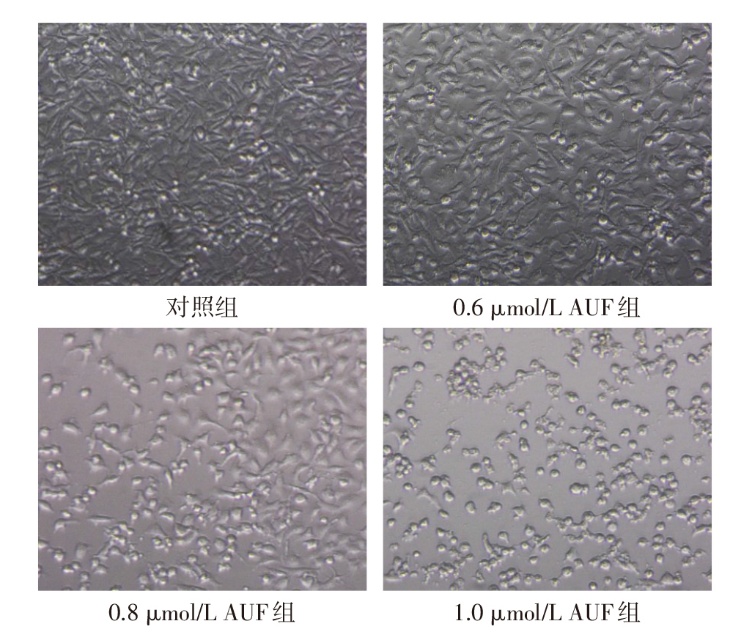
Fig.2 Microscopic cell growth following AUF treatment at varying concentrations (×100)
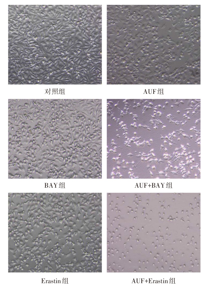
Fig.3 Microscopic morphological status of cells in various groups after combined treatment with AUF and other drugs (×100)
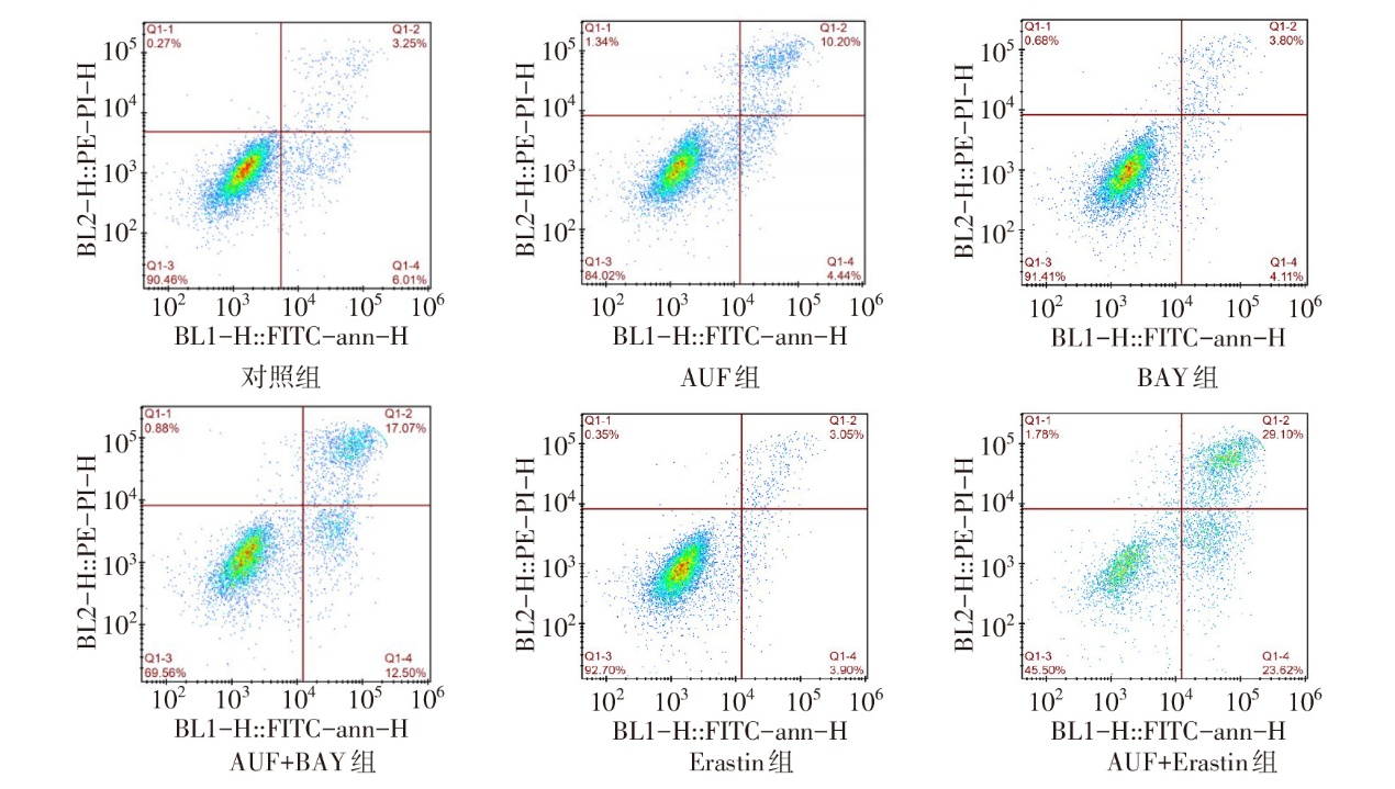
Fig.4 Apoptosis rates of different treatment groups following AUF and other drug treatments
| 组别 | 葡萄糖浓度/(n=4,mmol/L) | 铁含量/(n=3,ng/104 cell) | GSH含量/(n=4,μg/106 prot) |
|---|---|---|---|
| 对照组 | 8.60±1.64 | 3.92±0.49 | 1.32±0.02 |
| 0.6 μmol/L AUF组 | 13.23±2.58 | 6.86±1.77 | 0.89±0.01a |
| 0.8 μmol/L AUF组 | 23.44±3.71ab | 8.16±1.41a | 0.99±0.21a |
| 1.0 μmol/L AUF组 | 34.83±3.28abc | 11.59±2.26ab | 0.68±0.01ac |
| F | 64.160** | 11.590** | 24.810** |
Tab.2 Comparison of cell death metabolite levels between the four groups
| 组别 | 葡萄糖浓度/(n=4,mmol/L) | 铁含量/(n=3,ng/104 cell) | GSH含量/(n=4,μg/106 prot) |
|---|---|---|---|
| 对照组 | 8.60±1.64 | 3.92±0.49 | 1.32±0.02 |
| 0.6 μmol/L AUF组 | 13.23±2.58 | 6.86±1.77 | 0.89±0.01a |
| 0.8 μmol/L AUF组 | 23.44±3.71ab | 8.16±1.41a | 0.99±0.21a |
| 1.0 μmol/L AUF组 | 34.83±3.28abc | 11.59±2.26ab | 0.68±0.01ac |
| F | 64.160** | 11.590** | 24.810** |
| 组别 | GLUT1 | GPX4 | ACSL4 |
|---|---|---|---|
| 对照组 | 1.00±0.00 | 1.00±0.00 | 1.00±0.00 |
| 0.6 μmol/L AUF组 | 0.59±0.19a | 0.75±0.08a | 1.14±0.07 |
| 0.8 μmol/L AUF组 | 0.44±0.05a | 0.40±0.06ab | 1.48±0.05ab |
| 1.0 μmol/L AUF组 | 0.62±0.10a | 0.79±0.05ac | 1.71±0.17ab |
| F | 13.590** | 56.640** | 33.950** |
Tab.3 Comparison of expression levels of ferroptosis-related genes between the four groups of cells
| 组别 | GLUT1 | GPX4 | ACSL4 |
|---|---|---|---|
| 对照组 | 1.00±0.00 | 1.00±0.00 | 1.00±0.00 |
| 0.6 μmol/L AUF组 | 0.59±0.19a | 0.75±0.08a | 1.14±0.07 |
| 0.8 μmol/L AUF组 | 0.44±0.05a | 0.40±0.06ab | 1.48±0.05ab |
| 1.0 μmol/L AUF组 | 0.62±0.10a | 0.79±0.05ac | 1.71±0.17ab |
| F | 13.590** | 56.640** | 33.950** |
| 组别 | 2 d | 4 d | 6 d | ||||
|---|---|---|---|---|---|---|---|
| 对照组 | 337.6±63.9 | 412.9±162.2 | 461.9±184.5 | ||||
| AUF组 | 343.5±123.5 | 341.5±105.1 | 334.9±144.4 | ||||
| AUF+Erastin组 | 298.6±167.6 | 271.7±151.5 | 279.5±166.7 | ||||
| F | 0.113 | 0.744 | 0.952 | ||||
| 组别 | 8 d | 10 d | 12 d | 14 d | |||
| 对照组 | 552.8±143.8 | 608.9±228.9 | 649.8±269.3 | 721.2±235.7 | |||
| AUF组 | 361.1±118.9 | 369.6±167.3 | 378.8±152.0 | 411.4±138.5 | |||
| AUF+Erastin组 | 252.4±143.3 | 298.2±180.4 | 253.8±155.3 | 274.8±171.4a | |||
| F | 3.762 | 2.110 | 3.080 | 4.522 | |||
Tab.4 Tumor growth condition of ATC at different time points after gavage administration
| 组别 | 2 d | 4 d | 6 d | ||||
|---|---|---|---|---|---|---|---|
| 对照组 | 337.6±63.9 | 412.9±162.2 | 461.9±184.5 | ||||
| AUF组 | 343.5±123.5 | 341.5±105.1 | 334.9±144.4 | ||||
| AUF+Erastin组 | 298.6±167.6 | 271.7±151.5 | 279.5±166.7 | ||||
| F | 0.113 | 0.744 | 0.952 | ||||
| 组别 | 8 d | 10 d | 12 d | 14 d | |||
| 对照组 | 552.8±143.8 | 608.9±228.9 | 649.8±269.3 | 721.2±235.7 | |||
| AUF组 | 361.1±118.9 | 369.6±167.3 | 378.8±152.0 | 411.4±138.5 | |||
| AUF+Erastin组 | 252.4±143.3 | 298.2±180.4 | 253.8±155.3 | 274.8±171.4a | |||
| F | 3.762 | 2.110 | 3.080 | 4.522 | |||
| 组别 | 2 d | 4 d | 6 d | ||||
|---|---|---|---|---|---|---|---|
| 对照组 | 19.00±0.87 | 19.00±0.53 | 18.90±0.46 | ||||
| AUF组 | 17.73±0.64 | 18.23±0.91 | 17.93±0.98 | ||||
| AUF+Erastin组 | 19.47±3.23 | 20.13±2.58 | 19.90±2.17 | ||||
| F | 0.606 | 1.062 | 1.489 | ||||
| 组别 | 8 d | 10 d | 12 d | 14 d | |||
| 对照组 | 19.17±0.81 | 19.40±0.69 | 18.80±0.26 | 19.53±0.40 | |||
| AUF组 | 17.53±0.72 | 16.97±0.81 | 17.93±0.25 | 18.10±0.72 | |||
| AUF+Erastin组 | 20.33±1.82 | 20.37±2.21 | 20.40±2.11 | 20.30±2.17 | |||
| F | 3.983 | 4.606 | 3.055 | 2.089 | |||
Tab.5 Changes of mouse body weight at different time points after gavage administration in three groups
| 组别 | 2 d | 4 d | 6 d | ||||
|---|---|---|---|---|---|---|---|
| 对照组 | 19.00±0.87 | 19.00±0.53 | 18.90±0.46 | ||||
| AUF组 | 17.73±0.64 | 18.23±0.91 | 17.93±0.98 | ||||
| AUF+Erastin组 | 19.47±3.23 | 20.13±2.58 | 19.90±2.17 | ||||
| F | 0.606 | 1.062 | 1.489 | ||||
| 组别 | 8 d | 10 d | 12 d | 14 d | |||
| 对照组 | 19.17±0.81 | 19.40±0.69 | 18.80±0.26 | 19.53±0.40 | |||
| AUF组 | 17.53±0.72 | 16.97±0.81 | 17.93±0.25 | 18.10±0.72 | |||
| AUF+Erastin组 | 20.33±1.82 | 20.37±2.21 | 20.40±2.11 | 20.30±2.17 | |||
| F | 3.983 | 4.606 | 3.055 | 2.089 | |||
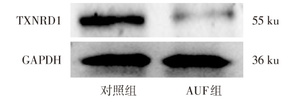
Fig.5 The expression level of TXNRD1 protein in ATC tissue of the AUF-treated group
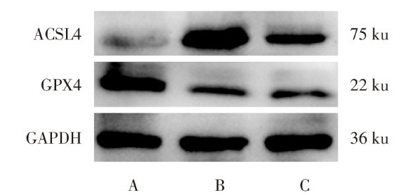
Fig.6 The expression levels of ferroptosis-related proteins in ATC tissue following AUF treatment
| 组别 | TXNRD1 | GPX4 | ACSL4 |
|---|---|---|---|
| 对照组 | 0.99±0.13 | 1.12±0.26 | 0.28±0.22 |
| AUF组 | 0.49±0.06 | 0.62±0.08a | 0.96±0.12a |
| AUF+Erastin组 | - | 0.36±0.08a | 0.73±0.22a |
| t或F | 5.938** | 16.810** | 9.727* |
Tab.6 Comparison of the expression levels of ferroptosis-related proteins between the three groups of ATC tissue
| 组别 | TXNRD1 | GPX4 | ACSL4 |
|---|---|---|---|
| 对照组 | 0.99±0.13 | 1.12±0.26 | 0.28±0.22 |
| AUF组 | 0.49±0.06 | 0.62±0.08a | 0.96±0.12a |
| AUF+Erastin组 | - | 0.36±0.08a | 0.73±0.22a |
| t或F | 5.938** | 16.810** | 9.727* |
| [1] | RAO S N, SMALLRIDGE R C. Anaplastic thyroid cancer:An update[J]. Best Pract Res Clin Endocrinol Metab, 2023, 37(1):101678. doi:10.1016/j.beem.2022.101678. |
| [2] | ALOBUIA W, GILLIS A, KEBEBEW E. Contemporary management of anaplastic thyroid cancer[J]. Curr Treat Options Oncol, 2020, 21(10):78. doi:10.1007/s11864-020-00776-2. |
| [3] | ZHOU Q, MENG Y, LI D, et al. Ferroptosis in cancer:from molecular mechanisms to therapeutic strategies[J]. Signal Transduct Target Ther, 2024, 9(1):55. doi:10.1038/s41392-024-01769-5. |
| [4] | LI D, LI Y. The interaction between ferroptosis and lipid metabolism in cancer[J]. Signal Transduct Target Ther, 2020, 5(1):108. doi:10.1038/s41392-020-00216-5. |
| [5] | CHEN H, PENG F, XU J, et al. Increased expression of GPX4 promotes the tumorigenesis of thyroid cancer by inhibiting ferroptosis and predicts poor clinical outcomes[J]. Aging, 2023, 15(1):230-245. doi:10.18632/aging.204473. |
| [6] | CHEN J, DING C, CHEN Y, et al. ACSL4 reprograms fatty acid metabolism in hepatocellular carcinoma via c-Myc/SREBP1 pathway[J]. Cancer Lett, 2021, 502:154-165. doi:10.1016/j.canlet.2020.12.019. |
| [7] | CHANDEL N S. Glycolysis[J]. Cold Spring Harb Perspect Biol, 2021, 13(5):a040535. doi:10.1101/cshperspect.a040535. |
| [8] | ANCEY P B, CONTAT C, BOIVIN G, et al. GLUT1 expression in tumor-associated neutrophils promotes lung cancer growth and resistance to radiotherapy[J]. Cancer Res, 2021, 81(9):2345-2357. doi:10.1158/0008-5472.Can-20-2870. |
| [9] | YAMASHITA M. Auranofin:past to present, and repurposing[J]. Int Immunopharmacol, 2021, 101(Pt B):108272. doi:10.1016/j.intimp.2021.108272. |
| [10] | YAN X, ZHANG X, WANG L, et al. Inhibition of thioredoxin/thioredoxin reductase induces synthetic lethality in lung cancers with compromised glutathione homeostasis[J]. Cancer Res, 2019, 79(1):125-132. doi:10.1158/0008-5472.Can-18-1938. |
| [11] | TIEDJE V, STUSCHKE M, WEBER F, et al. Anaplastic thyroid carcinoma:review of treatment protocols[J]. Endocr Relat Cancer, 2018, 25(3):R153-R161. doi:10.1530/erc-17-0435. |
| [12] | SAINI S, TULLA K, MAKER A V, et al. Therapeutic advances in anaplastic thyroid cancer:a current perspective[J]. Mol Cancer, 2018, 17(1):154. doi:10.1186/s12943-018-0903-0. |
| [13] | MADEIRA J M, GIBSON D L, KEAN W F, et al. The biological activity of auranofin:implications for novel treatment of diseases[J]. Inflammopharmacology, 2012, 20(6):297-306. doi:10.1007/s10787-012-0149-1. |
| [14] | GAMBERI T, CHIAPPETTA J, FIASCHI T, et al. Upgrade of an old drug:Auranofin in innovative cancer therapies to overcome drug resistance and to increase drug effectiveness[J]. Med Res Rev, 2022, 42(3):1111-1146. doi:10.1002/med.21872. |
| [15] | LIU C, ZHAO Y, WANG J, et al. FoxO3 reverses 5-fluorouracil resistance in human colorectal cancer cells by inhibiting the Nrf2/TR1 signaling pathway[J]. Cancer Lett, 2020, 470:29-42. doi:10.1016/j.canlet.2019.11.042. |
| [16] | ZHANG Z, LI X, YANG F, et al. DHHC9-mediated GLUT1 S-palmitoylation promotes glioblastoma glycolysis and tumorigenesis[J]. Nat Commun, 2021, 12(1):5872. doi:10.1038/s41467-021-26180-4. |
| [17] | CAI K, CHEN S, ZHU C, et al. FOXD1 facilitates pancreatic cancer cell proliferation,invasion,and metastasis by regulating GLUT1-mediated aerobic glycolysis[J]. Cell Death Dis, 2022, 13(9):765. doi:10.1038/s41419-022-05213-w. |
| [18] | WU Q, BA-ALAWI W, DEBLOIS G, et al. GLUT1 inhibition blocks growth of RB1-positive triple negative breast cancer[J]. Nat Commun, 2020, 11(1):4205. doi:10.1038/s41467-020-18020-8. |
| [19] | 李艳萍, 王协涛, 史立彬, 等. 白藜芦醇调控Nrf2-GPX4通路对H2O2诱导肺泡上皮细胞铁死亡的影响[J]. 天津医药, 2023, 51(6):568-572. |
| LI Y P, WANG X T, SHI L B, et al. Influence of resveratrol on H2O2-induced ferroptosis in alveolar epithelial cells by regulating the Nrf2-GPX4 pathway[J]. Tianjin Med J, 2023, 51(6):568-572. doi:10.11958/20221584. | |
| [20] | ZHANG W, LIU Y, LIAO Y, et al. GPX4,ferroptosis,and diseases[J]. Biomed Pharmacother, 2024, 174:116512. doi:10.1016/j.biopha.2024.116512. |
| [21] | CHEN X, LI J, KANG R, et al. Ferroptosis:machinery and regulation[J]. Autophagy, 2020, 17(9):2054-2081. doi:10.1080/15548627.2020.1810918. |
| [22] | CHEN C, YANG Y, GUO Y, et al. CYP1B1 inhibits ferroptosis and induces anti-PD-1 resistance by degrading ACSL4 in colorectal cancer[J]. Cell Death Dis, 2023, 14(4):271. doi:10.1038/s41419-023-05803-2. |
| [1] | ZHAO Lanjun, LI Lianghui, MA Xin, GONG Jiaojiao, ZHENG Chenhui, SHI Lin. The effect of andrographolide regulating STAT3/GPX4 pathway on proliferation and apoptosis of myeloma cells [J]. Tianjin Medical Journal, 2026, 54(1): 8-13. |
| [2] | SU Hongjian, ZHANG Chunyan, ZHANG Weidong, HAN Li, QIAO Yahong. Irisin affects the proliferation and migration of lung adenocarcinoma cells by regulating the EBF3/ALOX15 pathway [J]. Tianjin Medical Journal, 2025, 53(4): 337-342. |
| [3] | LI Bo, LIN Hong, HUANG Lanying, LI Lingyu. The relationship between HMOX1 and MAPK14 with the onset and prognosis of sepsis-induced acute lung injury in children [J]. Tianjin Medical Journal, 2025, 53(10): 1052-1056. |
| [4] | GAO Chen, QIAO Yunyang, JI Jialing, WANG E, HUO Ying, ZHANG Aiqing. The effect of bufalin on extracellular matrix synthesis in renal tubular epithelial cells induced by high glucose [J]. Tianjin Medical Journal, 2025, 53(10): 1009-1015. |
| [5] | ZHANG Xungong, YANG Guanghui, DU Zengli, XUE Pei, MA Zikun. Correlation between ferroptosis and post operative cognitive dysfunction in elderly patients with fractures [J]. Tianjin Medical Journal, 2025, 53(1): 47-51. |
| [6] | WU Bin, LIU Zhaoxiang, ZHANG Yuehong, WANG Changyao. The mechanism of excessive mechanical stress modulates Piezo1-mediated ferroptosis in chondrocytes [J]. Tianjin Medical Journal, 2025, 53(1): 14-18. |
| [7] | ZHANG Chunhong, HUANG Hongchao, LIU Yue, DU Lilong, XU Haiwei, LI Ning, LI Yongjin. Identification of key ferroptosis genes in paraspinal muscle degeneration based on RNA sequencing and bioinformatics analysis [J]. Tianjin Medical Journal, 2024, 52(9): 991-995. |
| [8] | WANG Xinshuang, AN Yajuan, GUAN Xiuju, LI Jiao, LIU Yue, WEI Liping, QI Xin. Study of magnesium isoglycyrrhizinate in ameliorating cisplatin induced myocardial injury in rats [J]. Tianjin Medical Journal, 2024, 52(8): 809-814. |
| [9] | LIU Bin, YANG Long, LI Wenli, SHAO Ningning, DONG Jinrui. Mechanism of microglia ferroptosis in smoke inhalation-induced brain injury [J]. Tianjin Medical Journal, 2024, 52(8): 791-797. |
| [10] | HUANG Xiaolei, GE Tingting, ZHAO Junsong, NI Zhihua. Study on the role of ginsenoside Rg1 in IL-6-induced neuronal ferroptosis in rats [J]. Tianjin Medical Journal, 2024, 52(11): 1137-1140. |
| [11] | LI Yanping, WANG Xietao, SHI Libin, LIU Qiong. Influence of resveratrol on H2O2-induced ferroptosis in alveolar epithelial cells by regulating the Nrf2-GPX4 pathway [J]. Tianjin Medical Journal, 2023, 51(6): 568-572. |
| [12] | HU Yingshan, WANG Jingjing, GAO Hongmei. Research progress in the pathogenesis of sepsis-related intestinal dysfunction [J]. Tianjin Medical Journal, 2023, 51(3): 333-336. |
| [13] | MAO Quanxi, LI Zuoxiao. Neuroprotective mechanism of edaravone dexborneol in rats with cerebral hemorrhage through ferroptosis-lipid peroxidation pathway [J]. Tianjin Medical Journal, 2023, 51(11): 1199-1204. |
| [14] | WANG Ke, YE Hanlu. Effect and its mechanism of emodin on the ferroptosis of chondrocytes in rats with knee osteoarthritis [J]. Tianjin Medical Journal, 2023, 51(10): 1090-1097. |
| [15] | XIONG Xicheng, WANG Yiping, WANG Gang, ZHANG Tian, BAO Yali, Di na·AINIWAER, SUN Zhan. Ferrostatin-1 delay D-gal induced cardiomyocyte senescence by inhibiting ferroptosis [J]. Tianjin Medical Journal, 2023, 51(1): 19-23. |
| Viewed | ||||||
|
Full text |
|
|||||
|
Abstract |
|
|||||
