Tianjin Medical Journal ›› 2023, Vol. 51 ›› Issue (12): 1382-1386.doi: 10.11958/20230513
• Clinical Research • Previous Articles Next Articles
GUO Zhenjiang( ), ZHAO Guangyuan, DU Liqiang, LIU Fangzhen()
Received:2023-04-17
Revised:2023-06-29
Published:2023-12-15
Online:2023-12-22
Contact:
△ E-mail:GUO Zhenjiang, ZHAO Guangyuan, DU Liqiang, LIU Fangzhen. Establishment and validation of a predictive nomogram model for advanced gastric cancer with lymphovascular invasion[J]. Tianjin Medical Journal, 2023, 51(12): 1382-1386.
CLC Number:
| 临床特征 | LVI阳性 (n=95) | LVI阴性 (n=151) | χ2或Z |
|---|---|---|---|
| 年龄 | |||
| <60岁 | 42(44.2) | 67(44.4) | 0.001 |
| ≥60岁 | 53(55.8) | 84(55.6) | |
| 性别 | |||
| 男 | 62(65.3) | 96(63.6) | 0.072 |
| 女 | 33(34.7) | 55(36.4) | |
| 肿瘤大小 | |||
| <5 cm | 34(35.8) | 84(55.6) | 9.196** |
| ≥5 cm | 61(64.2) | 67(44.4) | |
| 肿瘤位置 | |||
| 上部 | 21(22.1) | 34(22.5) | 0.524 |
| 中部 | 31(32.6) | 44(29.1) | |
| 下部 | 34(35.8) | 60(39.7) | |
| ≥2/3 | 9(9.5) | 13(8.6) | |
| Borrmann分型 | |||
| Ⅰ—Ⅱ | 20(21.1) | 51(33.8) | 4.597* |
| Ⅲ—Ⅳ | 75(78.9) | 100(66.2) | |
| 肿瘤分化 | |||
| 高-中分化 | 27(28.4) | 64(42.4) | 4.878* |
| 低-未分化 | 68(71.6) | 87(57.6) | |
| Lauren分型 | |||
| 肠型 | 18(18.9) | 51(33.8) | |
| 弥漫型 | 52(54.7) | 58(38.4) | 8.095* |
| 混合型 | 25(26.3) | 42(27.8) | |
| cT分期 | |||
| T2—3 | 33(34.7) | 84(55.6) | 10.205** |
| T4 | 62(65.3) | 67(44.4) | |
| cN分期 | |||
| N0 | 31(32.6) | 74(49.0) | 6.391* |
| N+ | 64(67.4) | 77(51.0) | |
| SII | 878.00 (649.50,1 351.00) | 682.00 (526.00,1 054.00) | 3.047** |
Tab.1 Univariate analysis of LVI positive influencing factors in gastric cancer patients with different clinical characteristics
| 临床特征 | LVI阳性 (n=95) | LVI阴性 (n=151) | χ2或Z |
|---|---|---|---|
| 年龄 | |||
| <60岁 | 42(44.2) | 67(44.4) | 0.001 |
| ≥60岁 | 53(55.8) | 84(55.6) | |
| 性别 | |||
| 男 | 62(65.3) | 96(63.6) | 0.072 |
| 女 | 33(34.7) | 55(36.4) | |
| 肿瘤大小 | |||
| <5 cm | 34(35.8) | 84(55.6) | 9.196** |
| ≥5 cm | 61(64.2) | 67(44.4) | |
| 肿瘤位置 | |||
| 上部 | 21(22.1) | 34(22.5) | 0.524 |
| 中部 | 31(32.6) | 44(29.1) | |
| 下部 | 34(35.8) | 60(39.7) | |
| ≥2/3 | 9(9.5) | 13(8.6) | |
| Borrmann分型 | |||
| Ⅰ—Ⅱ | 20(21.1) | 51(33.8) | 4.597* |
| Ⅲ—Ⅳ | 75(78.9) | 100(66.2) | |
| 肿瘤分化 | |||
| 高-中分化 | 27(28.4) | 64(42.4) | 4.878* |
| 低-未分化 | 68(71.6) | 87(57.6) | |
| Lauren分型 | |||
| 肠型 | 18(18.9) | 51(33.8) | |
| 弥漫型 | 52(54.7) | 58(38.4) | 8.095* |
| 混合型 | 25(26.3) | 42(27.8) | |
| cT分期 | |||
| T2—3 | 33(34.7) | 84(55.6) | 10.205** |
| T4 | 62(65.3) | 67(44.4) | |
| cN分期 | |||
| N0 | 31(32.6) | 74(49.0) | 6.391* |
| N+ | 64(67.4) | 77(51.0) | |
| SII | 878.00 (649.50,1 351.00) | 682.00 (526.00,1 054.00) | 3.047** |
| 变量 | 变量类型 | 变量赋值 |
|---|---|---|
| LVI | 因变量 | 阴性=0,阳性=1 |
| 肿瘤大小 | 自变量 | <5 cm=1,≥5 cm=2 |
| Borrmann分型 | 自变量 | Ⅰ—Ⅱ型=1,Ⅲ—Ⅳ型=2 |
| 肿瘤分化 | 自变量 | 高-中分化=1,低-未分化=2 |
| Lauren分型 | 自变量 | 肠型=1,弥漫型=2,混合型=3 |
| cT分期 | 自变量 | cT2—3期=1,cT4期=2 |
| cN分期 | 自变量 | cN0期=1,cN+期=2 |
| SII | 自变量 | 连续变量 |
Tab.2 Variable assignment table
| 变量 | 变量类型 | 变量赋值 |
|---|---|---|
| LVI | 因变量 | 阴性=0,阳性=1 |
| 肿瘤大小 | 自变量 | <5 cm=1,≥5 cm=2 |
| Borrmann分型 | 自变量 | Ⅰ—Ⅱ型=1,Ⅲ—Ⅳ型=2 |
| 肿瘤分化 | 自变量 | 高-中分化=1,低-未分化=2 |
| Lauren分型 | 自变量 | 肠型=1,弥漫型=2,混合型=3 |
| cT分期 | 自变量 | cT2—3期=1,cT4期=2 |
| cN分期 | 自变量 | cN0期=1,cN+期=2 |
| SII | 自变量 | 连续变量 |
| 变量 | β | SE | Waldχ2 | P | OR(95%CI) |
|---|---|---|---|---|---|
| 肿瘤大小 | 0.781 | 0.296 | 6.984 | 0.008 | 2.184(1.224~3.898) |
| Borrmann 分型 | 0.923 | 0.339 | 7.398 | 0.007 | 2.517(1.294~4.896) |
| Lauren 分型 | 5.129 | 0.077 | |||
| 弥漫型 | -0.659 | 0.41 | 2.582 | 0.108 | 0.518(0.232~1.156) |
| 混合型 | 0.159 | 0.346 | 0.213 | 0.645 | 1.173(0.596~2.309) |
| cT分期 | 0.620 | 0.294 | 4.456 | 0.035 | 1.860(1.045~3.308) |
| cN分期 | 0.597 | 0.302 | 3.896 | 0.048 | 1.816(1.004~3.285) |
| SII | 0.001 | <0.001 | 10.235 | 0.001 | 1.001(1.000~1.002) |
| 常数项 | -6.039 | 1.119 | 29.106 | <0.001 | 0.002 |
Tab.3 Multivariate analysis of preoperative predictors of LVI in gastric cancer
| 变量 | β | SE | Waldχ2 | P | OR(95%CI) |
|---|---|---|---|---|---|
| 肿瘤大小 | 0.781 | 0.296 | 6.984 | 0.008 | 2.184(1.224~3.898) |
| Borrmann 分型 | 0.923 | 0.339 | 7.398 | 0.007 | 2.517(1.294~4.896) |
| Lauren 分型 | 5.129 | 0.077 | |||
| 弥漫型 | -0.659 | 0.41 | 2.582 | 0.108 | 0.518(0.232~1.156) |
| 混合型 | 0.159 | 0.346 | 0.213 | 0.645 | 1.173(0.596~2.309) |
| cT分期 | 0.620 | 0.294 | 4.456 | 0.035 | 1.860(1.045~3.308) |
| cN分期 | 0.597 | 0.302 | 3.896 | 0.048 | 1.816(1.004~3.285) |
| SII | 0.001 | <0.001 | 10.235 | 0.001 | 1.001(1.000~1.002) |
| 常数项 | -6.039 | 1.119 | 29.106 | <0.001 | 0.002 |
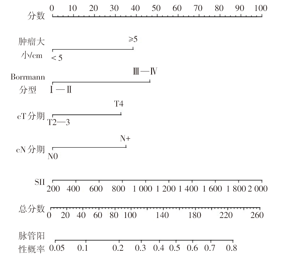
Fig.1 Prediction model of advanced gastric cancer LVI
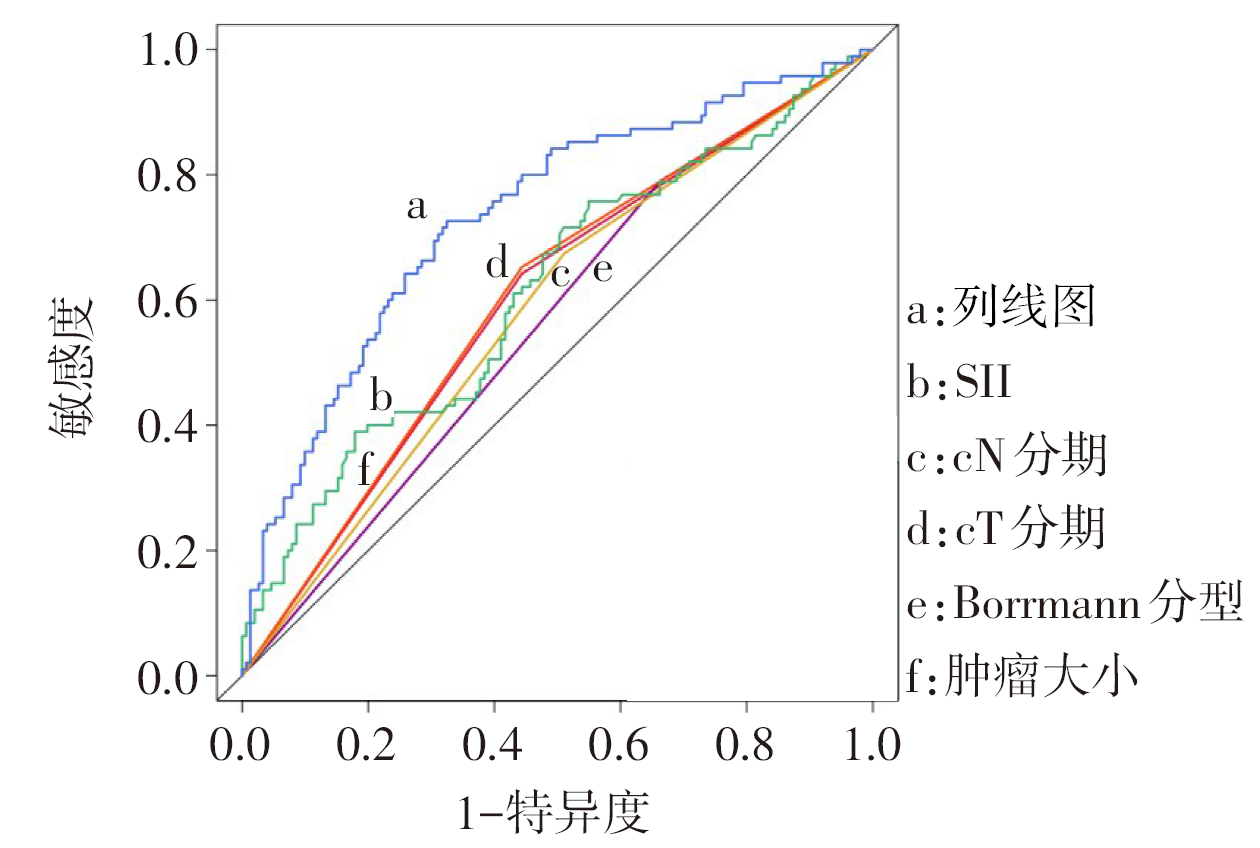
Fig.2 The prediction model and ROC curves for individual predictors
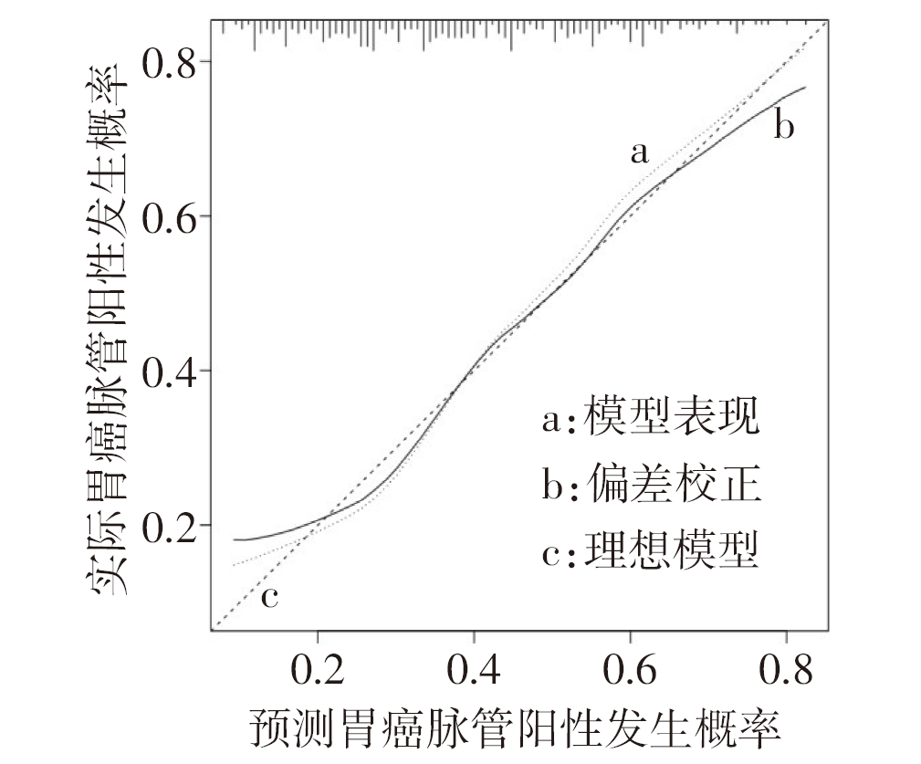
Fig.3 The calibration curve of the prediction model
| [1] | 赫捷, 陈万青, 李兆申, 等. 中国胃癌筛查与早诊早治指南(2022,北京)[J]. 中华肿瘤杂志,2022, 44(7):634-666. |
| HE J, CHEN W Q, LI Z S, et al. China guideline for the screening,early detection and early treatment of gastric cancer(2022,Beijing)[J]. Chin J Oncol,2022, 44(7):634-666. doi:10.3760/cma.j.cn112152-20220617-00430. | |
| [2] | ZHANG C D, NING F L, ZENG X T, et al. Lymphovascular invasion as a predictor for lymph node metastasis and a prognostic factor in gastric cancer patients under 70 years of age:a retrospective analysis[J]. Int J Surg, 2018, 53:214-220. doi:10.1016/j.ijsu.2018.03.073. |
| [3] | MEI D, ZHAO B C, ZHANG J L, et al. Impact of lymphovascular invasion on survival outcome in patients with gastric cancer[J]. Am J Clin Pathol, 2020, 153(6):833-841. doi:10.1093/ajcp/aqaa021. |
| [4] | WU L L, LIANG Y X, ZHANG C, et al. Prognostic significance of lymphovascular infiltration in overall survival of gastric cancer patients after surgery with curative intent[J]. Chin J Cancer Res, 2019, 31(5):785-796. doi:10.21147/j.issn.1000-9604.2019.05.08. |
| [5] | MA Z L, LIANG C H, HUANG Y Q, et al. Can lymphovascular invasion be predicted by preoperative multiphasic dynamic CT in patients with advanced gastric cancer?[J]. Eur Radiol, 2017, 27(8):3383-3391. doi:10.1007/s00330-016-4695-6. |
| [6] | XU R, XIAO S M, DING Z, et al. The value of the C-reactive protein-to-lymphocyte ratio for predicting lymphovascular invasion based on nutritional status in gastric cancer[J]. Technol Cancer Res Treat, 2022, 21:15330338221106517. doi:10.1177/15330338221106517. |
| [7] | DING P A, GUO H H, SUN C Y, et al. Combined systemic immune-inflammatory index (SII) and prognostic nutritional index (PNI) predicts chemotherapy response and prognosis in locally advanced gastric cancer patients receiving neoadjuvant chemotherapy with PD-1 antibody sintilimab and XELOX:a prospective study[J]. BMC Gastroenterol, 2022, 22(1):121. doi:10.1186/s12876-022-02199-9. |
| [8] | 王举, 窦忠霞, 姜洪伟, 等. 基于癌症基因组图谱构建胃癌预后评估模型[J]. 天津医药, 2018, 46(12):1262-1266. |
| WANG J, DOU Z X, JIANG H W, et al. Construction of prognostic predictive model of gastric cancer based on the cancer genome atlas[J]. Tianjin Med J, 2018, 46(12):1262-1266. doi:10.11958/20181254. | |
| [9] | DICKEN B J, GRAHAM K, HAMILTON S M, et al. Lymphovascular invasion is associated with poor survival in gastric cancer:an application of gene-expression and tissue array techniques[J]. Ann Surg, 2006, 243(1):64-73. doi:10.1097/01.sla.0000194087.96582.3e. |
| [10] | KIKUCHI E, MARGULIS V, KARAKIEWICZ P I, et al. Lymphovascular invasion predicts clinical outcomes in patients with node-negative upper tract urothelial carcinoma[J]. J Clin Oncol, 2009, 27(4):612-618. doi:10.1200/jco.2008.17.2361. |
| [11] | ZHU Z, GONG Y B, XU H M. Clinical and pathological staging of gastric cancer:current perspectives and implications[J]. Eur J Surg Oncol, 2020, 46(< W>10 Pt B):e14-e19. doi:10.1016/j.ejso.2020.06.006. |
| [12] | CHOI S, SONG J H, LEE S, et al. Lymphovascular invasion:traditional but vital and sensible prognostic factor in early gastric cancer[J]. Ann Surg Oncol, 2021, 28(13):8928-8935. doi:10.1245/s10434-021-10224-6. |
| [13] | FUJIKAWA H, KOUNORI K, WATANABE H, et al. The clinical significance of lymphovascular invasion in gastric cancer[J]. In Vivo, 2020, 34(3):1533-1539. doi:10.21873/invivo.11942. |
| [14] | LI P, HE H Q, ZHU C M, et al. The prognostic significance of lymphovascular invasion in patients with resectable gastric cancer:a large retrospective study from Southern China[J]. BMC Cancer, 2015, 15:370. doi:10.1186/s12885-015-1370-2. |
| [15] | CHEN X F, YANG Z Q, YANG J D, et al. Radiomics analysis of contrast-enhanced CT predicts vascular invasion and disease outcome in gastric cancer:a preliminary study[J]. Cancer Imaging, 2020, 20(1):24. doi:10.1186/s40644-020-00302-5. |
| [16] | 刘书豪, 侯新月, 张宪祥, 等. 进展期胃癌神经侵犯列线图预测模型的构建与验证[J]. 中华胃肠外科杂志, 2020, 23(11):1059-1066. |
| LIU S H, HOU X Y, ZHANG X X, et al. Establishment and validation of a predictive nomogram model for advanced gastric cancer with perineural invasion[J]. Chin J Gastrointest Surg, 2020, 23(11):1059-1066. doi:10.3760/cma.j.cn.441530-20200103-00004. | |
| [17] | GRESTA L T, RODRIGUES-JUNIOR I A, CASTRO L P, et al. Assessment of vascular invasion in gastric cancer:a comparative study[J]. World J Gastroenterol, 2013, 19(24):3761-3769. doi:10.3748/wjg.v19.i24.3761. |
| [18] | SEEVARATNAM R, CARDOSO R, MCGREGOR C, et al. How useful is preoperative imaging for tumor,node,metastasis (TNM) staging of gastric cancer ? A meta-analysis[J]. Gastric Cancer, 2012, 15 Suppl 1:S3-18. doi:10.1007/s10120-011-0069-6. |
| [19] | HIRAHARA N, MATSUBARA T, KAJI S, et al. Novel inflammation-combined prognostic index to predict survival outcomes in patients with gastric cancer[J]. Oncotarget, 2023, 14:71-82. doi:10.18632/oncotarget.28353. |
| [20] | ZHU Z Y, CONG X L, LI R, et al. Preoperative systemic immune-inflammation index (SII) for predicting the survival of patients with stage I-III gastric cancer with a signet-ring cell (SRC) component[J]. Biomed Res Int, 2020, 2020:5038217. doi:10.1155/2020/5038217. |
| Viewed | ||||||
|
Full text |
|
|||||
|
Abstract |
|
|||||