Tianjin Medical Journal ›› 2022, Vol. 50 ›› Issue (10): 1093-1097.doi: 10.11958/20220292
• Applied Research • Previous Articles Next Articles
ZHANG Lixia( ), LIU Haixia(), ZHANG Xuetao, CAO Zhen, WANG Guidong
Received:2022-03-08
Revised:2022-04-21
Published:2022-10-15
Online:2022-10-20
Contact:
LIU Haixia
E-mail:zhanglixia1522@126.com;15932518118@163.com
ZHANG Lixia, LIU Haixia, ZHANG Xuetao, CAO Zhen, WANG Guidong. The value of CT features in differentiating moderately and highly differentiated invasive adenocarcinoma in pure ground glass nodule[J]. Tianjin Medical Journal, 2022, 50(10): 1093-1097.
CLC Number:
| 组别 | n | 年龄(岁) | 性别(男/女) | 吸烟史 |
|---|---|---|---|---|
| 高分化组 | 45 | 57.1±9.2 | 14/31 | 12(26.7) |
| 中分化组 | 38 | 56.2±10.1 | 8/30 | 4(10.5) |
| t或χ² | 0.441 | 1.070 | 3.449 |
Tab. 1 Comparison of the clinical characteristics between the moderately differentiated group and the highly differentiated group
| 组别 | n | 年龄(岁) | 性别(男/女) | 吸烟史 |
|---|---|---|---|---|
| 高分化组 | 45 | 57.1±9.2 | 14/31 | 12(26.7) |
| 中分化组 | 38 | 56.2±10.1 | 8/30 | 4(10.5) |
| t或χ² | 0.441 | 1.070 | 3.449 |
| 组别 | n | 最大直径(cm) | 平均CT值(HU) | 分叶征 | 毛刺征 | 异常血管征 | 异常支气管征 | 空泡征 | 胸膜凹陷征 |
|---|---|---|---|---|---|---|---|---|---|
| 高分化组 | 46 | 1.35(1.10,1.53) | -573.0±55.8 | 28(60.9) | 14(30.4) | 20(43.5) | 16(34.8) | 14(30.4) | 13(28.3) |
| 中分化组 | 38 | 1.50(1.10,2.00) | -481.8±52.5 | 22(57.9) | 24(63.2) | 28(73.7) | 24(63.2) | 14(36.8) | 12(31.6) |
| Z、t或χ² | 1.969* | 7.656** | 0.076 | 8.995** | 7.753** | 6.717* | 0.384 | 0.110 |
Tab. 2 Comparison of CT features between the moderately differentiated group and the highly differentiated group
| 组别 | n | 最大直径(cm) | 平均CT值(HU) | 分叶征 | 毛刺征 | 异常血管征 | 异常支气管征 | 空泡征 | 胸膜凹陷征 |
|---|---|---|---|---|---|---|---|---|---|
| 高分化组 | 46 | 1.35(1.10,1.53) | -573.0±55.8 | 28(60.9) | 14(30.4) | 20(43.5) | 16(34.8) | 14(30.4) | 13(28.3) |
| 中分化组 | 38 | 1.50(1.10,2.00) | -481.8±52.5 | 22(57.9) | 24(63.2) | 28(73.7) | 24(63.2) | 14(36.8) | 12(31.6) |
| Z、t或χ² | 1.969* | 7.656** | 0.076 | 8.995** | 7.753** | 6.717* | 0.384 | 0.110 |
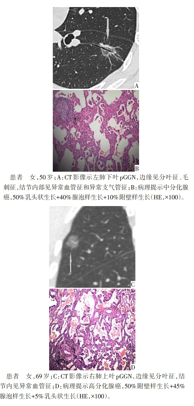
Fig.1 CT and pathological findings of typical moderately and highly differentiated invasive adenocarcinoma
| 变量 | β | SE | Wald χ2 | P | OR(95%CI) |
|---|---|---|---|---|---|
| 平均CT值 | 0.041 | 0.010 | 17.178 | <0.001 | 1.042(1.022~1.062) |
| 异常血管征 | 1.770 | 0.852 | 4.316 | 0.038 | 5.869(1.105~31.161) |
| 异常支气管征 | 0.027 | 0.848 | 0.001 | 0.974 | 1.028(0.195~5.420) |
| 最大直径 | 0.788 | 0.664 | 2.360 | 0.124 | 3.167(0.917~9.638) |
| 毛刺征 | 0.753 | 0.751 | 1.003 | 0.316 | 2.123(0.487~9.258) |
| 常数项 | 22.589 | 5.696 | 15.724 | <0.001 | 6 459 421 837.000 |
Tab. 3 Logistic regression analysis of moderately differentiated invasive adenocarcinoma
| 变量 | β | SE | Wald χ2 | P | OR(95%CI) |
|---|---|---|---|---|---|
| 平均CT值 | 0.041 | 0.010 | 17.178 | <0.001 | 1.042(1.022~1.062) |
| 异常血管征 | 1.770 | 0.852 | 4.316 | 0.038 | 5.869(1.105~31.161) |
| 异常支气管征 | 0.027 | 0.848 | 0.001 | 0.974 | 1.028(0.195~5.420) |
| 最大直径 | 0.788 | 0.664 | 2.360 | 0.124 | 3.167(0.917~9.638) |
| 毛刺征 | 0.753 | 0.751 | 1.003 | 0.316 | 2.123(0.487~9.258) |
| 常数项 | 22.589 | 5.696 | 15.724 | <0.001 | 6 459 421 837.000 |
| 指标 | AUC(95%CI) | 敏感度 | 特异度 | 约登 指数 | 截断值 |
|---|---|---|---|---|---|
| 联合诊断 | 0.903(0.842~0.964) | 0.870 | 0.789 | 0.659 | 0.541 |
| 平均CT值 | 0.883(0.814~0.952) | 0.739 | 0.895 | 0.634 | -531.5 HU |
| 异常血管征 | 0.651(0.533~0.769) | 0.737 | 0.565 | 0.302 | - |
Tab. 4 Combined diagnostic value of mean CT value and abnormal vascular signs in moderately differentiated IAC
| 指标 | AUC(95%CI) | 敏感度 | 特异度 | 约登 指数 | 截断值 |
|---|---|---|---|---|---|
| 联合诊断 | 0.903(0.842~0.964) | 0.870 | 0.789 | 0.659 | 0.541 |
| 平均CT值 | 0.883(0.814~0.952) | 0.739 | 0.895 | 0.634 | -531.5 HU |
| 异常血管征 | 0.651(0.533~0.769) | 0.737 | 0.565 | 0.302 | - |
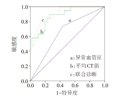
Fig.2 ROC curve analysis of combined diagnosis and mean CT value, abnormal vascular sign for the diagnosis of moderately differentiated IAC
| [1] | 胡小琴, 黄原义, 康彤, 等. 大于5 mm亚实性结节的CT影像特征在预测肺腺癌浸润程度中的价值[J]. 实用放射学杂志, 2021, 37(5):741-745. |
| HU X Q, HUANG Y Y, KANG T, et al. The value of CT imaging features of subsolid nodules larger than 5 mm in predicting invasion degree of lung adenocarcinoma[J]. J Pract Radiol, 2021, 37(5):741-745. doi: 10.3969/j.issn.1002-1671.2021.05.012. | |
| [2] | 张磊, 谢晓东, 沈文荣, 等. CT征象在预测肺纯磨玻璃结节侵袭风险中的价值[J]. 实用放射学杂志, 2020, 36(2):207-210,213. |
| ZHANG L, XIE X D, SHEN W R, et al. The value of CT features in predicting the invasiveness of PGGN[J]. J Pract Radiol, 2020, 36(2):207-210,213. doi: 10.3969/j.issn.1002-1671.2020.02.009. | |
| [3] | 李媛, 谢惠康, 武春燕. WHO胸部肿瘤分类(第5版)中肺肿瘤部分解读[J]. 中国癌症杂志, 2021, 31(7):574-580. |
| LI Y, XIE H K, WU S Y. Interpretation of lung tumours in the WHO classification of thoracic tumours(5th edition)[J]. China Oncology, 2021, 31(7):574-580. doi: 10.19401/j.cnki.1007-3639.2021.07.003. | |
| [4] | 北京医学会胸外科分会, 中国医疗保健国际交流促进会胸外科分会. 基于高分辨CT影像学指导≤2 cm磨玻璃结节肺癌手术方式胸外科专家共识(2019版)[J]. 中国胸心血管外科临床杂志, 2020, 27(4):395-400. |
| Beijing Association for Thoracic Surgery, Thoracic Surgery Branch of China International Exchange Association and Promotive Association for Medical and Health Care. Expert consensus on surgical methods of lung cancer with ground glass nodules ≤2 cm based on high-resolution CT imaging guidance(2019 edition)[J]. Chinese Journal of Clinical Thoracic and Cardiovascular Surgery, 2020, 27(4):395-400. doi:10.7507/1007-4848.201912078. | |
| [5] | CHU Z G, LI W J, FU B J, et al. CT characteristics for predicting invasiveness in pulmonary pure ground-glass nodules[J]. AJR Am J Roentgenol, 2020, 15(2):351-358. doi: 10.2214/AJR.19.22381. |
| [6] | HU F Y, HUANG H H, JIANG Y Y, et al. Discriminating invasive adenocarcinoma among lung pure ground-glass nodules:A multi-parameter prediction model[J]. J Thorac Dis, 2021, 13(9):5383-5394. doi: 10.21037/jtd-21-786. |
| [7] | CHENG X, ZHENG D, LI Y, et al. Tumor histology predicts mediastinal nodal status and may be used to guide limited lymphadenectomy in patients with clinical stage I non-small cell lung cancer[J]. J Thorac Cardiovasc Surg, 2018, 155(6):2648-2656. doi: 10.1016/j.jtcvs.2018.02.010. |
| [8] | 姜格宁, 陈昶, 朱余明, 等. 上海市肺科医院磨玻璃结节早期肺腺癌的诊疗共识(第一版)[J]. 中国肺癌杂志, 2018, 21(3):147-159. |
| JIANG G N, CHEN C, ZHU Y M, et al. Shanghai pulmonary hospital experts consensus on the management of ground glass nodules suspected as lung adenocarcinoma(Version 1)[J]. Chin J Lung Cancer, 2018, 21(3):147-159. doi: 10.3779/j.issn.1009-3419.2018.03.05. | |
| [9] | 中华医学会肿瘤学分会, 中华医学会杂志社. 中华医学会肿瘤学分会肺癌临床诊疗指南(2021版)[J]. 中华医学杂志, 2021, 101(23):1725-1757. |
| Oncology Branch of Chinese Medical Association,Chinese Medical Journals Publishing House. Clinical diagnosis and treatment guidelines for lung cancer of Oncology Branch of Chinese Medical Association (2021 edition)[J]. Natl Med J China, 2021, 101(23):1725-1757. doi: 10.3760/cma.j.cn112137-20210207-00377. | |
| [10] | 裴蕾, 潘伟, 纪清源, 等. HRCT影像学表现对浸润性肺腺癌病理亚型的预测价值[J]. 中国现代医生, 2021, 59(31):127-130. |
| PEI L, PAN W, JI Q Y, et al. Predictive value of HRCT imaging performance on pathological subtypes of invasive lung adenocarcinoma[J]. China Modern Doctor, 2021, 59(31):127-130. | |
| [11] | 刘丽, 于婷, 乔红艳, 等. 肺浸润性腺癌各病理亚型的临床资料及CT征象分析[J]. 东南大学学报(医学版), 2020, 39(6):759-763. |
| LIU L, YU T, QIAO H Y, et al. Analysis of clinical data and CT signs of various pathological subtypes of invasive lung adenocarcinoma[J]. J Southeast Univ(Med Sci Edi),2020, 39(6):759-763. doi: 10.3969/j.issn.1671-6264.2020.06.011. | |
| [12] | 单文莉, 周围, 孔丹, 等. 肺腺癌CT征象与病理组织学亚型及分化程度的对照研究[J]. 临床放射学杂志, 2019, 38(12): |
| 2312-2316. SHAN W L, ZHOU W, KONG D, et al. Control study between CT signs and pathological subtypes and differentiation degree in lung adenocarcinoma[J]. Journal of Clinical Radiology, 2019, 38(12):2312-2316. doi: 10.1343/j.cnki.jcr.2019.12.020. | |
| [13] | 顾鑫蕾, 刘展, 邵为朋, 等. 肺结节CT特征对腺癌病理亚型的预测价值[J/OL]. 中国胸心血管外科临床杂志:1-9[2021-12-29]. . |
| GU X L, LIU Z, SHAO W P, et al. CT features of pulmonary nodules predictive value of histological subtypes of adenocarcinoma[J/OL]. Chinese Journal of Clinical Thoracic and Cardiovascular Surgery:1- 9 [2021-12-29]. https://kns.cnki.net/kcms/detail/51.1492.R.20211228.1214.030.html. doi: 10.7507/1007-4848.202106072. | |
| [14] | 闵旭红, 宋奇隆, 余永强, 等. 三维CT定量联合定性参数的logistic回归模型对纯磨玻璃结节侵袭程度的临床预测价值[J]. 中华放射学杂志, 2021, 55(1):34-39. |
| MIN X H, SONG Q L, YU Y Q, et al. The clinical value of the logistic regression model with a combination of three-dimensional CT quantitative and qualitative parameters in predicting the invasiveness of pure ground glass nodules[J]. Chin J Radiol, 2021, 55(1):34-39. doi: 10.3760/cma.j.cn112149-20200318-00416. | |
| [15] | 刘莉, 吴宁, 周丽娜, 等. 亚实性结节血管及支气管异常与肺腺癌类病变侵袭性的相关性分析[J]. 中华放射学杂志, 2019, 53(11):987-991. |
| LIU L, WU N, ZHOU L N, et al. The correlations between vascular and bronchial abnormality on high resolution CT and the invasiveness of lung adenocarcinoma in subsolid nodules[J]. Chin J Radiol, 2019, 53(11):987-991. doi: 10.3760/cma.j.issn.1005-1201.2019.11.011. | |
| [16] | 肖瑜, 杨娅, 蒋锐, 等. 亚实性肺结节CT征象与肺腺癌浸润程度及其组织学亚型关系分析[J]. 生物医学工程与临床, 2022, 26(1):40-44. |
| XIAO Y, YANG Y, JIANG R, et al. Correlation between CT signs of subsolid lung nodules and lung adenocarcinoma invasion,histological subtypes[J]. BME & Clin Med, 2022, 26(1):40-44. doi: 10.13339/j.cnki.sglc.20211217.019. | |
| [17] | 张鹏举, 李天然, 陶雪敏, 等. 磨玻璃结节早期贴壁生长为主型浸润性肺腺癌与其他病理亚型的CT特征分析[J]. 中华放射学杂志, 2021, 55(7):739-744. |
| ZHANG P J, LI T R, TAO X M, et al. Analysis of CT features of lepidic predominant subtype and other pathological subtypes in early-stage invasive lung adenocarcinoma appearing as ground-glass nodule[J]. Chin J Radiol, 2021, 55(7):739-744. doi: 10.3760/cma.j.cn112149-20200924-01110. | |
| [18] | 赵鎏, 刘馨, 杜思瑶, 等. 定量诊断模型对表现为磨玻璃密度结节浸润性肺腺癌的诊断[J]. 中国临床医学影像杂志, 2020, 31(7):474-477. |
| ZHAO L, LIU X, DU S Y, et al. A quantitative diagnosis model for lung invasive adenocarcinoma with ground glass node[J]. J Chin Clin Med Imaging, 2020, 31(7):474-477. doi: 10.12117/jccmi.2020.07.005. | |
| [19] | 马东, 姜加学, 杨小庆, 等. 肺部磨玻璃结节的CT影像特征评估肺腺癌浸润性的价值[J]. 临床肺科杂志, 2019, 24(8):1470-1473. |
| MA D, JIANG J X, YANG X Q, et al. Evaluating value of CT scanning grinding glass nodules in invasive lung adenocarcinoma[J]. Journal of Clinical Pulmonary Medicine, 2019, 24(8):1470-1473. doi: 10.3969/j.issn.1009-6663.2019.08.028. | |
| [20] | 黎佳维, 伍建林, 李凤. 贴壁型与非贴壁型肺腺癌的HRCT表现及鉴别诊断[J]. 湖北科技学院学报:医学版, 2018, 32(3):4. |
| LI J W, WU J L, LI F, et al. HRCT features and diagnosis of lung lepidic and non-lepidic predominant adenocarcinoma[J]. Journal of Hubei University of Science and Technology(Medical Sciences),2018, 32(3):4. doi: 10.16751/j.cnki.2095-4646.2018.03.0198. | |
| [21] | 刘芯言. 肺纯磨玻璃结节浸润性腺癌的CT特征判定贴壁成分占比的初步研究[D]. 大连: 大连医科大学, 2019. |
| LIU X Y. Assessing the proportion of lepidic growth in invasive adenocarcinoma with pure ground glass nodule using CT characteristics:a pilot study[D]. Dalian: Dalian medical university, 2019. |
| Viewed | ||||||
|
Full text |
|
|||||
|
Abstract |
|
|||||