Tianjin Medical Journal ›› 2022, Vol. 50 ›› Issue (11): 1146-1152.doi: 10.11958/20220727
• Experimental Research • Previous Articles Next Articles
ZHANG You( ), JIN Ziyan, YIN Yalong, WU Xingui△()
Received:2022-05-11
Revised:2022-06-24
Published:2022-11-15
Online:2022-11-11
Contact:
WU Xingui
E-mail:843757818@qq.com;wxingui200061@aliyun.com
ZHANG You, JIN Ziyan, YIN Yalong, WU Xingui. The effect of electroacupuncture at "Neiguan" and "Zusanli" points on inhibiting mTOR signaling pathway in alleviating cerebral ischemic injury in rats[J]. Tianjin Medical Journal, 2022, 50(11): 1146-1152.
CLC Number:
| 基因名称 | 引物序列(5'→3') | 产物大小(bp) |
|---|---|---|
| LC3B | 上游:AACACAGCCACCTCTCGACCT | 125 |
| 下游:ACACAACCCACACACGGCAG | ||
| Beclin-1 | 上游:AGGAGTTGCCGTTGTACTGTTCT | 178 |
| 下游:GTGTCTTCAATCTTGCCTTTCTCC | ||
| Bax | 上游:GGGTGGTTGCCCTTTTCTACTT | 104 |
| 下游:GAAGTCCAGTGTCCAGCCCAT | ||
| β-actin | 上游:TGCTATGTTGCCCTAGACTTCG | 240 |
| 下游:GTTGGCATAGAGGTCTTTACGG |
Tab.1 Primer sequences
| 基因名称 | 引物序列(5'→3') | 产物大小(bp) |
|---|---|---|
| LC3B | 上游:AACACAGCCACCTCTCGACCT | 125 |
| 下游:ACACAACCCACACACGGCAG | ||
| Beclin-1 | 上游:AGGAGTTGCCGTTGTACTGTTCT | 178 |
| 下游:GTGTCTTCAATCTTGCCTTTCTCC | ||
| Bax | 上游:GGGTGGTTGCCCTTTTCTACTT | 104 |
| 下游:GAAGTCCAGTGTCCAGCCCAT | ||
| β-actin | 上游:TGCTATGTTGCCCTAGACTTCG | 240 |
| 下游:GTTGGCATAGAGGTCTTTACGG |
| 组别 | 术后1 d | 术后3 d | 术后7 d |
|---|---|---|---|
| Sham组 | 0.00±0.00 | 0.00±0.00 | 0.00±0.00 |
| MCAO模型组 | 2.70±0.48a | 2.50±0.52a | 2.30±0.48a |
| EA干预组 | 2.80±0.42a | 2.60±0.51a | 1.60±0.51b |
| F | 184.135** | 119.571** | 83.400** |
Tab.2 Comparison of neurological deficit scores at different time points after ischemic brain injury between the three groups of rats
| 组别 | 术后1 d | 术后3 d | 术后7 d |
|---|---|---|---|
| Sham组 | 0.00±0.00 | 0.00±0.00 | 0.00±0.00 |
| MCAO模型组 | 2.70±0.48a | 2.50±0.52a | 2.30±0.48a |
| EA干预组 | 2.80±0.42a | 2.60±0.51a | 1.60±0.51b |
| F | 184.135** | 119.571** | 83.400** |
| 组别 | 脑梗死面积百分比(%) | 神经元数量(个/视野) |
|---|---|---|
| Sham组 | — | 542.00±43.13 |
| MCAO模型3 d组 | 68.32±5.78 | 245.27±48.84a |
| EA干预3 d组 | 52.91±0.47b | 356.13±82.28b |
| MCAO模型7 d组 | 60.25±2.86 | 406.27±39.40a |
| EA干预7 d组 | 36.15±1.66c | 500.87±36.23c |
| F | 84.053** | 75.273** |
Tab.3 Comparison of percentage of cerebral infarction area and number of intact neurons between the five groups of rats
| 组别 | 脑梗死面积百分比(%) | 神经元数量(个/视野) |
|---|---|---|
| Sham组 | — | 542.00±43.13 |
| MCAO模型3 d组 | 68.32±5.78 | 245.27±48.84a |
| EA干预3 d组 | 52.91±0.47b | 356.13±82.28b |
| MCAO模型7 d组 | 60.25±2.86 | 406.27±39.40a |
| EA干预7 d组 | 36.15±1.66c | 500.87±36.23c |
| F | 84.053** | 75.273** |
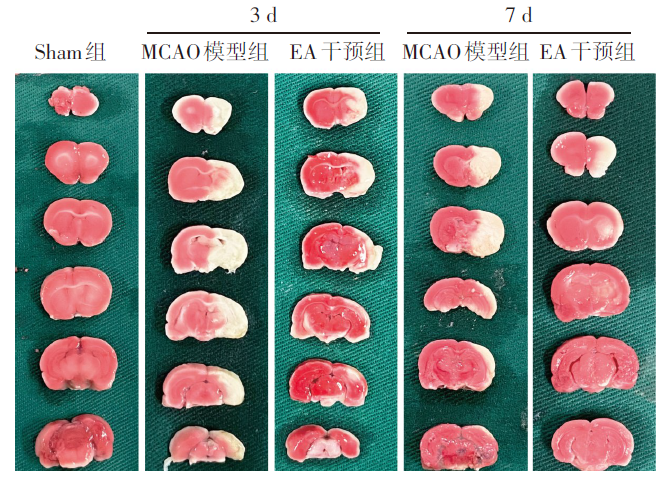
Fig.1 TTC staining of cerebral infarction area in each group
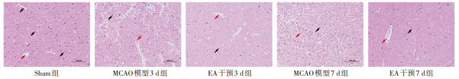
Fig.2 Comparison of pathomorphological changes of cortical areas on the infarct side of rats between the five groups (HE staining, ×200)
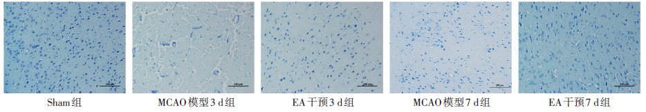
Fig.3 Comparison of neuronal morphology in the cortical area of the infarct side of rats between the five groups (Nissl staining, ×200)
| 组别 | AKT | p-mTOR/t-mTOR | t-mTOR |
|---|---|---|---|
| Sham组 | 2.07±1.14 | 1.15±0.40 | 0.87±0.32 |
| MCAO模型3 d组 | 2.89±0.70 | 0.65±0.22a | 1.40±0.26 |
| EA干预3 d组 | 2.33±0.73 | 0.69±0.30 | 1.10±0.33 |
| MCAO模型7 d组 | 2.74±0.59 | 1.49±0.40b | 1.02±0.93 |
| EA干预7 d组 | 2.11±0.90 | 0.63±0.21c | 0.97±0.32 |
| F | 0.972 | 7.015** | 2.470 |
Tab.4 Comparison of relative expression levels of AKT, p-mTOR and t-mTOR proteins in rat brain tissues between the five groups
| 组别 | AKT | p-mTOR/t-mTOR | t-mTOR |
|---|---|---|---|
| Sham组 | 2.07±1.14 | 1.15±0.40 | 0.87±0.32 |
| MCAO模型3 d组 | 2.89±0.70 | 0.65±0.22a | 1.40±0.26 |
| EA干预3 d组 | 2.33±0.73 | 0.69±0.30 | 1.10±0.33 |
| MCAO模型7 d组 | 2.74±0.59 | 1.49±0.40b | 1.02±0.93 |
| EA干预7 d组 | 2.11±0.90 | 0.63±0.21c | 0.97±0.32 |
| F | 0.972 | 7.015** | 2.470 |
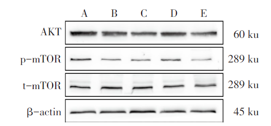
Fig.4 Western blot analysis showed the expressions of AKT, p-mTOR and t-mTOR in the five groups
| 组别 | LC3B | Beclin-1 | Bax |
|---|---|---|---|
| Sham组 | 1.00±0.00 | 1.00±0.00 | 1.00±0.00 |
| MCAO模型3 d组 | 0.21±0.00a | 0.21±0.01a | 0.57±0.08a |
| EA干预3 d组 | 0.19±0.02 | 0.16±0.00 | 0.53±0.06 |
| MCAO模型7 d组 | 0.62±0.23a | 0.17±0.01a | 1.58±0.21a |
| EA干预7 d组 | 1.68±0.09c | 0.76±0.03c | 1.30±0.10c |
| F | 142.452** | 2 229.827** | 78.799** |
Tab.5 Comparison of LC3B, Beclin-1 and Bax mRNA expressions in rat brain tissue at different time points between the five groups
| 组别 | LC3B | Beclin-1 | Bax |
|---|---|---|---|
| Sham组 | 1.00±0.00 | 1.00±0.00 | 1.00±0.00 |
| MCAO模型3 d组 | 0.21±0.00a | 0.21±0.01a | 0.57±0.08a |
| EA干预3 d组 | 0.19±0.02 | 0.16±0.00 | 0.53±0.06 |
| MCAO模型7 d组 | 0.62±0.23a | 0.17±0.01a | 1.58±0.21a |
| EA干预7 d组 | 1.68±0.09c | 0.76±0.03c | 1.30±0.10c |
| F | 142.452** | 2 229.827** | 78.799** |
| [1] | WANG Y, HAN S, QIN H, et al. Chinese Stroke Association guidelines for clinical management of cerebrovascular disorders:executive summary and 2019 update of the management of high-risk population[J]. Stroke Vasc Neurol, 2020, 5(3):270-278. doi:10.1136/svn-2020-000385. |
| [2] | 张游, 尹亚龙, 吴新贵. 基于EA疗法对脑梗死后神经功能恢复的研究进展[J]. 国际神经病学神经外科学杂志, 2022, 49(1):79-86. |
| ZHANG Y, YIN Y L, WU X G. Research progress of neurological function recovery after cerebral infarction based on electroacupuncture[J]. Journal of International Neurology and Neurosurgery, 2022, 49(1):79-86. doi:10.16636/j.cnki.jinn.1673-2642.2022.01.017. | |
| [3] | SHINTANI T, KLIONSKY D J. Autophagy in health and disease:A double-edged sword[J]. Science, 2004, 306(5698):990-995. doi:10.1126/science.1099993. |
| [4] | XU Q, MA G H, LI D D, et al. lncRNA C2dat2 facilitates autophagy and apoptosis via the miR-30d-5p/DDIT4/mTOR axis in cerebral ischemia-reperfusion injury[J]. Aging (Albany NY), 2021, 13(8):11315-11335. doi:10.18632/aging.202824. |
| [5] | MAIESE K, CHONG Z Z, SHANG Y C, et al. mTOR:on target for novel therapeutic strategies in the nervous system[J]. Trends Mol Med, 2013, 19(1):51-60. doi:10.1016/j.molmed.2012.11.001. |
| [6] | YAN H J, ZHANG X N, HU W W, et al. Histamine H3 receptors aggravate cerebral ischaemic injury by histamine-independent mechanisms[J]. Nat Commun, 2014, 5:3334. doi:10.1038/ncomms4334. |
| [7] | SHARIFIFAR S, SHUSTER J J, BISHOP M D. Adding electrical stimulation during standard rehabilitation after stroke to improve motor function. A systematic review and meta-analysis[J]. Ann Phys Rehabil Med, 2018, 61(5):339-344. doi:10.1016/j.rehab.2018.06.005. |
| [8] | BEDERSON J B, PITTS L H, TSUJI M, et al. Rat middle cerebral artery occlusion:evaluation of the model and development of a neurologic examination[J]. Stroke, 1986, 17:472-476. doi:10.1161/01.str.17.3.472. |
| [9] | LACKLAND D T, ROCCELLA E J, DEUTSCH A F, et al. Factors influencing the decline in stroke mortality:a statement from the American Heart Association/American Stroke Association[J]. Stroke, 2014, 45(1):315-353. doi:10.1161/01.str.0000437068.30550.cf. |
| [10] | LI W L, YU S P, OGLE M E, et al. Enhanced neurogenesis and cell migration following focal ischemia and peripheral stimulation in mice[J]. Dev Neurobiol, 2008, 68(13):1474-1486. doi:10.1002/dneu.20674. |
| [11] | 何敏, 刘志强, 蒋玲霞, 等. 电针疗法治疗急性脑梗死的研究进展[J]. 中国当代医药, 2019, 26(36):16-20. |
| HE M, LIU Z Q, JIANG L X, et al. Research progress of electro-acupuncture therapy in the treatment of acute cerebral infraction[J]. China Modern Medicine, 2019, 26(36):16-20. | |
| [12] | LONGHURST J C. Defining meridians:a modern basis of understanding[J]. J Acupunct Meridian Stud, 2010, 3(2):67-74. doi:10.1016/S2005-2901(10)60014-3. |
| [13] | 葛云鹏, 嵇波, 苏杭, 等. 艾灸足三里穴的现代研究进展[J]. 中医药导报, 2019, 25(22):95-99. |
| GE Y P, JI B, SU H, et al. Modern research progress of moxibustion at Zusanli (ST36) points[J]. Guiding Journal of Traditional Chinese Medicine and Pharmacy, 2019, 25(22):95-99. doi:10.13862/j.cnki.cn43-1446/r.2019.22.027. | |
| [14] | 向丽莉, 黄伟, 张小蕾, 等. 探意内关[J]. 中华中医药杂志, 2021, 36(7):3890-3892. |
| XIANG L L, HUANG W, ZHANG X L, et al. Exploration of Neiguan(PC 6)[J]. China Journal of Traditional Chinese Medicine and Pharmacy, 2021, 36(7):3890-3892. doi:CNKI:SUN:BXYY.0.2021-07-028. | |
| [15] | LI S. Spasticity,motor recovery,and neural plasticity after stroke[J]. Front Neurol, 2017, 8:120. doi:10.3389/fneur.2017.00120. |
| [16] | TIAN G H, TAO S S, CHEN M T, et al. Electroacupuncture treatment alleviates central poststroke pain by inhibiting brain neuronal apoptosis and aberrant astrocyte activation[J]. Neural Plast, 2016, 2016:1437148. doi:10.1155/2016/1437148. |
| [17] | DONG L L, QIU M M, LIU Y L, et al. Attenuation of histone H4 lysine 16 acetylation(H4K16ac)elicits a neuroprotection against ischemic stroke by alleviating the autophagic/lysosomal dysfunction in neurons at the penumbra[J]. Brain Res Bull, 2022, 184:24-33. doi:10.1016/j.brainresbull.2022.03.013. |
| [18] | WINDEN K D, EBRAHIMI F D, SAHIN M. Abnormal mTOR activation in autism[J]. Annu Rev Neurosci, 2018, 41:1-23. doi:10.1146/annurev-neuro-080317-061747. |
| [19] | SUN F, PARK K K, BELIN S, et al. Sustained axon regeneration induced by co-deletion of PTEN and SOCS3[J]. Nature, 2011, 480(7377):372-375. doi:10.1038/nature10594. |
| [20] | SHEHATA M, MATSUMURA H, OKUBO S R, et al. Neuronal stimulation induces autophagy in hippocampal neurons that is involved in AMPA receptor degradation after chemical long-term depression[J]. J Neurosci, 2012, 32(30):10413-10422. doi:10.1523/JNEUROSCI.4533-11.2012. |
| [21] | SAXTON R A, SABATINI D M. mTOR signaling in growth,metabolism,and disease[J]. Cell, 2017, 168(6):960-976. doi:10.1016/j.cell.2017.02.004 |
| [22] | ZHAO H, SHIMOHATA T, WANG J Q, et al. Akt contributes to neuroprotection by hypothermia against cerebral ischemia in rats[J]. J Neurosci, 2005, 25(42):9794-9806. doi:10.1523/JNEUROSCI.3163-05.2005. |
| [23] | HOSOKAWA N, HARA T, KAIZUKA T, et al. Nutrient-dependent mTORC1 association with the ULK1-Atg13-FIP200 complex required for autophagy[J]. Mol Biol Cell, 2009, 20(7):1981-1991. doi:10.1091/mbc.e08-12-1248. |
| [24] | HWANG J Y, GERTNER M, PONTARELLI F, et al. Global ischemia induces lysosomal-mediated degradation of mTOR and activation of autophagy in hippocampal neurons destined to die[J]. Cell Death Differ, 2017, 24(2):317-329. doi:10.1038/cdd.2016.140. |
| [25] | KULBE J R, MULCAHY L J M, COULTRAP S J, et al. Excitotoxic glutamate insults block autophagic flux in hippocampal neurons[J]. Brain Res, 2014, 1542:12-19. doi:10.1016/j.brainres.2013.10.032. |
| [26] | YUAN J Y. Neuroprotective strategies targeting apoptotic and necrotic cell death for stroke[J]. Apoptosis, 2009, 14(4):469-477. doi:10.1007/s10495-008-0304-8. |
| [27] | FU C, ZHANG X Y, LU Y, et al. Geniposide inhibits NLRP3 inflammasome activation via autophagy in BV-2 microglial cells exposed to oxygen-glucose deprivation/reoxygenation[J]. Int Immunopharmacol, 2020, 84:106547. doi:10.1016/j.intimp.2020.106547. |
| [28] | SCORRANO L, KORSMEYER S J. Mechanisms of cytochrome c release by proapoptotic BCL-2 family members[J]. Biochem Biophys Res Commun, 2003, 304(3):437-444. doi:10.1016/s0006-291x(03)00615-6. |
| [29] | WASAN H, SINGH D, JOSHI B, et al. Post stroke safinamide treatment attenuates neurological damage by modulating autophagy and apoptosis in experimental model of stroke in rats[J]. Mol Neurobiol, 2021, 58(12):6121-6135. doi:10.1007/s12035-021-02523-6. |
| [1] | HUANG Huiqi, WU Qiuyuan, ZHANG Kun, LI Peixian, XIONG Yaming, YE Guolin, ZHOU Dan. Research on the anti-tumor mechanism of toosendanin combined with olaparib in triple negative breast cancer [J]. Tianjin Medical Journal, 2025, 53(9): 897-902. |
| [2] | LIN Yiwei, WEI Tanjun, CHEN Fei, XIAO Cheng, YUAN Lie, WANG Yi. The in vivo and in vitro effects of Eriodictyol on metabolic dysfunction-associated steatotic liver disease by regulating UBA52 expression [J]. Tianjin Medical Journal, 2025, 53(9): 916-922. |
| [3] | MA Chunmei, YU Peng, ZHANG Qicheng, YANG Lei, LI Dihua, TAN Jian, MENG Zhaowei. Mechanism study of benzyl isothiocyanate combined with sorafenib in the treatment of anaplastic thyroid cancer [J]. Tianjin Medical Journal, 2025, 53(5): 449-455. |
| [4] | LI Bingxin, XU Junying, ZHANG Yaru, ZHOU Xiaobing. Effect of Cordyceps sinensis on podocyte damage induced by high glucose by regulating the AMPK/mTOR pathway [J]. Tianjin Medical Journal, 2025, 53(3): 225-229. |
| [5] | FANG Jie, HUANG Rui, ZHENG Honghui, JIA Qianqian, BAO Jing. miR-9-5p-induced autophagy and apoptosis in multiple myeloma cells by targeting TIMP2 [J]. Tianjin Medical Journal, 2024, 52(8): 785-790. |
| [6] | LI Daqiang, LI Jian, LU Zheming, CAO Yang. Effects of calycosin on neuronal autophagy and apoptosis in rats with spinal cord injury [J]. Tianjin Medical Journal, 2024, 52(8): 798-803. |
| [7] | WANG Junyi, LI Chen, WU Xinyue, DING Xinyu, WAN Chunxiao. Effect and mechanism of early exercise intervention on cerebral nerve myelin in rats with cerebral ischemia [J]. Tianjin Medical Journal, 2024, 52(6): 589-594. |
| [8] | ZHANG Lihong, LI Ruiqing, WANG Yiying, MEI Jinjin, SU Kaiqi, GU Changyu, HUANG Mengling. The mechanism of Du meridian electroacupuncture regulating cystine/glutamate reverse transporter to improve limb spasm after stroke [J]. Tianjin Medical Journal, 2024, 52(5): 463-468. |
| [9] | WANG Ke, YE Hanlu. Impacts of cryptotanshinone on autophagy and apoptosis of chondrocytes in rabbit model of knee osteoarthritis by regulating HIF-1α/BNIP3 signaling pathway [J]. Tianjin Medical Journal, 2024, 52(4): 372-378. |
| [10] | HE Ying, ZHANG Guanghua, TIAN Lidong, YU Yonghao. Hydrogen-rich saline treated neuropathic pain in rats by increasing autophagy [J]. Tianjin Medical Journal, 2024, 52(3): 261-265. |
| [11] | WANG Aihua, ZHANG Feizhong, WANG Hongying. Impacts of muscone on malignant progression of ovarian cancer cells by regulating SHH mediated autophagy [J]. Tianjin Medical Journal, 2024, 52(2): 142-147. |
| [12] | ZHAO Yuanyuan, WU Xiaohua. The effect of LINC00173 regulating autophagy of PCOS granulosa cells based on PI3K/Akt/mTOR signaling pathway [J]. Tianjin Medical Journal, 2024, 52(11): 1121-1126. |
| [13] | OUYANG Jie, ZHAO Haiqian, KONG Yun, NIU Qin, CHEN Ying, SI Yongyu. The effect of electroacupuncture on paclitaxel-induced neuropathic pain in rats [J]. Tianjin Medical Journal, 2024, 52(11): 1141-1145. |
| [14] | ZHANG Rui, CHEN Sisi, WANG Tongdan, YU Pei. Krüppel-like factor 4 alleviated cholesterol deposition in macrophages by promoting autophagy at high glucose concentration [J]. Tianjin Medical Journal, 2024, 52(10): 1014-1019. |
| [15] | QIAO Na, TIAN Ying, CHEN Yang, HAO Jing. Impacts of lncRNA MALAT1 on apoptosis, autophagy of granulosa cells and PI3K/Akt/mTOR pathway in polycystic ovary syndrome [J]. Tianjin Medical Journal, 2024, 52(10): 1020-1024. |
| Viewed | ||||||
|
Full text |
|
|||||
|
Abstract |
|
|||||