Tianjin Medical Journal ›› 2023, Vol. 51 ›› Issue (9): 977-982.doi: 10.11958/20221801
• Experimental Research • Previous Articles Next Articles
ZHANG Jinwu( ), XIE Dingling, CHEN Li△()
Received:2022-11-14
Revised:2023-01-12
Published:2023-09-15
Online:2023-09-13
Contact:
△E-mail: ZHANG Jinwu, XIE Dingling, CHEN Li. The effect of securinine on neurological function recovery after cerebral ischemia-reperfusion injury in rats[J]. Tianjin Medical Journal, 2023, 51(9): 977-982.
CLC Number:
| 组别 | n | 神经功能缺损评分/分 | ||
|---|---|---|---|---|
| 24 h | 48 h | 72 h | ||
| Sham组 | 8 | 0.00±0.00 | 0.00±0.00 | 0.00±0.00 |
| CIRI组 | 8 | 2.75±0.39a | 2.70±0.38a | 2.46±0.40a |
| SE低剂量组 | 8 | 2.71±0.38 | 2.46±0.43 | 2.33±0.44 |
| SE中剂量组 | 8 | 2.42±0.35 | 2.25±0.43b | 1.95±0.38bc |
| SE高剂量组 | 8 | 2.21±0.40b | 1.83±0.44b | 1.33±0.40bcd |
| F | 3.687* | 6.249* | 12.625* | |
Tab.1 Comparison of neurological function scores after CIRI between the five groups of rats
| 组别 | n | 神经功能缺损评分/分 | ||
|---|---|---|---|---|
| 24 h | 48 h | 72 h | ||
| Sham组 | 8 | 0.00±0.00 | 0.00±0.00 | 0.00±0.00 |
| CIRI组 | 8 | 2.75±0.39a | 2.70±0.38a | 2.46±0.40a |
| SE低剂量组 | 8 | 2.71±0.38 | 2.46±0.43 | 2.33±0.44 |
| SE中剂量组 | 8 | 2.42±0.35 | 2.25±0.43b | 1.95±0.38bc |
| SE高剂量组 | 8 | 2.21±0.40b | 1.83±0.44b | 1.33±0.40bcd |
| F | 3.687* | 6.249* | 12.625* | |
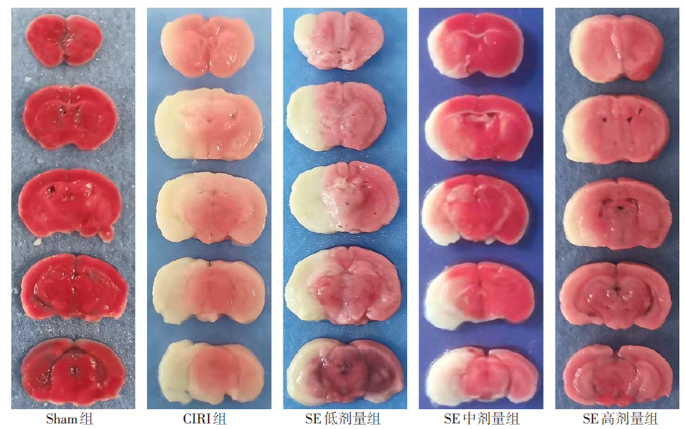
Fig.1 Percentage of cerebral infarction volume after CIRI in each group of rats (TTC staining)
| 组别 | n | 脑梗死体积百分比 | n | 脑组织含水量 |
|---|---|---|---|---|
| Sham组 | 5 | 0.00±0.00 | 6 | 66.54±2.37 |
| CIRI组 | 5 | 42.53±3.00a | 6 | 82.49±7.30a |
| SE低剂量组 | 5 | 39.18±3.27 | 6 | 79.18±7.76 |
| SE中剂量组 | 5 | 27.02±2.45bc | 6 | 73.17±8.63b |
| SE高剂量组 | 5 | 16.91±2.17bcd | 6 | 68.78±8.33bc |
| F | 90.629* | 5.204* |
Tab.2 Comparison of the percentage of cerebral infarct volume and brain tissue water content after CIRI between the five groups of rats
| 组别 | n | 脑梗死体积百分比 | n | 脑组织含水量 |
|---|---|---|---|---|
| Sham组 | 5 | 0.00±0.00 | 6 | 66.54±2.37 |
| CIRI组 | 5 | 42.53±3.00a | 6 | 82.49±7.30a |
| SE低剂量组 | 5 | 39.18±3.27 | 6 | 79.18±7.76 |
| SE中剂量组 | 5 | 27.02±2.45bc | 6 | 73.17±8.63b |
| SE高剂量组 | 5 | 16.91±2.17bcd | 6 | 68.78±8.33bc |
| F | 90.629* | 5.204* |
| 组别 | TLR4/ β-actin | p-NF-κB p65/ t-NF-κB p65 | p-NF-κB/ β-actin | Iba-1/ β-actin |
|---|---|---|---|---|
| Sham组 | 0.36±0.03 | 0.66±0.05 | 0.45±0.05 | 0.29±0.05 |
| CIRI组 | 0.57±0.04a | 1.19±0.06a | 0.86±0.07a | 0.76±0.06a |
| SE低剂量组 | 0.55±0.04 | 1.08±0.07 | 0.76±0.07 | 0.68±0.06 |
| SE中剂量组 | 0.40±0.04bc | 0.73±0.05bc | 0.49±0.05bc | 0.39±0.06bc |
| SE高剂量组 | 0.39±0.04bc | 0.67±0.06bc | 0.47±0.05bc | 0.29±0.05bcd |
| F | 45.248* | 101.488* | 61.862* | 92.760* |
Tab.3 Comparison of TLR4, NF-κB p65 and Iba-1 protein expression levels of brain tissue after CIRI between the five groups of rats
| 组别 | TLR4/ β-actin | p-NF-κB p65/ t-NF-κB p65 | p-NF-κB/ β-actin | Iba-1/ β-actin |
|---|---|---|---|---|
| Sham组 | 0.36±0.03 | 0.66±0.05 | 0.45±0.05 | 0.29±0.05 |
| CIRI组 | 0.57±0.04a | 1.19±0.06a | 0.86±0.07a | 0.76±0.06a |
| SE低剂量组 | 0.55±0.04 | 1.08±0.07 | 0.76±0.07 | 0.68±0.06 |
| SE中剂量组 | 0.40±0.04bc | 0.73±0.05bc | 0.49±0.05bc | 0.39±0.06bc |
| SE高剂量组 | 0.39±0.04bc | 0.67±0.06bc | 0.47±0.05bc | 0.29±0.05bcd |
| F | 45.248* | 101.488* | 61.862* | 92.760* |
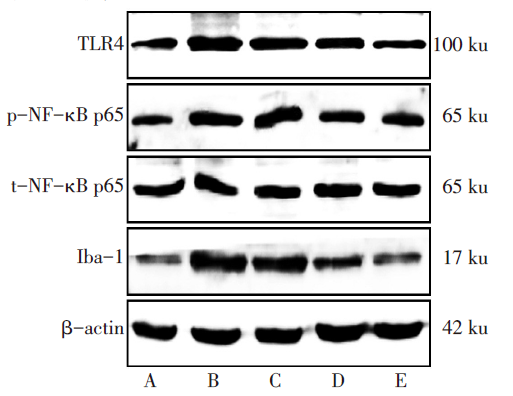
Fig.2 Expression levels of TLR4, NF-κB p65 and Iba-1 after CIRI in each group detected by Western blot assay
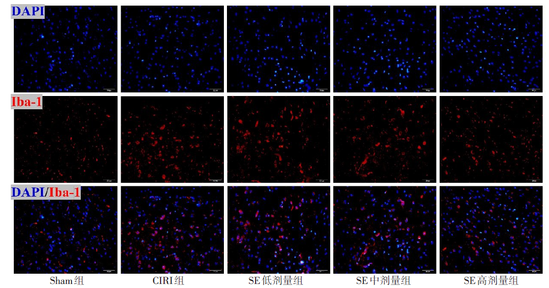
Fig.3 Expression levels of Iba-1 after CIRI in each group (immunofluorescent staining,×400)
| 组别 | n | IL-1β | TNF-α | IL-6 |
|---|---|---|---|---|
| Sham组 | 5 | 30.20±4.67 | 9.19±0.87 | 0.43±0.11 |
| CIRI组 | 5 | 85.67±7.39a | 18.61±1.15a | 2.64±0.32a |
| SE低剂量组 | 5 | 81.64±6.87 | 17.27±1.31 | 2.42±0.24 |
| SE中剂量组 | 5 | 55.32±6.32bc | 13.86±1.25bc | 1.64±0.21bc |
| SE高剂量组 | 5 | 45.64±5.82bcd | 12.05±1.14bcd | 0.91±1.14bcd |
| F | 85.261* | 66.366* | 116.747* |
Tab.4 Comparison of IL-1β, TNF-α and IL-6 levels of brain tissue after CIRI between the five groups of rats
| 组别 | n | IL-1β | TNF-α | IL-6 |
|---|---|---|---|---|
| Sham组 | 5 | 30.20±4.67 | 9.19±0.87 | 0.43±0.11 |
| CIRI组 | 5 | 85.67±7.39a | 18.61±1.15a | 2.64±0.32a |
| SE低剂量组 | 5 | 81.64±6.87 | 17.27±1.31 | 2.42±0.24 |
| SE中剂量组 | 5 | 55.32±6.32bc | 13.86±1.25bc | 1.64±0.21bc |
| SE高剂量组 | 5 | 45.64±5.82bcd | 12.05±1.14bcd | 0.91±1.14bcd |
| F | 85.261* | 66.366* | 116.747* |

Fig.4 The number of neuronal survival after CIRI in each group (immunofluorescent staining,×400)
| [1] | BOOT E, EKKER M S, PUTAALA J, et al. Ischaemic stroke in young adults:a global perspective[J]. J Neurol Neurosurg Psychiatry, 2020, 91(4):411-417. doi:10.1136/jnnp-2019-322424. |
| [2] | DONG X, WANG L, SONG G, et al. Physcion protects rats against cerebral ischemia-reperfusion injury via inhibition of TLR4/NF-κB signaling pathway[J]. Drug Des Devel Ther, 2021, 15:277-287. doi:10.2147/DDDT.S267856. |
| [3] | ZHOU X, WANG Z, XU B, et al. Long non-coding RNA NORAD protects against cerebral ischemia/reperfusion injury induced brain damage,cell apoptosis,oxidative stress and inflammation by regulating miR-30a-5p/YWHAG[J]. Bioengineered, 2021, 12(2):9174-9188. doi:10.1080/21655979.2021.1995115. |
| [4] | XIE X, WANG F, LI X. Inhibition of TRIM14 protects cerebral ischemia/reperfusion injury through regulating NF-κB/NLRP3 pathway-mediated inflammation and apoptosis[J]. J Recept Signal Transduct Res, 2022, 42(2):197-205. doi:10.1080/10799893.2021.1887218. |
| [5] | ZHENG K, ZHANG Y, ZHANG C, et al. PRMT8 attenuates cerebral ischemia/reperfusion injury via modulating microglia activation and polarization to suppress neuroinflammation by upregulating Lin28a[J]. ACS Chem Neurosci, 2022, 13(7):1096-1104. doi:10.1021/acschemneuro.2c00096. |
| [6] | CHIRKIN E, ATKATLIAN W, POREE F H. The securinega alkaloids[J]. Chem Biol, 2015, 74:1-120. doi:10.1016/bs.alkal.2014.11.001. |
| [7] | BEUTLER J A, KARBON E W, BRUBAKER A N, et al. Securinine alkaloids:a new class of GABA receptor antagonist[J]. Brain Res, 1985, 330(1):135-140. doi:10.1016/0006-8993(85)90014-9. |
| [8] | BURAVTSEVA G R. Result of application of securinine in acute poliomyelitis[J]. Farmakol Toksikol, 1958, 21(5):7-12. doi:10.1371/journal.pone.0021203. |
| [9] | COPPERMAN R, COPPERMAN G, DER MARDEROSIAN A. From Asia securinine:a central nervous stimulant is used in treatment of amytrophic lateral sclerosis[J]. Pa Med, 1973, 76(1):36-41. |
| [10] | LIN X, JUN T Z. Neuroprotection by D-securinine against neurotoxicity induced by beta-amyloid(25-35)[J]. Neurol Res, 2004, 26(7):792-796. doi:10.1179/016164104225014148. |
| [11] | LEONOUDAKIS D, RANE A, ANGELI S, et al. Anti-inflammatory and neuroprotective role of natural product securinine in activated glial cells:Implications for Parkinson's disease[J]. Mediators Inflamm, 2017, 2017:8302636. doi:10.1155/2017/8302636. |
| [12] | LONGA E Z, WEINSTEIN P R, CARLSON S, et al. Reversible middle cerebral artery occlusion without craniectomy in rats[J]. Stroke, 1989, 20(1):84-91. doi:10.1161/01.str.20.1.84. |
| [13] | YE Y, JIN T, ZHANG X, et al. Meisoindigo protects against focal cerebral ischemia-reperfusion injury by inhibiting NLRP3 inflammasome activation and regulating microglia/macrophage polarization via TLR4/NF-kappaB signaling pathway[J]. Front Cell Neurosci, 2019, 13:553. doi:10.3389/fncel.2019.00553. |
| [14] | WU M Y, YIANG G T, LIAO W T, et al. Current mechanistic concepts in ischemia and reperfusion injury[J]. Cell Physiol Biochem, 2018, 46(4):1650-1667. doi:10.1159/000489241. |
| [15] | CUI Y, ZHANG N N, WANG D, et al. Modified citrus pectin alleviates cerebral ischemia/reperfusion injury by inhibiting NLRP3 inflammasome activation via TLR4/NF-κB signaling pathway in microglia[J]. J Inflamm Res, 2022, 15:3369-3385. doi:10.2147/JIR.S366927. |
| [16] | NEGANOVA M E, KLOCHKOV S G, AFANASIEVA S V, et al. Neuroprotective effects of the securinine-analogues:Identification of allomargaritarine as a lead compound[J]. CNS Neurol Disord Drug Targets, 2016, 15(1):102-107. doi:10.2174/1871527314666150821111812. |
| [17] | XIAO H, ZHANG Q, ZHONG P, et al. Securinine promotes neuronal development and exhibits antidepressant-like effects via mtor activation[J]. ACS Chem Neurosci, 2021, 12(19):3650-3661. doi:10.1021/acschemneuro.1c00381. |
| [18] | LIU Q, ZHANG Y. PRDX1 enhances cerebral ischemia-reperfusion injury through activation of TLR4-regulated inflammation and apoptosis[J]. Biochem Biophys Res Commun, 2019, 519(3):453-461. doi:10.1016/j.bbrc.2019.08.077. |
| [19] | ZHAO D, JI J, LI S, et al. Skullcapflavone II protects neuronal damage in cerebral ischemic rats via inhibiting NF-κB and promoting angiogenesis[J]. Microvasc Res, 2022, 141:104318. doi:10.1016/j.mvr.2022.104318. |
| [20] | TIAN X Y, XIE L, WANG W Y, et al. Pomelo peel volatile oil alleviates neuroinflammation on focal cerebral ischemia reperfusion injury rats via inhibiting TLR4/NF-κB signaling pathway[J]. Curr Pharm Biotechnol, 2021, 22(14):1878-1890. doi:10.2174/1389201022666201231114403. |
| [21] | LIU J, MA W, ZANG C H, et al. Salidroside inhibits NLRP3 inflammasome activation and apoptosis in microglia induced by cerebral ischemia/reperfusion injury by inhibiting the TLR4/NF-κB signaling pathway[J]. Ann Transl Med, 2021, 9(22):1694. doi:10.21037/atm-21-5752. |
| Viewed | ||||||
|
Full text |
|
|||||
|
Abstract |
|
|||||