Tianjin Medical Journal ›› 2022, Vol. 50 ›› Issue (9): 921-926.doi: 10.11958/20220034
• Experimental Research • Previous Articles Next Articles
LONG Guangwen( ), ZHANG Qian, YANG Xiulin, JI Chunling, DONG Yukang
Received:2022-01-06
Revised:2022-02-22
Published:2022-09-15
Online:2022-09-05
LONG Guangwen, ZHANG Qian, YANG Xiulin, JI Chunling, DONG Yukang. The effect and mechanism of inhibiting miR-33 expression on pulmonary fibrosis in rats with acute respiratory distress syndrome[J]. Tianjin Medical Journal, 2022, 50(9): 921-926.
CLC Number:
| 基因 名称 | 引物序列(5'→3') | 产物大小(bp) |
|---|---|---|
| miR-33 | 上游:GATCCTCAGTGCATTGTAGTTGC | 69 |
| 下游:CTCTGTCTCTCGTCTTGTTGGTAT | ||
| U6 | 上游:CCTGCTTCGGCAG CACA | 96 |
| 下游:AACGCTTCACGAATTTGCGT | ||
| TGF-β1 | 上游:GTGGAAATCAACGGGATCAG | 188 |
| 下游:CGCACACAGCAGTTCTTCTC | ||
| CollagenⅠ | 上游:TTGCTTCCCAGATGTCCTATG | 108 |
| 下游:CTTCCCCATCATCTCCATTCT | ||
| CollagenⅢ | 上游:CAGACGGGA GTTTCTCCTCGGACGT | 210 |
| 下游:GACCAGGAGGACCAGGAAGTCCACGT | ||
| GAPDH | 上游:AACTGCTTAGCACCCCTGGC | 92 |
| 下游:ATGACCTTGCCCA CACAGCCTT |
Tab.1 Primer sequence for qPCR
| 基因 名称 | 引物序列(5'→3') | 产物大小(bp) |
|---|---|---|
| miR-33 | 上游:GATCCTCAGTGCATTGTAGTTGC | 69 |
| 下游:CTCTGTCTCTCGTCTTGTTGGTAT | ||
| U6 | 上游:CCTGCTTCGGCAG CACA | 96 |
| 下游:AACGCTTCACGAATTTGCGT | ||
| TGF-β1 | 上游:GTGGAAATCAACGGGATCAG | 188 |
| 下游:CGCACACAGCAGTTCTTCTC | ||
| CollagenⅠ | 上游:TTGCTTCCCAGATGTCCTATG | 108 |
| 下游:CTTCCCCATCATCTCCATTCT | ||
| CollagenⅢ | 上游:CAGACGGGA GTTTCTCCTCGGACGT | 210 |
| 下游:GACCAGGAGGACCAGGAAGTCCACGT | ||
| GAPDH | 上游:AACTGCTTAGCACCCCTGGC | 92 |
| 下游:ATGACCTTGCCCA CACAGCCTT |
| 组别 | p(O2) | OI |
|---|---|---|
| Sham组 | 97.73±7.49 | 459.30±21.27 |
| Model组 | 68.11±1.99a | 158.94±18.48a |
| antagomir-NC组 | 69.60±1.55a | 157.19±13.64a |
| antagomir组 | 80.24±1.91abc | 268.27±22.52abc |
| F | 33.845** | 162.902** |
Tab.2 Comparison of p(O2) and OI between the four groups of rats
| 组别 | p(O2) | OI |
|---|---|---|
| Sham组 | 97.73±7.49 | 459.30±21.27 |
| Model组 | 68.11±1.99a | 158.94±18.48a |
| antagomir-NC组 | 69.60±1.55a | 157.19±13.64a |
| antagomir组 | 80.24±1.91abc | 268.27±22.52abc |
| F | 33.845** | 162.902** |
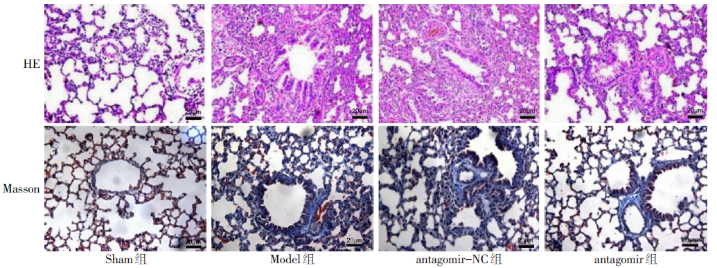
Fig.1 HE staining and Masson staining of lung tissue (×200)
| 组别 | IL-1β | IL-6 | TNF-α |
|---|---|---|---|
| Sham组 | 28.70±8.30 | 41.84±10.71 | 107.51±19.57 |
| Model组 | 61.56±11.72a | 158.18±24.13a | 431.92±23.58a |
| antagomir-NC组 | 60.79±8.85a | 157.15±35.72a | 430.37±16.17a |
| antagomir组 | 39.83±2.23bc | 88.64±12.92abc | 288.11±23.74abc |
| F | 10.859** | 18.017** | 160.420** |
Tab.3 Comparison of inflammatory cytokinesin in lung tissue between the four groups of rats (n=15,ng/L,$\bar{x}±s$)
| 组别 | IL-1β | IL-6 | TNF-α |
|---|---|---|---|
| Sham组 | 28.70±8.30 | 41.84±10.71 | 107.51±19.57 |
| Model组 | 61.56±11.72a | 158.18±24.13a | 431.92±23.58a |
| antagomir-NC组 | 60.79±8.85a | 157.15±35.72a | 430.37±16.17a |
| antagomir组 | 39.83±2.23bc | 88.64±12.92abc | 288.11±23.74abc |
| F | 10.859** | 18.017** | 160.420** |
| 组别 | miR-33 | TGF-β1 | CollagenⅠ | CollagenⅢ |
|---|---|---|---|---|
| Sham组 | 1.00±0.27 | 1.01±0.05 | 1.00±0.07 | 1.00±0.09 |
| Model组 | 2.21±0.26a | 2.18±0.24a | 1.80±0.17a | 1.97±0.21a |
| antagomir-NC组 | 2.21±0.20a | 2.15±0.21a | 1.80±0.15a | 1.94±0.12a |
| antagomir组 | 0.08±0.02abc | 1.38±0.17abc | 1.46±0.06abc | 1.35±0.11abc |
| F | 71.948** | 30.299** | 29.265** | 34.497** |
Tab.4 Comparison of the expression level of miR-33 and mRNA expression levels of TGF-β1, CollagenⅠand Collagen Ⅲ between the four groups of rats
| 组别 | miR-33 | TGF-β1 | CollagenⅠ | CollagenⅢ |
|---|---|---|---|---|
| Sham组 | 1.00±0.27 | 1.01±0.05 | 1.00±0.07 | 1.00±0.09 |
| Model组 | 2.21±0.26a | 2.18±0.24a | 1.80±0.17a | 1.97±0.21a |
| antagomir-NC组 | 2.21±0.20a | 2.15±0.21a | 1.80±0.15a | 1.94±0.12a |
| antagomir组 | 0.08±0.02abc | 1.38±0.17abc | 1.46±0.06abc | 1.35±0.11abc |
| F | 71.948** | 30.299** | 29.265** | 34.497** |
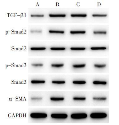
Fig.2 Western blot assay of TGF-β1, Smad2, p-Smad2, Smad3, p-Smad3 and α-SMA in lung tissue of rat in each group
| 组别 | TGF-β1 | p-Smad2/ Smad2 | p-Smad3/ Smad3 | α-SMA |
|---|---|---|---|---|
| Sham组 | 0.31±0.05 | 0.32±0.03 | 0.41±0.06 | 0.21±0.02 |
| Model组 | 0.75±0.07a | 0.87±0.10a | 0.79±0.06a | 0.49±0.05a |
| antagomir-NC组 | 0.74±0.05a | 0.87±0.11a | 0.79±0.07a | 0.50±0.07a |
| antagomir组 | 0.37±0.02bc | 0.56±0.10abc | 0.61±0.06abc | 0.30±0.04bc |
| F | 61.722** | 26.354* * | 23.452* * | 22.852* * |
Tab.5 Comparison of protein expression levels of TGF-β1, p-Smad2/Smad2, p-Smad3/Smad3 and α-SMA in lung tissue between the four groups of rats
| 组别 | TGF-β1 | p-Smad2/ Smad2 | p-Smad3/ Smad3 | α-SMA |
|---|---|---|---|---|
| Sham组 | 0.31±0.05 | 0.32±0.03 | 0.41±0.06 | 0.21±0.02 |
| Model组 | 0.75±0.07a | 0.87±0.10a | 0.79±0.06a | 0.49±0.05a |
| antagomir-NC组 | 0.74±0.05a | 0.87±0.11a | 0.79±0.07a | 0.50±0.07a |
| antagomir组 | 0.37±0.02bc | 0.56±0.10abc | 0.61±0.06abc | 0.30±0.04bc |
| F | 61.722** | 26.354* * | 23.452* * | 22.852* * |
| [1] | STEVENS J P, LAW A, GIANNAKOULIS J. Acute respiratory distress syndrome[J]. JAMA, 2018, 319(7):732. doi: 10.1001/jama.2018.0483. |
| [2] | SHAH J, RANA S S. Acute respiratory distress syndrome in acute pancreatitis[J]. Indian J Gastroenterol, 2020, 39(2):123-132. doi: 10.1007/s12664-020-01016-z. |
| [3] | MATTHAY M A, ALDRICH J M, GOTTS J E. Treatment for severe acute respiratory distress syndrome from COVID-19[J]. Lancet Respir Med, 2020, 8(5):433-434. doi: 10.1016/S2213-2600(20)30127-2. |
| [4] | SHIMBORI C, BELLAYE P S, XIA J, et al. Fibroblast growth factor-1 attenuates TGF-β1-induced lung fibrosis[J]. J Pathol, 2016, 240(2):197-210. doi: 10.1002/path.4768. |
| [5] | NITHIANANTHAN S, CRAWFORD A, KNOCK J C, et al. Physiological fluid flow moderates fibroblast responses to TGF-β1[J]. J Cell Biochem, 2017, 118(4):878-890. doi: 10.1002/jcb.25767. |
| [6] | HU X, HUANG X. Alleviation of inflammatory response of pulmonary fibrosis in acute respiratory distress syndrome by puerarin via transforming growth factor(TGF-β1)[J]. Med Sci Monit, 2019, 25:6523-6531. doi: 10.12659/MSM.915570. |
| [7] | PRICE N L, MIGUEL V, DING W, et al. Genetic deficiency or pharmacological inhibition of miR-33 protects from kidney fibrosis[J]. JCI Insight, 2019, 4(22):e131102. doi: 10.1172/jci.insight.131102. |
| [8] | CHEN Z, DING H S, GUO X, et al. MiR-33 promotes myocardial fibrosis by inhibiting MMP16 and stimulating p38 MAPK signaling[J]. Oncotarget, 2018, 9(31):22047-22057. doi: 10.18632/oncotarget.25173. |
| [9] | NISHIGA M, HORIE T, KUWABARA Y, et al. MicroRNA-33 controls adaptive fibrotic response in the remodeling heart by preserving lipid raft cholesterol[J]. Circ Res, 2017, 120(5):835-847. doi: 10.1161/CIRCRESAHA.116.309528. |
| [10] | 马绍磊, 王宇杰, 左祥荣, 等. 内毒素诱导ARDS对大鼠右心功能的影响[J]. 中华危重病急救医学, 2018, 30(3):204-208. |
| MA S L, WANG Y J, ZUO X R, et al. Effects of acute respiratory distress syndrome induced by endotoxin on the right ventricular function in rats[J]. Chin Crit Care Med, 2018, 30(3):204-208. doi: 10.3760/cma.j.issn.2095-4352.2018.03.003. | |
| [11] | GAO J, CHU W, DUAN J, et al. Six-month outcomes of post-ARDS pulmonary fibrosis in patients with H1N1 pneumonia[J]. Front Mol Biosci, 2021, 8:640763. doi: 10.3389/fmolb.2021.640763. |
| [12] | HUPPERT L A, MATTHAY M A, WARE L B. Pathogenesis of acute respiratory distress syndrome[J]. Semin Respir Crit Care Med, 2019, 40(1):31-39. doi: 10.1055/s-0039-1683996. |
| [13] | SANCHEZ M L. Mechanical ventilation in patients subjected to extracorporeal membrane oxygenation(ECMO)[J]. Med Intensiva, 2017, 41(8):491-496. doi: 10.1016/j.medin.2016.12.007. |
| [14] | 赵振宇. miR-33表达与炎症反应的关系[J]. 医学信息, 2020, 33(2):73-75. |
| ZHAO Z Y. Relationship between miR-33 expression and inflammatory response[J]. Medical Information, 2020, 33(2):73-75. doi: 10.3969/j.issn.1006-1959.2020.02.020. | |
| [15] | 秦永亭, 张晓蕾, 方小霞, 等. miR-33-5p对糖尿病肾脏疾病大鼠肾纤维化影响机制的研究[J]. 中国糖尿病杂志, 2021, 29(5):378-383. |
| QIN Y T, ZHANG X L, FANG X X, et al. Effect and mechanism of miR-33-5p on renal fibrosis in diabetic kidney disease rats[J]. Chinese Journal of Diabetes, 2021, 29(5):378-383. doi: 10.3969/j.issn.1006-6187.2021.05.011. | |
| [16] | TOMITA K, TERATANI T, SUZUKI T, et al. Free cholesterol accumulation in hepatic stellate cells:mechanism of liver fibrosis aggravation in nonalcoholic steatohepatitis in mice[J]. Hepatology, 2014, 59(1):154-169. doi: 10.1002/hep.26604. |
| [17] | YU B, LI W, AL F, et al. MicroRNA-33a deficiency inhibits proliferation and fibrosis through inactivation of TGF-β/Smad pathway in human cardiac fibroblasts[J]. Pharmazie, 2017, 72(8):456-460. doi: 10.1691/ph.2017.7561. |
| [18] | KIM K K, SHEPPARD D, CHAPMAN H A. TGF-β1 signaling and tissue fibrosis[J]. Cold Spring Harb Perspect Biol, 2018, 10(4):a022293. doi: 10.1101/cshperspect.a022293. |
| [19] | 陈希琦, 张晓双, 周永坤, 等. TGF-β1/Smads信号通路在纤维化疾病中的研究进展[J]. 中国中西医结合外科杂志, 2021, 27(2):351-354. |
| CHEN X Q, ZHANG X S, ZHOU Y K, et al. Research progress of TGF-β1/Smads signaling pathway in fibrotic diseases[J]. Chinese Journal of Surgery of Integrated Traditional and Western Medicine, 2021, 27(2):351-354. doi: 10.3969/j.issn.1007-6948.2021.02.037. | |
| [20] | WANG L, LIU J, XIE W, et al. miR-425 reduction causes aberrant proliferation and collagen synthesis through modulating TGF-β/Smad signaling in acute respiratory distress syndrome[J]. Int J Clin Exp Pathol, 2019, 12(7):2604-2612. |
| [21] | CAO Y, LIU Y, PING F, et al. miR-200b/c attenuates lipopolysaccharide-induced early pulmonary fibrosis by targeting ZEB1/2 via p38 MAPK and TGF-β/smad3 signaling pathways[J]. Lab Invest, 2018, 98(3):339-359. doi: 10.1038/labinvest.2017.123. |
| [22] | MU E, DING R, AN X, et al. Heparin attenuates lipopolysaccharide-induced acute lung injury by inhibiting nitric oxide synthase and TGF-β/Smad signaling pathway[J]. Thromb Res, 2012, 129(4):479-485. doi: 10.1016/j.thromres.2011.10.003. |
| [23] | ZHANG Y Q, LIU Y J, MAO Y F, et al. Resveratrol ameliorates lipopolysaccharide-induced epithelial mesenchymal transition and pulmonary fibrosis through suppression of oxidative stress and transforming growth factor-β1 signaling[J]. Clin Nutr, 2015, 34(4):752-760. doi: 10.1016/j.clnu.2014.08.014. |
| [1] | HUANG Wei, WANG Jianjian, HUANG Ying, YANG Jun. The effect of compound Kushen Injection combined with chemotherapy and bevacizumab on short-term efficacy of patients with ovarian cancer [J]. Tianjin Medical Journal, 2026, 54(1): 88-92. |
| [2] | YAN Lingxin, LI Sen, GUO Gaili, MENG Wanqiu, XU Chao. Isovitexin regulates proliferation, migration and invasion of pancreatic cancer cells via the miR-339-5p/HSPA8 axis [J]. Tianjin Medical Journal, 2025, 53(3): 230-235. |
| [3] | HU Lao, ZHANG Cheng, HU Zhijun. The evaluation value of serum TRPV1, TIMP4 and TGF-β1 levels in predicting recurrence of benign paroxysmal positional vertigo [J]. Tianjin Medical Journal, 2025, 53(3): 267-271. |
| [4] | LIU Wenjie, WU Fan, ZHAO Nana, SHEN Ying, QI Haiyan. Analysis of serum levels of EDN, IL-13, TGF-β1 and risk factors in children with recurrent wheezing of mycoplasma pneumoniae infection [J]. Tianjin Medical Journal, 2025, 53(2): 151-155. |
| [5] | LI Xin, LI Xue, WANG An. Effects of chrysotile on expression of Wnt5a, p16 and p21 in endothelial cells [J]. Tianjin Medical Journal, 2024, 52(7): 679-682. |
| [6] | LIU Qingqing, LI Yiqiang, SHI Yushi, LU Haisong, CHENG Weimin. Research progress on the mechanism of the TGF-β signaling pathway in myelodysplastic syndrome [J]. Tianjin Medical Journal, 2024, 52(7): 781-784. |
| [7] | ZHANG Linlin, ZHAO Tangming, HUANG Chan, LI Shanwen, GAN Weihua. Effects and mechanism of AMPP2 on mesangial cell proliferation induced by TGF-β1 [J]. Tianjin Medical Journal, 2024, 52(1): 50-55. |
| [8] | ZHANG Liqun, WURI Jimusi, ZHENG Xiaoming, WANG Lin, HAN Yuxiu, ZHANG Wei, YAN Tao. The mechanisms of circFAT1 on the biological process of GBM cells [J]. Tianjin Medical Journal, 2023, 51(8): 797-802. |
| [9] | CHEN Lixi, CHEN Yuanliang, ZHUO Zeming, WANG Hejie. Effects of linezolid on bacterial load and bone repair in rats with chronic osteomyelitis induced by MRSA infection [J]. Tianjin Medical Journal, 2023, 51(7): 729-733. |
| [10] | LONG Guangwen, ZHANG Qian, YANG Xiulin, SUN Hongpeng, JI Chunling. Impacts of miR-141-3p on pulmonary fibrosis in rats with acute respiratory distress syndrome by regulating Keap1-NRF2/ARE signaling pathway [J]. Tianjin Medical Journal, 2023, 51(12): 1300-1306. |
| [11] | LIU Hongwei, ZHU Yi, XIANG Lingbao, XIONG Hong, CHEN Ruiqi. MiR-338-5p can promote the proliferation, migration and invasion of bladder cancer cells by targeting TSHZ3 [J]. Tianjin Medical Journal, 2023, 51(10): 1025-1031. |
| [12] | HAN Jiao, WANG Huabing, XU Lingwen, DONG Fang. The role of γ-secretase inhibitor in pulmonary fibrosis epithelial-mesenchymal transition [J]. Tianjin Medical Journal, 2022, 50(9): 917-920. |
| [13] | CHEN Lixu, XIONG Jia, XIE Kun, ZHU Tingde, ZHONG Zhiying, GUAN Liang, PAN Yongping. Study on the mechanism of Fuzheng Huayu prescription drug-containing serum affecting the activation of activinA/smad signaling pathway in hepatic stellate cells [J]. Tianjin Medical Journal, 2022, 50(7): 707-712. |
| [14] | HUANG Bin, ZHANG Jun, ZHENG Jinxu△, DING Manling, WU Yan. The study on the mechanism of circ_0007762 regulating autophagy of lung fibroblasts through miR-18a-5p [J]. Tianjin Medical Journal, 2022, 50(6): 571-578. |
| [15] | ZOU Lin, ZHANG Xin, LI Li. Effects of pirfenidone on myocardial fibrosis in rats by regulating TGF-β/Smad pathway through miRNA-425-5p [J]. Tianjin Medical Journal, 2022, 50(10): 1037-1042. |
| Viewed | ||||||
|
Full text |
|
|||||
|
Abstract |
|
|||||