Tianjin Medical Journal ›› 2024, Vol. 52 ›› Issue (5): 469-474.doi: 10.11958/20231226
• Experimental Research • Previous Articles Next Articles
CHEN Huigang( ), CHI Xiaofeng, FENG Di, MI Yali△()
Received:2023-08-14
Revised:2023-10-31
Published:2024-05-15
Online:2024-05-09
Contact:
△ E-mail:CHEN Huigang, CHI Xiaofeng, FENG Di, MI Yali. Astragaloside inhibits Fas/FasL signaling pathway to reduce neural dysfunction and neuronal apoptosis in traumatic brain injury of rats[J]. Tianjin Medical Journal, 2024, 52(5): 469-474.
CLC Number:
| 基因名称 | 引物序列(5'→3') | 产物大小/bp |
|---|---|---|
| Fas | 上游:TGCACAGAATTGAAGGAGTA | 216 |
| 下游:ATGGTTTCACGACTGGAGGT | ||
| FasL | 上游:GACAGCAGTGCCACTTCATC | 159 |
| 下游:TTAAGGCTTTGGTTGGTGAA | ||
| GAPDH | 上游:GGGAATAGTAGCTGTCAAATT | 91 |
| 下游:AATGACAGCTACCAGTCCCTT |
Tab.1 QRT-PCR primer sequence
| 基因名称 | 引物序列(5'→3') | 产物大小/bp |
|---|---|---|
| Fas | 上游:TGCACAGAATTGAAGGAGTA | 216 |
| 下游:ATGGTTTCACGACTGGAGGT | ||
| FasL | 上游:GACAGCAGTGCCACTTCATC | 159 |
| 下游:TTAAGGCTTTGGTTGGTGAA | ||
| GAPDH | 上游:GGGAATAGTAGCTGTCAAATT | 91 |
| 下游:AATGACAGCTACCAGTCCCTT |
| 组别 | 逃避潜伏期/s | 穿越平台次数/次 | ||||
|---|---|---|---|---|---|---|
| 第1天 | 第2天 | 第3天 | 第4天 | 第5天 | ||
| 假手术组 | 49.28±9.51 | 43.76±8.62 | 38.52±7.73 | 31.75±6.59 | 28.64±5.67 | 7.25±1.34 |
| 模型组 | 83.71±16.08a | 77.68±15.26a | 71.54±14.36a | 65.29±13.54a | 51.37±10.63a | 3.58±0.73a |
| 黄芪甲苷组 | 53.66±10.64b | 48.72±9.58b | 41.65±8.31b | 35.94±7.24b | 27.86±5.29b | 5.34±1.06b |
| Fas沉默组 | 52.63±10.59b | 46.28±9.27b | 43.16±8.51b | 35.22±6.98b | 27.06±4.32b | 5.55±1.29b |
| 黄芪甲苷+Fas沉默组 | 46.59±9.35 | 41.20±8.27 | 34.59±6.85 | 28.61±5.54 | 22.36±4.15 | 6.52±1.27 |
| F | 17.217** | 20.025** | 23.807** | 30.490** | 31.068** | 14.318** |
Tab.2 Comparison of Morris water maze test results between five groups of rats
| 组别 | 逃避潜伏期/s | 穿越平台次数/次 | ||||
|---|---|---|---|---|---|---|
| 第1天 | 第2天 | 第3天 | 第4天 | 第5天 | ||
| 假手术组 | 49.28±9.51 | 43.76±8.62 | 38.52±7.73 | 31.75±6.59 | 28.64±5.67 | 7.25±1.34 |
| 模型组 | 83.71±16.08a | 77.68±15.26a | 71.54±14.36a | 65.29±13.54a | 51.37±10.63a | 3.58±0.73a |
| 黄芪甲苷组 | 53.66±10.64b | 48.72±9.58b | 41.65±8.31b | 35.94±7.24b | 27.86±5.29b | 5.34±1.06b |
| Fas沉默组 | 52.63±10.59b | 46.28±9.27b | 43.16±8.51b | 35.22±6.98b | 27.06±4.32b | 5.55±1.29b |
| 黄芪甲苷+Fas沉默组 | 46.59±9.35 | 41.20±8.27 | 34.59±6.85 | 28.61±5.54 | 22.36±4.15 | 6.52±1.27 |
| F | 17.217** | 20.025** | 23.807** | 30.490** | 31.068** | 14.318** |
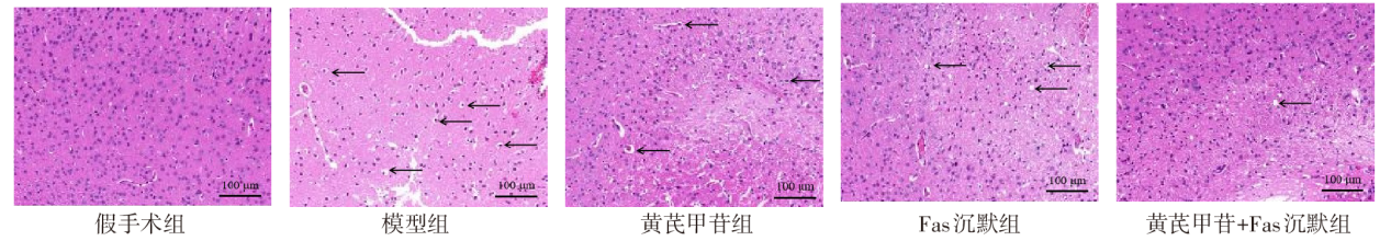
Fig.1 Pathological changes of brain tissue of rats in each group (HE staining, ×200)
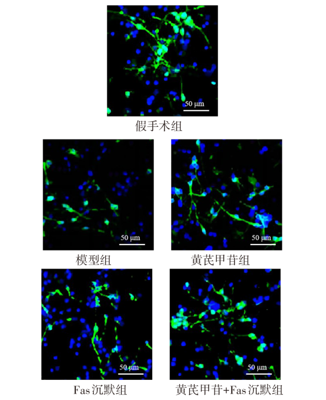
Fig.2 Results of Tuj1 immunofluorescence staining in brain tissue of rats in each group (×200)
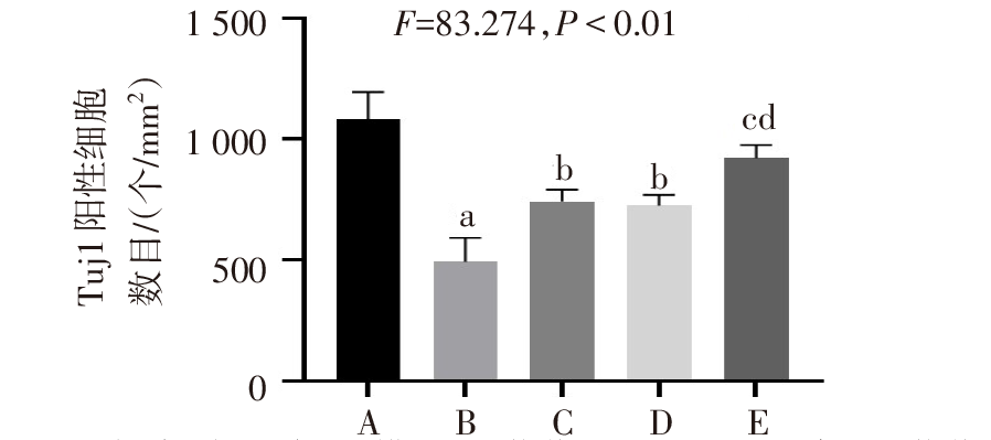
Fig.3 Comparison of the number of Tuj1 positive cells in brain tissue between five groups of rats
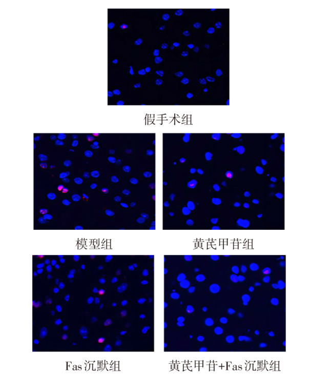
Fig.4 Apoptosis of neurons in brain tissue of rats in each group (TUNEL staining, ×200)
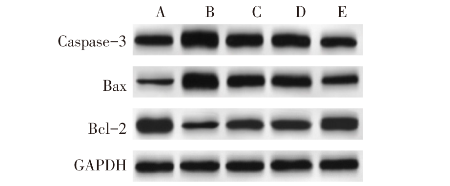
Fig.5 Expression levels of Caspase-3, Bax and Bcl-2 protein in brain tissue of rats in each group
| 组别 | 凋亡率/% | 凋亡相关蛋白表达 | ||
|---|---|---|---|---|
| Caspase-3 | Bax | Bcl-2 | ||
| 假手术组 | 1.91±0.37 | 1.24±0.26 | 0.67±0.13 | 2.05±0.40 |
| 模型组 | 16.52±3.06a | 3.06±0.61a | 2.38±0.46a | 0.53±0.11a |
| 黄芪甲苷组 | 10.57±2.43b | 1.80±0.37b | 1.46±0.27b | 0.94±0.22b |
| Fas沉默组 | 10.63±2.55b | 1.83±0.32b | 1.39±0.22b | 0.96±0.20b |
| 黄芪甲苷+ Fas沉默组 | 6.72±0.83cd | 1.05±0.19cd | 0.88±0.16cd | 1.45±0.34cd |
| F | 64.597** | 43.080** | 58.528** | 45.138** |
Tab.3 Comparison of neuronal apoptosis rate and apoptosis-related protein expression in brain tissue of rats between five groups
| 组别 | 凋亡率/% | 凋亡相关蛋白表达 | ||
|---|---|---|---|---|
| Caspase-3 | Bax | Bcl-2 | ||
| 假手术组 | 1.91±0.37 | 1.24±0.26 | 0.67±0.13 | 2.05±0.40 |
| 模型组 | 16.52±3.06a | 3.06±0.61a | 2.38±0.46a | 0.53±0.11a |
| 黄芪甲苷组 | 10.57±2.43b | 1.80±0.37b | 1.46±0.27b | 0.94±0.22b |
| Fas沉默组 | 10.63±2.55b | 1.83±0.32b | 1.39±0.22b | 0.96±0.20b |
| 黄芪甲苷+ Fas沉默组 | 6.72±0.83cd | 1.05±0.19cd | 0.88±0.16cd | 1.45±0.34cd |
| F | 64.597** | 43.080** | 58.528** | 45.138** |
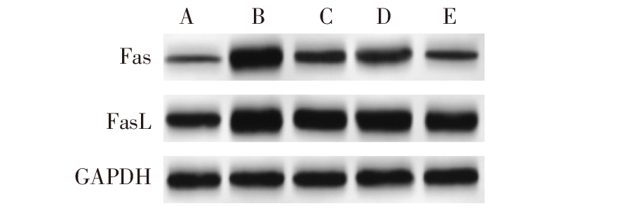
Fig.6 The expression of Fas and FasL protein in brain tissue of rats in each group
| 组别 | Fas | FasL | ||
|---|---|---|---|---|
| mRNA | 蛋白 | mRNA | 蛋白 | |
| 假手术组 | 1.00±0.04 | 0.25±0.04 | 1.00±0.03 | 0.94±0.17 |
| 模型组 | 3.58±0.72a | 1.56±0.30a | 5.21±1.02a | 2.03±0.43a |
| 黄芪甲苷组 | 2.23±0.46b | 0.79±0.16b | 3.39±0.66b | 1.57±0.32b |
| Fas沉默组 | 2.17±0.46b | 0.77±0.13b | 3.22±0.60b | 1.55±0.31b |
| 黄芪甲苷+ Fas沉默组 | 1.32±0.25cd | 0.43±0.08cd | 2.24±0.45cd | 1.21±0.26cd |
| F | 49.998** | 89.858** | 59.218** | 21.098** |
Tab.4 Comparison of Fas and FasL mRNA and protein expression in brain tissue of rats between five groups
| 组别 | Fas | FasL | ||
|---|---|---|---|---|
| mRNA | 蛋白 | mRNA | 蛋白 | |
| 假手术组 | 1.00±0.04 | 0.25±0.04 | 1.00±0.03 | 0.94±0.17 |
| 模型组 | 3.58±0.72a | 1.56±0.30a | 5.21±1.02a | 2.03±0.43a |
| 黄芪甲苷组 | 2.23±0.46b | 0.79±0.16b | 3.39±0.66b | 1.57±0.32b |
| Fas沉默组 | 2.17±0.46b | 0.77±0.13b | 3.22±0.60b | 1.55±0.31b |
| 黄芪甲苷+ Fas沉默组 | 1.32±0.25cd | 0.43±0.08cd | 2.24±0.45cd | 1.21±0.26cd |
| F | 49.998** | 89.858** | 59.218** | 21.098** |
| [1] | CENTE M, MATYASOVA K, CSICSATKOVA N, et al. Traumatic microRNAs:deconvolving the signal after severe traumatic brain injury[J]. Cell Mol Neurobiol, 2023, 43(3):1061-1075. doi:10.1007/s10571-022-01254-z. |
| [2] | ZHANG C, CHEN S. Role of TREM2 in the development of neurodegenerative diseases after traumatic brain injury[J]. Mol Neurobiol, 2023, 60(1):342-354. doi:10.1007/s12035-022-03094-w. |
| [3] | MI L, MIN X, SHI M, et al. Neutrophil extracellular traps aggravate neuronal endoplasmic reticulum stress and apoptosis via TLR9 after traumatic brain injury[J]. Cell Death Dis, 2023, 14(6):374-385. doi:10.1038/s41419-023-05898-7. |
| [4] | 李沅洋, 周湘忠, 雷向红, 等. 黄芪甲苷调控线粒体自噬减轻5-Fu诱导老龄大鼠心肌毒性的实验研究[J]. 天津医药, 2021, 49(4):378-384. |
| LI Y Y, ZHOU X Z, LEI X H, et al. The experimental study on astragaloside Ⅳ regulating mitochondrial autophagy to reduce myocardial toxicity induced by 5-Fu in aging rats[J]. Tianjin Med J, 2021, 49(4):378-384. doi:10.11958/20202229. | |
| [5] | WANG L, LIU C, WANG L, et al. Astragaloside IV mitigates cerebral ischaemia-reperfusion injury via inhibition of P62/Keap1/Nrf2 pathway-mediated ferroptosis[J]. Eur J Pharmacol, 2023, 944:175516. doi:10.1016/j.ejphar.2023.175516. |
| [6] | LAGUNAS-RANGEL F A.Fas (CD95)/FasL (CD178) system during ageing[J]. Cell Biol Int, 2023, 47(8):1295-1313. doi:10.1002/cbin.12032. |
| [7] | RUAN S, ZHAI L, WU S, et al. SCFAs promote intestinal double-negative T cells to regulate the inflammatory response mediated by NLRP3 inflammasome[J]. Aging (Albany NY), 2021, 13(17):21470-21482. doi:10.18632/aging.203487. |
| [8] | 杨琪, 安鹏飞, 王瑞辉, 等. 不同时期电针对创伤性颅脑损伤大鼠脑组织Fas/FasL表达的影响[J]. 针刺研究, 2020, 45(9):714-719. |
| YANG Q, AN P F, WANG R H, et al. Effect of electroacupuncture at different stages on the expression of Fas and FasL in brain tissue of rats with traumatic brain injury[J]. Acupuncture Research, 2020, 45(9):714-719. doi:10.13702/j.1000-0607.190863. | |
| [9] | 张怡, 张彐宁, 周晓红, 等. 黄芪甲苷缓解大脑中动脉阻塞/再灌注大鼠脑组织损伤的作用及机制[J]. 时珍国医国药, 2021, 32(11):2636-2639. |
| ZHANG Y, Zhang J N, ZHOU X H, et al. Effect and mechanism of astragaloside on brain tissue injury induced by middle cerebral artery occlusion/reperfusion in rats[J]. Lishizhen Medicine and Materia Medica Research, 2021, 32(11):2636-2639. doi:10.3969/j.issn.1008-0805.2021.11.18. | |
| [10] | 邹婷婷, 马莉, 潘文静, 等. 重型颅脑创伤并发颅内感染危险因素分析及列线图预测模型构建[J]. 中国现代神经疾病杂志, 2023, 23(6):496-502. |
| ZOU T T, MA L, PAN W J, et al. Analysis of risk factors of secondary intracranial infection in patients with severe traumatic brain injury and construction of a nomogram prediction model[J]. Chinese Journal of Contemporary Neurology and Neurosurgery, 2023, 23(6):496-502. doi:10.3969/j.issn.1672-6731.2023.06.005. | |
| [11] | SHRESTHA A, PAUDEL N, ADHIKARI G, et al. Traumatic brain injury among patients admitted in neurosurgical unit in a tertiary care centre:a descriptive cross-sectional study[J]. JNMA J Nepal Med Assoc, 2023, 61(262):514-518. doi:10.31729/jnma.8197. |
| [12] | KIM M S, KIM Y H, KIM M S, et al. Efficacy and safety of early anti-inflammatory drug therapy for secondary injury in traumatic brain injury[J]. World Neurosurg, 2023, 172(1):646-654. doi:0.1016/j.wneu.2023.01.110. |
| [13] | TANG X, LI X, ZHANG D, et al. Astragaloside-IV alleviates high glucose-induced ferroptosis in retinal pigment epithelial cells by disrupting the expression of miR-138-5p/Sirt1/Nrf2[J]. Bioengineered, 2022, 13(4):8240-8254. doi:10.1080/21655979.2022.2049471. |
| [14] | ZHANG D, LI Z, GAO Y, et al. Astragaloside IV improves renal function and alleviates renal damage and inflammation in rats with chronic glomerulonephritis[J]. Turk J Biol, 2022, 47(1):61-73. doi:10.55730/1300-0152.2641. |
| [15] | YIN F, ZHOU H F, FANG Y C, et al. Astragaloside IV alleviates ischemia reperfusion-induced apoptosis by inhibiting the activation of key factors in death receptor pathway and mitochondrial pathway[J]. J Ethnopharmacolo, 2020, 248:112319. doi:10.1016/j.jep.2019.112319. |
| [16] | WANG Y L, CHIO C C, KUO S C, et al. Exercise rehabilitation and/or Astragaloside attenuate amyloid-beta pathology by reversing BDNF/TrkB signaling deficits and mitochondrial dysfunction[J]. Mol Neurobiol, 2022, 59(5):3091-3109. doi:10.1007/s12035-022-02728-3. |
| [17] | ABOU SHOUSHA S, BAHEEG S, GHONEIM H, et al. The effect of Fas/FasL pathway blocking on apoptosis and stemness within breast cancer tumor microenvironment (preclinical study)[J]. Breast Dis, 2023, 42(1):163-176. doi:10.3233/BD-220077. |
| [18] | PIETRZAK B A, WNUK A, PRZEPIÓRSKA K, et al. Posttreatment with ospemifene attenuates hypoxia- and ischemia-induced apoptosis in primary neuronal cells via selective modulation of estrogen receptors[J]. Neurotox Res, 2023, 41(4):362-379. doi:10.1007/s12640-023-00644-5. |
| [19] | SUN H, YANG Y, GU M, et al. The role of Fas-FasL-FADD signaling pathway in arsenic-mediated neuronal apoptosis in vivo and in vitro[J]. Toxicol Lett, 2022, 356(1):143-150. doi:10.1016/j.toxlet.2021.11.012. |
| [20] | WEN S, WANG L, ZOU H, et al. Puerarin attenuates cadmium-induced neuronal injury via stimulating cadmium excretion, inhibiting oxidative stress and apoptosis[J]. Biomolecules, 2021, 11(7):978. doi:10.3390/biom11070978. |
| Viewed | ||||||
|
Full text |
|
|||||
|
Abstract |
|
|||||