Tianjin Medical Journal ›› 2023, Vol. 51 ›› Issue (1): 24-29.doi: 10.11958/20220433
• Cell and Molecular Biology • Previous Articles Next Articles
SONG Zhengfeng( ), LIU Yuanyuan(), QI Peng, TAN Xianxing, MA Lei
Received:2022-03-25
Revised:2022-06-21
Published:2023-01-15
Online:2023-01-17
Contact:
LIU Yuanyuan
E-mail:szf1979s@163.com;123hp@163.com
SONG Zhengfeng, LIU Yuanyuan, QI Peng, TAN Xianxing, MA Lei. The effect and mechanism of miR-15b gene interference on cerebral ischemia-reperfusion injury[J]. Tianjin Medical Journal, 2023, 51(1): 24-29.
CLC Number:

Fig.1 Identification of astrocytes in rat cerebral cortex (immunochemical staining,×200)

Fig.2 Cell morphology and transfection efficiency of each group (×100)
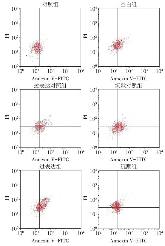
Fig.3 Analysis of apoptosis rate in each group
| 组别 | 存活率 | 细胞活力 | 凋亡率 |
|---|---|---|---|
| 对照组 | 95.62±6.39 | 1.85±0.33 | 4.55±0.81 |
| 空白组 | 29.51±5.84a | 52.27±9.02a | 40.26±7.04a |
| 过表达对照组 | 28.94±5.73a | 49.58±9.23a | 42.55±7.30a |
| 沉默对照组 | 26.45±6.86a | 51.14±9.55a | 41.93±7.55a |
| 过表达组 | 9.12±1.02abc | 77.25±11.78abc | 64.54±10.06abc |
| 沉默组 | 55.23±8.04abd | 27.36±5.15abd | 21.18±2.03abd |
| F | 76.586** | 28.127** | 28.700** |
Tab.1 Analysis of cell survival rate, cell viability and apoptosis rate in each group
| 组别 | 存活率 | 细胞活力 | 凋亡率 |
|---|---|---|---|
| 对照组 | 95.62±6.39 | 1.85±0.33 | 4.55±0.81 |
| 空白组 | 29.51±5.84a | 52.27±9.02a | 40.26±7.04a |
| 过表达对照组 | 28.94±5.73a | 49.58±9.23a | 42.55±7.30a |
| 沉默对照组 | 26.45±6.86a | 51.14±9.55a | 41.93±7.55a |
| 过表达组 | 9.12±1.02abc | 77.25±11.78abc | 64.54±10.06abc |
| 沉默组 | 55.23±8.04abd | 27.36±5.15abd | 21.18±2.03abd |
| F | 76.586** | 28.127** | 28.700** |
| 组别 | miR-15b | Bcl-2 | Caspase-3 | Caspase-9 |
|---|---|---|---|---|
| 对照组 | 1.03±0.18 | 1.01±0.20 | 0.98±0.17 | 1.00±0.19 |
| 空白组 | 1.78±0.31a | 0.52±0.09a | 2.05±0.38a | 1.62±0.28a |
| 过表达对照组 | 1.85±0.34a | 0.50±0.09a | 2.01±0.35a | 1.59±0.26a |
| 沉默对照组 | 1.82±0.33a | 0.47±0.08a | 1.99±0.36a | 1.65±0.30a |
| 过表达组 | 2.98±0.52abc | 0.18±0.03abc | 3.26±0.57abc | 2.74±0.51abc |
| 沉默组 | 1.11±0.19bd | 0.87±0.16abd | 1.24±0.21bd | 1.07±0.16bd |
| F | 16.952** | 30.145** | 21.377** | 14.108** |
Tab.2 Analysis of relative expression levels of miR-15b, Bcl-2, Caspase-3 and Caspase-9 mRNA in cells of each group
| 组别 | miR-15b | Bcl-2 | Caspase-3 | Caspase-9 |
|---|---|---|---|---|
| 对照组 | 1.03±0.18 | 1.01±0.20 | 0.98±0.17 | 1.00±0.19 |
| 空白组 | 1.78±0.31a | 0.52±0.09a | 2.05±0.38a | 1.62±0.28a |
| 过表达对照组 | 1.85±0.34a | 0.50±0.09a | 2.01±0.35a | 1.59±0.26a |
| 沉默对照组 | 1.82±0.33a | 0.47±0.08a | 1.99±0.36a | 1.65±0.30a |
| 过表达组 | 2.98±0.52abc | 0.18±0.03abc | 3.26±0.57abc | 2.74±0.51abc |
| 沉默组 | 1.11±0.19bd | 0.87±0.16abd | 1.24±0.21bd | 1.07±0.16bd |
| F | 16.952** | 30.145** | 21.377** | 14.108** |
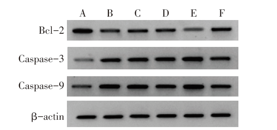
Fig.4 The expression levels of Bcl-2, Caspase-3 and Caspase-9 protein in cells of each group
| 组别 | Bcl-2 | Caspase-3 | Caspase-9 |
|---|---|---|---|
| 对照组 | 1.71±0.27 | 0.52±0.08 | 0.48±0.07 |
| 空白组 | 0.79±0.12a | 1.51±0.25a | 1.15±0.21a |
| 过表达对照组 | 0.76±0.12a | 1.55±0.28a | 1.22±0.22a |
| 沉默对照组 | 0.80±0.13a | 1.46±0.24a | 1.06±0.18a |
| 过表达组 | 0.30±0.08abc | 2.31±0.37abc | 1.85±0.31abc |
| 沉默组 | 1.26±0.18abd | 0.78±0.14bd | 0.51±0.12bd |
| F | 26.973** | 20.052** | 19.406** |
Tab.3 Analysis of relative expression levels of Bcl-2, Caspase-3 and Caspase-9 proteins in cells of each group
| 组别 | Bcl-2 | Caspase-3 | Caspase-9 |
|---|---|---|---|
| 对照组 | 1.71±0.27 | 0.52±0.08 | 0.48±0.07 |
| 空白组 | 0.79±0.12a | 1.51±0.25a | 1.15±0.21a |
| 过表达对照组 | 0.76±0.12a | 1.55±0.28a | 1.22±0.22a |
| 沉默对照组 | 0.80±0.13a | 1.46±0.24a | 1.06±0.18a |
| 过表达组 | 0.30±0.08abc | 2.31±0.37abc | 1.85±0.31abc |
| 沉默组 | 1.26±0.18abd | 0.78±0.14bd | 0.51±0.12bd |
| F | 26.973** | 20.052** | 19.406** |
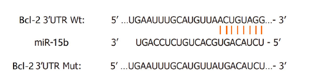
Fig.5 Binding site of miR-15b in Bcl-2
| [1] | MA C, WANG X, XU T, et al. Qingkailing injection ameliorates cerebral ischemia-reperfusion injury and modulates the AMPK/NLRP3 Inflammasome Signalling pathway[J]. BMC Complement Altern Med, 2019, 19(1):320. doi:10.1186/s12906-019-2703-5. |
| [2] | FRANKE M, BIEBER M, KRAFT P, et al. The NLRP3 inflammasome drives inflammation in ischemia/reperfusion injury after transient middle cerebral artery occlusion in mice[J]. Brain Behav Immun, 2021, 92(1):223-233. doi:10.1016/j.bbi.2020.12.009. |
| [3] | CHEN X, CHENG C, ZUO X, et al. Astragalin alleviates cerebral ischemia-reperfusion injury by improving anti-oxidant and anti-inflammatory activities and inhibiting apoptosis pathway in rats[J]. BMC Complement Med Ther, 2020, 20(1):120. doi:10.1186/s12906-020-02902-x. |
| [4] | LI G C, QIN X L, SONG H H, et al. Upregulated microRNA-15b alleviates ovarian cancer through inhitbition of the PI3K/Akt pathway by targeting LPAR3[J]. J Cell Physiol, 2019, 234(12):22331-22342. doi:10.1002/jcp.28799. |
| [5] | WANG P, SUN J, LV S, et al. Apigenin alleviates myocardial reperfusion injury in rats by downregulating miR-15b[J]. Med Sci Monit, 2019, 25(2):2764-2776. doi:10.12659/MSM.912014. |
| [6] | 胆迎, 李晨. 氧糖剥夺再灌注后星形胶质细胞活化、损伤、凋亡、自噬变化及意义[J]. 山东医药, 2020, 60(8):48-51. |
| DAN Y, LI C. Activation,injury,apoptosis and autophagy of astrocytes under OGD/R[J]. Shangdong Med J, 2020, 60(8):48-51. doi:10.3969/j.issn.1002-266X.2020.08.012. | |
| [7] | 廖鸿雁, 刘杰, 刘菁, 等. 白藜芦醇对氧糖剥夺/再复氧损伤后小胶质细胞系N9活化的影响[J]. 解剖学报, 2019, 50(2):137-144. |
| LIAO H Y, LIU J, LIU J, et al. Effect of resveratrol on activation of microglia cell line N9 after oxygen-glucose deprivation /reoxygenation injury in vitro[J]. Acta Anatomica Sinica, 2019, 50(2):137-144. doi:10.16098/j.issn.0529-1356.2019.02.001. | |
| [8] | LI Z, CAI B, ABDALLA B A, et al. LncIRS1 controls muscle atrophy via sponging miR-15 family to activate IGF1-PI3K/AKT pathway[J]. J Cachexia Sarcopenia Muscle, 2019, 10(2):391-410. doi:10.1002/jcsm.12374. |
| [9] | ZHOU Y, FAN R G, QIN C L, et al. LncRNA-H19 activates CDC42/PAK1 pathway to promote cell proliferation,migration and invasion by targeting miR-15b in hepatocellular carcinoma[J]. Genomics, 2019, 111(6):1862-1872. doi:10.1016/j.ygeno.2018.12.009. |
| [10] | YUAN C, ZHANG Y, TU W, et al. Integrated miRNA profiling and bioinformatics analyses reveal upregulated miRNAs in gastric cancer[J]. Oncol Lett, 2019, 18(2):1979-1988. doi:10.3892/ol.2019.10495. |
| [11] | 王红灵, 刘舒萍, 赵冀安. 东莨菪碱下调miR-15b的表达对缺氧/复氧损伤H9C2心肌细胞功能的影响[J]. 毒理学杂志, 2020, 34(6):486-491. |
| WANG H L, LIU S P, ZHAO J A, et al. Effects of scopolamine on the function of H9C2 cardiomyocytes injured by hypoxia /reoxygenation by down-regulating the expression of miR-15b[J]. J Toxicol, 2020, 34(6):486-491. doi:10.16421/j.cnki.1002-3127.2020.06.012. | |
| [12] | NAN L, XIE Q, CHEN Z, et al. Involvement of PARP-1/AIF signaling pathway in protective effects of gualou guizhi decoction against ischemia-reperfusion injury-induced apoptosis[J]. Neurochem Res, 2020, 45(2):278-294. doi:10.1007/s11064-019-02912-3. |
| [13] | LIU H, LIU S, TIAN X, et al. Bexarotene attenuates focal cerebral ischemia-reperfusion injury via the suppression of JNK/Caspase-3 signaling pathway[J]. Neurochem Res, 2019, 44(12):2809-2820. doi:10.1007/s11064-019-02902-5. |
| [14] | 李虎, 李健蕊, 刘楠楠, 等. 双环醇通过抑制线粒体凋亡途径缓解TNF-α/D-GalN诱导的爆发性肝损伤[J]. 中国药理学通报, 2021, 37(4):478-484. |
| LI H, LI J R, LIU N N, et al. Bicyclol alleviates TNF-α/D-GalN-induced fulminant liver injury by inhibiting mitochondrial apoptotic pathway[J]. Chin Pharmaco Bull, 2021, 37(4):478-484. doi:10.3969/j.issn.1001-1978.2021.04.008. | |
| [15] | ARAL K, ARAL C A, KAPILA Y. The role of caspase-8,caspase-9,and apoptosis inducing factor in periodontal disease[J]. J Periodontol, 2019, 90(3):288-294. doi:10.1002/JPER.17-0716. |
| [16] | 丁实, 赵学荣, 李宝群, 等. 基于Bcl-2/Beclin-1复合体探讨黄连素对脑缺血再灌注损伤大鼠模型的保护作用[J]. 中国实验动物学报, 2019, 27(5):651-657. |
| DING S, ZHAO X R, LI B Q, et al. Protective effect of berberine on the cerebral ischemia-reperfusion injury in rats based on Bcl-2/ Beclin-1 complex[J]. Acta Lab Anim Sci Sin, 2019, 27(5):651-657. doi:10.3969/j.issn.1005-4847.2019.05.016. | |
| [17] | ALI SYEDA Z, LANGDEN S S S, Munkhzul C, et al. Regulatory mechanism of microRNA expression in cancer[J]. Int J Mol Sci, 2020, 21(5):1723. doi:10.3390/ijms21051723. |
| [18] | LI L, SHAO Y, ZHENG H, et al. Kaempferol regulates miR-15b/Bcl-2/TLR4 to alleviate OGD-induced injury in H9c2 Cells[J]. Int Heart J, 2020, 61(3):585-594. doi:10.1536/ihj.19-359. |
| Viewed | ||||||
|
Full text |
|
|||||
|
Abstract |
|
|||||