Tianjin Medical Journal ›› 2023, Vol. 51 ›› Issue (11): 1199-1204.doi: 10.11958/20221777
• Experimental Research • Previous Articles Next Articles
MAO Quanxi( ), LI Zuoxiao△()
Received:2022-11-07
Revised:2023-03-13
Published:2023-11-15
Online:2023-11-07
Contact:
△E-mail:MAO Quanxi, LI Zuoxiao. Neuroprotective mechanism of edaravone dexborneol in rats with cerebral hemorrhage through ferroptosis-lipid peroxidation pathway[J]. Tianjin Medical Journal, 2023, 51(11): 1199-1204.
CLC Number:
| 组别 | n | 神经功能评分 | F | |||
|---|---|---|---|---|---|---|
| 1 d | 3 d | 7 d | 14 d | |||
| 假手术组 | 8 | 17.75±0.46 | 17.75±0.46 | 17.88±0.35 | 18.00±0.00 | 0.828 |
| 脑出血组 | 8 | 11.88±1.13a | 9.50±0.93aA | 13.38±0.92aAB | 14.75±0.89aABC | 43.110* |
| 依达拉奉组 | 8 | 13.25±1.28b | 11.63±1.06bA | 14.75±1.04bAB | 16.00±0.76bABC | 25.950* |
| 依达拉奉右莰醇组 | 8 | 14.25±0.89bc | 13.38±0.74bcA | 15.88±0.64bcAB | 17.50±0.53bcABC | 52.234* |
| F | 55.514* | 142.848* | 47.331* | 42.507* | ||
Tab.1 Comparison of neurological function scores between the four groups of rats
| 组别 | n | 神经功能评分 | F | |||
|---|---|---|---|---|---|---|
| 1 d | 3 d | 7 d | 14 d | |||
| 假手术组 | 8 | 17.75±0.46 | 17.75±0.46 | 17.88±0.35 | 18.00±0.00 | 0.828 |
| 脑出血组 | 8 | 11.88±1.13a | 9.50±0.93aA | 13.38±0.92aAB | 14.75±0.89aABC | 43.110* |
| 依达拉奉组 | 8 | 13.25±1.28b | 11.63±1.06bA | 14.75±1.04bAB | 16.00±0.76bABC | 25.950* |
| 依达拉奉右莰醇组 | 8 | 14.25±0.89bc | 13.38±0.74bcA | 15.88±0.64bcAB | 17.50±0.53bcABC | 52.234* |
| F | 55.514* | 142.848* | 47.331* | 42.507* | ||
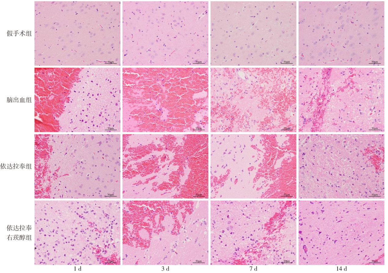
Fig.1 Histopathological changes of rat brain in each group (HE staining, ×400)
| 组别 | ROS含量 | F | |||
|---|---|---|---|---|---|
| 1 d | 3 d | 7 d | 14 d | ||
| 假手术组 | 401.13±34.85 | 419.31±12.77 | 469.35±50.88 | 423.93±40.35 | 3.006 |
| 脑出血组 | 2 627.93±191.86a | 4 843.82±248.58aA | 4 259.69±252.67aAB | 2 301.21±162.10aABC | 161.608* |
| 依达拉奉组 | 2 575.42±232.30 | 4 208.64±151.22bA | 2 780.60±307.92bB | 1 353.92±130.66bBC | 145.064* |
| 依达拉奉右莰醇组 | 2 378.65±193.52b | 2 850.56±271.71bcA | 1 560.46±181.28bcAB | 648.05±29.06bcABC | 129.150* |
| F | 176.405* | 438.943* | 273.577* | 311.623* | |
Tab.2 Changes of ROS content in brain tissue around hematoma of rats in each group
| 组别 | ROS含量 | F | |||
|---|---|---|---|---|---|
| 1 d | 3 d | 7 d | 14 d | ||
| 假手术组 | 401.13±34.85 | 419.31±12.77 | 469.35±50.88 | 423.93±40.35 | 3.006 |
| 脑出血组 | 2 627.93±191.86a | 4 843.82±248.58aA | 4 259.69±252.67aAB | 2 301.21±162.10aABC | 161.608* |
| 依达拉奉组 | 2 575.42±232.30 | 4 208.64±151.22bA | 2 780.60±307.92bB | 1 353.92±130.66bBC | 145.064* |
| 依达拉奉右莰醇组 | 2 378.65±193.52b | 2 850.56±271.71bcA | 1 560.46±181.28bcAB | 648.05±29.06bcABC | 129.150* |
| F | 176.405* | 438.943* | 273.577* | 311.623* | |
| 组别 | GSH含量 | F | |||
|---|---|---|---|---|---|
| 1 d | 3 d | 7 d | 14 d | ||
| 假手术组 | 15.44±1.19 | 15.19±1.27 | 15.16±1.18 | 16.04±0.40 | 0.725 |
| 脑出血组 | 5.54±0.60a | 0.90±0.76aA | 2.14±0.86aAB | 7.28±1.18aABC | 56.782* |
| 依达拉奉组 | 6.54±1.10 | 1.27±0.98A | 6.11±0.95bB | 9.8±0.79bABC | 67.218* |
| 依达拉奉右莰醇组 | 6.92±0.71b | 2.95±1.04bcA | 7.55±0.94bcB | 14.89±1.34bcABC | 116.679* |
| F | 121.032* | 219.383* | 152.153* | 86.897* | |
Tab.3 Changes of GSH content in brain tissue around hematoma of rats in each group
| 组别 | GSH含量 | F | |||
|---|---|---|---|---|---|
| 1 d | 3 d | 7 d | 14 d | ||
| 假手术组 | 15.44±1.19 | 15.19±1.27 | 15.16±1.18 | 16.04±0.40 | 0.725 |
| 脑出血组 | 5.54±0.60a | 0.90±0.76aA | 2.14±0.86aAB | 7.28±1.18aABC | 56.782* |
| 依达拉奉组 | 6.54±1.10 | 1.27±0.98A | 6.11±0.95bB | 9.8±0.79bABC | 67.218* |
| 依达拉奉右莰醇组 | 6.92±0.71b | 2.95±1.04bcA | 7.55±0.94bcB | 14.89±1.34bcABC | 116.679* |
| F | 121.032* | 219.383* | 152.153* | 86.897* | |
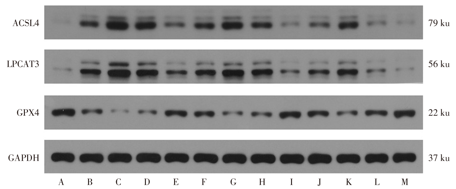
Fig.2 Changes in expression of ferroptosis-related proteins in brain tissue around hematoma of rats in each group
| 组别 | ACSL4 | F | |||
|---|---|---|---|---|---|
| 1 d | 3 d | 7 d | 14 d | ||
| 假手术组 | 0.06±0.01 | 0.06±0.01 | 0.06±0.01 | 0.06±0.01 | 0.000 |
| 脑出血组 | 0.43±0.03a | 0.72±0.03aA | 0.59±0.02aAB | 0.28±0.04aABC | 209.196* |
| 依达拉奉组 | 0.38±0.04b | 0.65±0.04bA | 0.48±0.03bAB | 0.18±0.03bABC | 164.729* |
| 依达拉奉右莰醇组 | 0.30±0.03bc | 0.49±0.05bcA | 0.19±0.04bcAB | 0.09±0.01bcABC | 125.193* |
| F | 162.535* | 383.899* | 451.068* | 76.947* | |
| 组别 | LPCAT3 | F | |||
| 1 d | 3 d | 7 d | 14 d | ||
| 假手术组 | 0.08±0.01 | 0.08±0.01 | 0.08±0.01 | 0.08±0.01 | 0.000 |
| 脑出血组 | 0.59±0.03a | 0.84±0.03aA | 0.71±0.04aAB | 0.39±0.02aABC | 194.560* |
| 依达拉奉组 | 0.55±0.06 | 0.74±0.07bA | 0.57±0.03bB | 0.27±0.03bABC | 76.592* |
| 依达拉奉右莰醇组 | 0.41±0.03bc | 0.54±0.04bcA | 0.24±0.04bcAB | 0.12±0.04bcABC | 120.285* |
| F | 191.531* | 338.113* | 366.208* | 128.922* | |
| 组别 | GPX4 | F | |||
| 1 d | 3 d | 7 d | 14 d | ||
| 假手术组 | 0.70±0.03 | 0.70±0.03 | 0.70±0.03 | 0.70±0.03 | 0.000 |
| 脑出血组 | 0.41±0.04a | 0.15±0.04aA | 0.25±0.03aAB | 0.49±0.03aABC | 84.135* |
| 依达拉奉组 | 0.49±0.04b | 0.27±0.03bA | 0.32±0.03bAB | 0.58±0.02bABC | 111.808* |
| 依达拉奉右莰醇组 | 0.56±0.04bc | 0.35±0.03bcA | 0.49±0.06bcAB | 0.66±0.03bcABC | 50.498* |
| F | 491.616* | 865.259* | 749.863* | 134.870* | |
Tab.4 Changes of ACSL4, LPCAT3 and GPX4 expression in brain tissue around hematoma of rats in each group
| 组别 | ACSL4 | F | |||
|---|---|---|---|---|---|
| 1 d | 3 d | 7 d | 14 d | ||
| 假手术组 | 0.06±0.01 | 0.06±0.01 | 0.06±0.01 | 0.06±0.01 | 0.000 |
| 脑出血组 | 0.43±0.03a | 0.72±0.03aA | 0.59±0.02aAB | 0.28±0.04aABC | 209.196* |
| 依达拉奉组 | 0.38±0.04b | 0.65±0.04bA | 0.48±0.03bAB | 0.18±0.03bABC | 164.729* |
| 依达拉奉右莰醇组 | 0.30±0.03bc | 0.49±0.05bcA | 0.19±0.04bcAB | 0.09±0.01bcABC | 125.193* |
| F | 162.535* | 383.899* | 451.068* | 76.947* | |
| 组别 | LPCAT3 | F | |||
| 1 d | 3 d | 7 d | 14 d | ||
| 假手术组 | 0.08±0.01 | 0.08±0.01 | 0.08±0.01 | 0.08±0.01 | 0.000 |
| 脑出血组 | 0.59±0.03a | 0.84±0.03aA | 0.71±0.04aAB | 0.39±0.02aABC | 194.560* |
| 依达拉奉组 | 0.55±0.06 | 0.74±0.07bA | 0.57±0.03bB | 0.27±0.03bABC | 76.592* |
| 依达拉奉右莰醇组 | 0.41±0.03bc | 0.54±0.04bcA | 0.24±0.04bcAB | 0.12±0.04bcABC | 120.285* |
| F | 191.531* | 338.113* | 366.208* | 128.922* | |
| 组别 | GPX4 | F | |||
| 1 d | 3 d | 7 d | 14 d | ||
| 假手术组 | 0.70±0.03 | 0.70±0.03 | 0.70±0.03 | 0.70±0.03 | 0.000 |
| 脑出血组 | 0.41±0.04a | 0.15±0.04aA | 0.25±0.03aAB | 0.49±0.03aABC | 84.135* |
| 依达拉奉组 | 0.49±0.04b | 0.27±0.03bA | 0.32±0.03bAB | 0.58±0.02bABC | 111.808* |
| 依达拉奉右莰醇组 | 0.56±0.04bc | 0.35±0.03bcA | 0.49±0.06bcAB | 0.66±0.03bcABC | 50.498* |
| F | 491.616* | 865.259* | 749.863* | 134.870* | |
| [1] | KLEBE D, INIAGHE L, BURCHELL S, et al. Intracerebral hemorrhage in mice[J]. Methods Mol Biol, 2018, 1717:83-91. doi:10.1007/978-1-4939-7526-6_7. |
| [2] | YAO M Y, LIU T, ZHANG L, et al. Role of ferroptosis in neurological diseases[J]. Neurosci Lett, 2021, 747:135614. doi:10.1016/j.neulet.2020.135614. |
| [3] | MIYAKE S, SHINDO R, NAKANO H. The molecular mechanisms and the functions of new types of regulated cell death including necroptosis,ferroptosis,and pyroptosis[J]. Clin Calcium, 2019, 29(2):248-253. doi:10.20837/4201902248. |
| [4] | XU J, WANG A, MENG X, et al. Edaravone dexborneol versus edaravone alone for the treatment of acute ischemic stroke:A phase Ⅲ,randomized,double-blind,comparative trial[J]. Stroke, 2021, 52(3):772-780. doi:10.1161/STROKEAHA.120.031197. |
| [5] | 李秋畅, 闫顺昌, 蒙亚珍, 等. Nrf2-GPX4介导的铁死亡通路参与右美托咪定对脑出血大鼠神经保护作用的机制研究[J]. 天津医药, 2022, 50(8):817-821. |
| LI Q C, YAN S C, MENG Y Z, et al. Neuroprotective effects of dexmedetomidine on intracerebral hemorrhage of rats by Nrf2-GPX4 mediated iron death pathway[J]. Tianjin Med J, 2022, 50(8):817-821. doi:10.11958/20212825. | |
| [6] | GARCIA J H, WAGNER S, LIU K F, et al. Neurological deficit and extent of neuronal necrosis attributable to middle cerebral artery occlusion in rats. Statistical validation[J]. Stroke, 1995, 26(4):627-634; discussion635. doi:10.1161/01.str.26.4.627. |
| [7] | AL-KAWAZ M N, HANLEY D F, ZIAI W. Advances in therapeutic approaches for spontaneous intracerebral hemorrhage[J]. Neurotherapeutics, 2020, 17(4):1757-1767. doi:10.1007/s13311-020-00902-w. |
| [8] | ZILLE M, KARUPPAGOUNDER S S, CHEN Y X, et al. Neuronal death after hemorrhagic stroke in vitro and in vivo shares features of ferroptosis and necroptosis[J]. Stroke, 2017, 48(4):1033-1043. doi:10.1161/STROKEAHA.116.015609. |
| [9] | BAI Q, LIU J, WANG G. Ferroptosis,a regulated neuronal cell death type after intracerebral hemorrhage[J]. Front Cell Neurosci, 2020, 14:591874. doi:10.3389/fncel.2020.591874. |
| [10] | BAO W D, ZHOU X T, ZHOU L T, et al. Targeting miR-124/Ferroportin signaling ameliorated neuronal cell death through inhibiting apoptosis and ferroptosis in aged intracerebral hemorrhage murine model[J]. Aging Cell, 2020, 19(11):e13235. doi:10.1111/acel.13235. |
| [11] | AGMON E, SOLON J, BASSEREAU P, et al. Modeling the effects of lipid peroxidation during ferroptosis on membrane properties[J]. Sci Rep, 2018, 8(1):5155. doi:10.1038/s41598-018-23408-0. |
| [12] | BOURGEOIS T, JALIL A, THOMAS C, et al. Deletion of lysophosphatidylcholine acyltransferase 3 in myeloid cells worsens hepatic steatosis after a high-fat diet[J]. J Lipid Res, 2021, 62:100013. doi:10.1194/jlr.RA120000737. |
| [13] | KUWATA H, NAKATANI E, SHIMBARA-MATSUBAYASHI S, et al. Long-chain acyl-CoA synthetase 4 participates in the formation of highly unsaturated fatty acid-containing phospholipids in murine macrophages[J]. Biochim Biophys Acta Mol Cell Biol Lipids, 2019, 1864(11):1606-1618. doi:10.1016/j.bbalip.2019.07.013. |
| [14] | REICHERT C O, DE FREITAS F A, SAMPAIO-SILVA J, et al. Ferroptosis mechanisms involved in neurodegenerative diseases[J]. Int J Mol Sci, 2020, 21(22):8765. doi:10.3390/ijms21228765. |
| [15] | URSINI F, MAIORINO M. Lipid peroxidation and ferroptosis:the role of GSH and GPx4[J]. Free Radic Biol Med, 2020, 152:175-185. doi:10.1016/j.freeradbiomed.2020.02.027. |
| [16] | STOCKWELL B R, JIANG X J, GU W. Emerging mechanisms and disease relevance of ferroptosis[J]. Trends Cell Biol, 2020, 30(6):478-490. doi:10.1016/j.tcb.2020.02.009. |
| [17] | SU L J, ZHANG J H, GOMEZ H, et al. Reactive oxygen species-induced lipid peroxidation in apoptosis,autophagy,and ferroptosis[J]. Oxid Med Cell Longev, 2019, 2019:5080843. doi:10.1155/2019/5080843. |
| [18] | FUJIWARA N, SOM A T, PHAM L D D, et al. A free radical scavenger edaravone suppresses systemic inflammatory responses in a rat transient focal ischemia model[J]. Neurosci Lett, 2016, 633:7-13. doi:10.1016/j.neulet.2016.08.048. |
| [19] | ZHANG M, TENG C H, WU F F, et al. Edaravone attenuates traumatic brain injury through anti-inflammatory and anti-oxidative modulation[J]. Exp Ther Med, 2019, 18(1):467-474. doi:10.3892/etm.2019.7632. |
| [20] | DANG L, DONG X, YANG J. Influence of nanoparticle-loaded edaravone on postoperative effects in patients with cerebral hemorrhage[J]. J Nanosci Nanotechnol, 2021, 21(2):1202-1211. doi:10.1166/jnn.2021.18668. |
| [21] | ZHANG J B, SHI X, CHEN Z, et al. Edaravone reduces iron-mediated Hydrocephalus and behavioral disorder in rat by activating the Nrf2/HO-1 pathway[J]. J Stroke Cerebrovasc Dis, 2018, 27(12):3511-3520. doi:10.1016/j.jstrokecerebrovasdis.2018.08.019. |
| [22] | HOMMA T, KOBAYASHI S, SATO H, et al. Edaravone,a free radical scavenger,protects against ferroptotic cell death in vitro[J]. Exp Cell Res, 2019, 384(1):111592. doi:10.1016/j.yexcr.2019.111592. |
| [23] | LIU R, ZHANG L, LAN X, et al. Protection by borneol on cortical neurons against oxygen-glucose deprivation/reperfusion:involvement of anti-oxidation and anti-inflammation through nuclear transcription factor κappaB signaling pathway[J]. Neuroscience, 2011, 176:408-419. doi:10.1016/j.neuroscience.2010.11.029. |
| [24] | WU H Y, TANG Y, GAO L Y, et al. The synergetic effect of edaravone and borneol in the rat model of ischemic stroke[J]. Eur J Pharmacol, 2014, 740:522-531. doi:10.1016/j.ejphar.2014.06.035. |
| [25] | HUA Y, ZHOU L M, YANG W D, et al. Y-2 reduces oxidative stress and inflammation and improves neurological function of collagenase-induced intracerebral hemorrhage rats[J]. Eur J Pharmacol, 2021, 910:174507. doi:10.1016/j.ejphar.2021.174507. |
| [1] | ZHAO Lanjun, LI Lianghui, MA Xin, GONG Jiaojiao, ZHENG Chenhui, SHI Lin. The effect of andrographolide regulating STAT3/GPX4 pathway on proliferation and apoptosis of myeloma cells [J]. Tianjin Medical Journal, 2026, 54(1): 8-13. |
| [2] | ZHONG Wenwen, ZOU Zhengshou, XIANG Qingwei. Protective effect of baicalin on nerve injury in rats with cerebral microbleeds through PI3K/AKT/eNOS pathway [J]. Tianjin Medical Journal, 2025, 53(5): 468-473. |
| [3] | SU Hongjian, ZHANG Chunyan, ZHANG Weidong, HAN Li, QIAO Yahong. Irisin affects the proliferation and migration of lung adenocarcinoma cells by regulating the EBF3/ALOX15 pathway [J]. Tianjin Medical Journal, 2025, 53(4): 337-342. |
| [4] | WANG Xiaoya, ZHANG Min, HE Xiaogang. Clinical study of pentoxifylline combined with edaravone dextrocamphenol in the treatment of acute cerebral infarction [J]. Tianjin Medical Journal, 2025, 53(2): 208-212. |
| [5] | GAO Chen, QIAO Yunyang, JI Jialing, WANG E, HUO Ying, ZHANG Aiqing. The effect of bufalin on extracellular matrix synthesis in renal tubular epithelial cells induced by high glucose [J]. Tianjin Medical Journal, 2025, 53(10): 1009-1015. |
| [6] | LI Bo, LIN Hong, HUANG Lanying, LI Lingyu. The relationship between HMOX1 and MAPK14 with the onset and prognosis of sepsis-induced acute lung injury in children [J]. Tianjin Medical Journal, 2025, 53(10): 1052-1056. |
| [7] | WU Bin, LIU Zhaoxiang, ZHANG Yuehong, WANG Changyao. The mechanism of excessive mechanical stress modulates Piezo1-mediated ferroptosis in chondrocytes [J]. Tianjin Medical Journal, 2025, 53(1): 14-18. |
| [8] | ZHANG Xungong, YANG Guanghui, DU Zengli, XUE Pei, MA Zikun. Correlation between ferroptosis and post operative cognitive dysfunction in elderly patients with fractures [J]. Tianjin Medical Journal, 2025, 53(1): 47-51. |
| [9] | ZHANG Chunhong, HUANG Hongchao, LIU Yue, DU Lilong, XU Haiwei, LI Ning, LI Yongjin. Identification of key ferroptosis genes in paraspinal muscle degeneration based on RNA sequencing and bioinformatics analysis [J]. Tianjin Medical Journal, 2024, 52(9): 991-995. |
| [10] | LIU Bin, YANG Long, LI Wenli, SHAO Ningning, DONG Jinrui. Mechanism of microglia ferroptosis in smoke inhalation-induced brain injury [J]. Tianjin Medical Journal, 2024, 52(8): 791-797. |
| [11] | WANG Xinshuang, AN Yajuan, GUAN Xiuju, LI Jiao, LIU Yue, WEI Liping, QI Xin. Study of magnesium isoglycyrrhizinate in ameliorating cisplatin induced myocardial injury in rats [J]. Tianjin Medical Journal, 2024, 52(8): 809-814. |
| [12] | WU Xiaoxiao, HU Yukun, WU Jiang. Protective effect of asiatic acid on brain injury after experimental subarachnoidhemorrhage in rats [J]. Tianjin Medical Journal, 2024, 52(3): 285-289. |
| [13] | ZHANG Wenchao, YANG Xuehui, YIN Tao, WANG Ruijian, ZHANG Mengmeng. The relationship between plasma sCD163/sTWEAK ratio and prognosis in patients with spontaneous acute cerebral hemorrhage [J]. Tianjin Medical Journal, 2024, 52(3): 297-301. |
| [14] | HUANG Xiaolei, GE Tingting, ZHAO Junsong, NI Zhihua. Study on the role of ginsenoside Rg1 in IL-6-induced neuronal ferroptosis in rats [J]. Tianjin Medical Journal, 2024, 52(11): 1137-1140. |
| [15] | NIE Yameng, ZHANG Xiaoqiang, FENG Pengzhan, DING Yaqian. Effects of edaravone dexborneol combined with rt-PA intravenous thrombolysis therapy on efficacy and prognosis of ultra-early acute cerebral infarction [J]. Tianjin Medical Journal, 2023, 51(7): 766-770. |
| Viewed | ||||||
|
Full text |
|
|||||
|
Abstract |
|
|||||