Tianjin Medical Journal ›› 2024, Vol. 52 ›› Issue (4): 337-345.doi: 10.11958/20230825
• Cell and Molecular Biology • Next Articles
HUANG Yu1,2( ), HE Ruiying3, LIU Sen1, CHEN Kaiyuan1, LI Meiyun1, CHENG Jianye1, WU Yan1,△()
Received:2023-06-14
Revised:2023-07-28
Published:2024-04-15
Online:2024-04-19
Contact:
△E-mail:HUANG Yu, HE Ruiying, LIU Sen, CHEN Kaiyuan, LI Meiyun, CHENG Jianye, WU Yan. Study on the effect of Chlorella extract on promoting skin wound healing in diabetic mice[J]. Tianjin Medical Journal, 2024, 52(4): 337-345.
CLC Number:
| 基因名称 | 引物序列(5′→3′) | 产物大小/bp |
|---|---|---|
| CD86 | 上游:ACGGAGTCAATGAAGATTTCCT | 151 |
| 下游:GATTCGGCTTCTTGTGACATAC | ||
| IL-1β | 上游:CTCCATGAGCTTTGTACAAGG | 245 |
| 下游:TGCTGATGTACCAGTTGGGG | ||
| TNF-α | 上游:CCCTCACACTCAGATCATCTTCT | 61 |
| 下游:GCTACGACGTGGGCTACAG | ||
| CD206 | 上游:CCTATGAAAATTGGGCTTACGG | 131 |
| 下游:CTGACAAATCCAGTTGTTGAGG | ||
| IL-10 | 上游:GTAGAAGTGATGCCCCAGGC | 187 |
| 下游:CACCTTGGTCTTGGAGCTTATT | ||
| ARG-1 | 上游:TTGGGTGGATGCTCACACTG | 67 |
| 下游:TTGCCCATGCAGATTCCC | ||
| β-actin | 上游:CTTTGCAGCTCCTTCGTTGC | 150 |
| 下游:ACGATGGAGGGGAATACAGC |
Tab.1 Primer sequences for CD86, IL-1β, TNF-α, CD206, IL-10, ARG-1 and β-actin
| 基因名称 | 引物序列(5′→3′) | 产物大小/bp |
|---|---|---|
| CD86 | 上游:ACGGAGTCAATGAAGATTTCCT | 151 |
| 下游:GATTCGGCTTCTTGTGACATAC | ||
| IL-1β | 上游:CTCCATGAGCTTTGTACAAGG | 245 |
| 下游:TGCTGATGTACCAGTTGGGG | ||
| TNF-α | 上游:CCCTCACACTCAGATCATCTTCT | 61 |
| 下游:GCTACGACGTGGGCTACAG | ||
| CD206 | 上游:CCTATGAAAATTGGGCTTACGG | 131 |
| 下游:CTGACAAATCCAGTTGTTGAGG | ||
| IL-10 | 上游:GTAGAAGTGATGCCCCAGGC | 187 |
| 下游:CACCTTGGTCTTGGAGCTTATT | ||
| ARG-1 | 上游:TTGGGTGGATGCTCACACTG | 67 |
| 下游:TTGCCCATGCAGATTCCC | ||
| β-actin | 上游:CTTTGCAGCTCCTTCGTTGC | 150 |
| 下游:ACGATGGAGGGGAATACAGC |
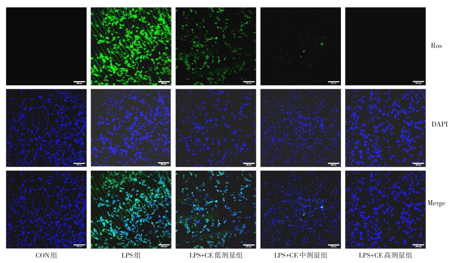
Fig.1 ROS results detected by DCFH-DA fluorescent probe assay in NIH-3T3 cells in each group (×200)
| 组别 | CD86 | IL-1β | TNF-α |
|---|---|---|---|
| CON组 | 0.98±0.02 | 0.97±0.02 | 0.98±0.01 |
| LPS组 | 2.60±0.08a | 1.79±0.02a | 1.76±0.03a |
| LPS+CE低剂量组 | 2.23±0.06b | 1.68±0.03b | 1.55±0.02b |
| LPS+CE中剂量组 | 1.87±0.09bc | 1.50±0.03bc | 1.49±0.07bc |
| LPS+CE高剂量组 | 1.36±0.03bcd | 1.40±0.02bcd | 1.17±0.02bcd |
| F | 322.100** | 548.400** | 220.700** |
| 组别 | CD206 | IL-10 | Arg-1 |
| CON组 | 0.96±0.02 | 0.98±0.01 | 0.97±0.01 |
| LPS组 | 0.56±0.03a | 0.73±0.04a | 0.69±0.03a |
| LPS+CE低剂量组 | 0.63±0.02ab | 0.76±0.01b | 0.77±0.01b |
| LPS+CE中剂量组 | 0.72±0.02bc | 0.81±0.01bc | 0.81±0.03bc |
| LPS+CE高剂量组 | 0.89±0.03bcd | 0.87±0.01bcd | 0.92±0.03bcd |
| F | 151.200** | 94.250** | 72.950** |
Tab.2 Comparison of expression levels of CD86, IL-1β, TNF-α, CD206, IL-10 and Arg-1 in macrophages between the five groups
| 组别 | CD86 | IL-1β | TNF-α |
|---|---|---|---|
| CON组 | 0.98±0.02 | 0.97±0.02 | 0.98±0.01 |
| LPS组 | 2.60±0.08a | 1.79±0.02a | 1.76±0.03a |
| LPS+CE低剂量组 | 2.23±0.06b | 1.68±0.03b | 1.55±0.02b |
| LPS+CE中剂量组 | 1.87±0.09bc | 1.50±0.03bc | 1.49±0.07bc |
| LPS+CE高剂量组 | 1.36±0.03bcd | 1.40±0.02bcd | 1.17±0.02bcd |
| F | 322.100** | 548.400** | 220.700** |
| 组别 | CD206 | IL-10 | Arg-1 |
| CON组 | 0.96±0.02 | 0.98±0.01 | 0.97±0.01 |
| LPS组 | 0.56±0.03a | 0.73±0.04a | 0.69±0.03a |
| LPS+CE低剂量组 | 0.63±0.02ab | 0.76±0.01b | 0.77±0.01b |
| LPS+CE中剂量组 | 0.72±0.02bc | 0.81±0.01bc | 0.81±0.03bc |
| LPS+CE高剂量组 | 0.89±0.03bcd | 0.87±0.01bcd | 0.92±0.03bcd |
| F | 151.200** | 94.250** | 72.950** |
| 组别 | 金黄色葡萄球菌存活率 | 大肠杆菌存活率 |
|---|---|---|
| CON组 | 100.00±0.00 | 100.00±0.00 |
| CE低剂量组 | 34.83±1.14a | 24.57±0.70a |
| CE中剂量组 | 23.07±1.88ab | 15.70±0.40ab |
| CE高剂量组 | 0.00±0.00abc | 0.00±0.00abc |
| F | 4 578.000** | 36 314.000** |
Tab.3 Comparison of survival rate of Staphylococcus aureus and Escherichia coli between the four group
| 组别 | 金黄色葡萄球菌存活率 | 大肠杆菌存活率 |
|---|---|---|
| CON组 | 100.00±0.00 | 100.00±0.00 |
| CE低剂量组 | 34.83±1.14a | 24.57±0.70a |
| CE中剂量组 | 23.07±1.88ab | 15.70±0.40ab |
| CE高剂量组 | 0.00±0.00abc | 0.00±0.00abc |
| F | 4 578.000** | 36 314.000** |
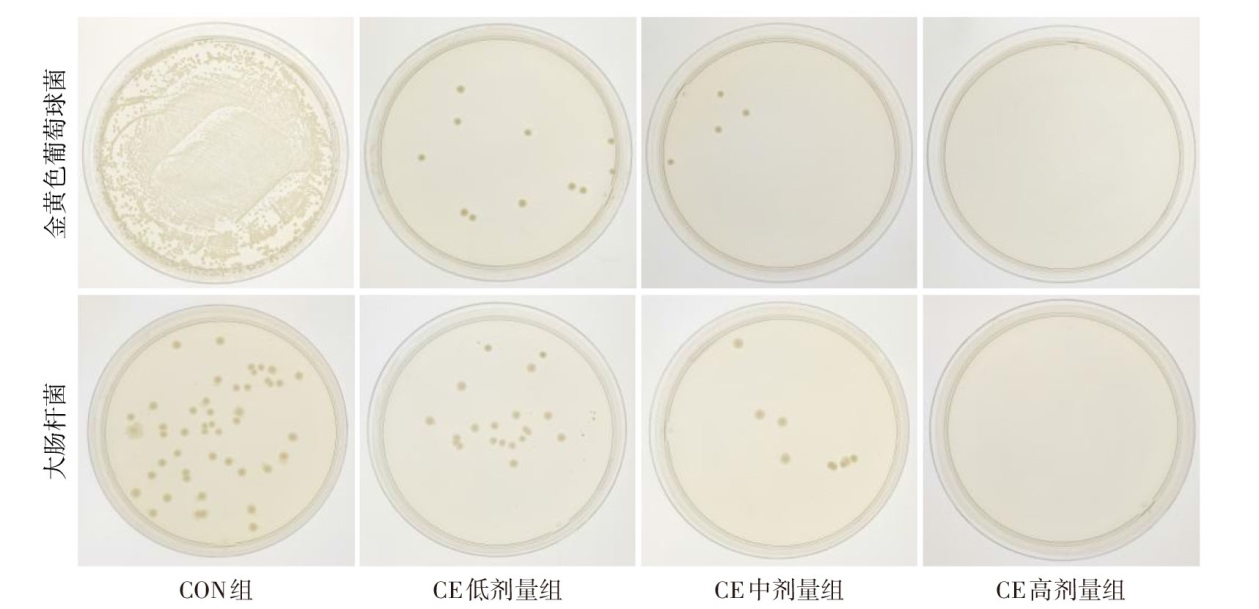
Fig.2 Bacteriostatic experiment of Chlorella extract against Staphylococcus aureus and Escherichia coli
| 组别 | 第7天 | 第14天 |
|---|---|---|
| CON组 | 30.57±1.19 | 54.08±1.64 |
| CE低剂量组 | 34.34±1.13a | 61.55±1.04a |
| CE中剂量组 | 45.70±2.06ab | 74.08±1.43ab |
| CE高剂量组 | 55.89±1.71abc | 97.23±1.35abc |
| F | 160.900** | 563.100** |
Tab.4 Comparison of wound healing rate at different time points after skin injury between the four groups
| 组别 | 第7天 | 第14天 |
|---|---|---|
| CON组 | 30.57±1.19 | 54.08±1.64 |
| CE低剂量组 | 34.34±1.13a | 61.55±1.04a |
| CE中剂量组 | 45.70±2.06ab | 74.08±1.43ab |
| CE高剂量组 | 55.89±1.71abc | 97.23±1.35abc |
| F | 160.900** | 563.100** |
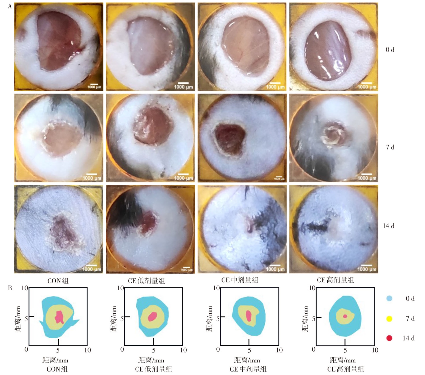
Fig.3 Morphological observation of wound skin in mice
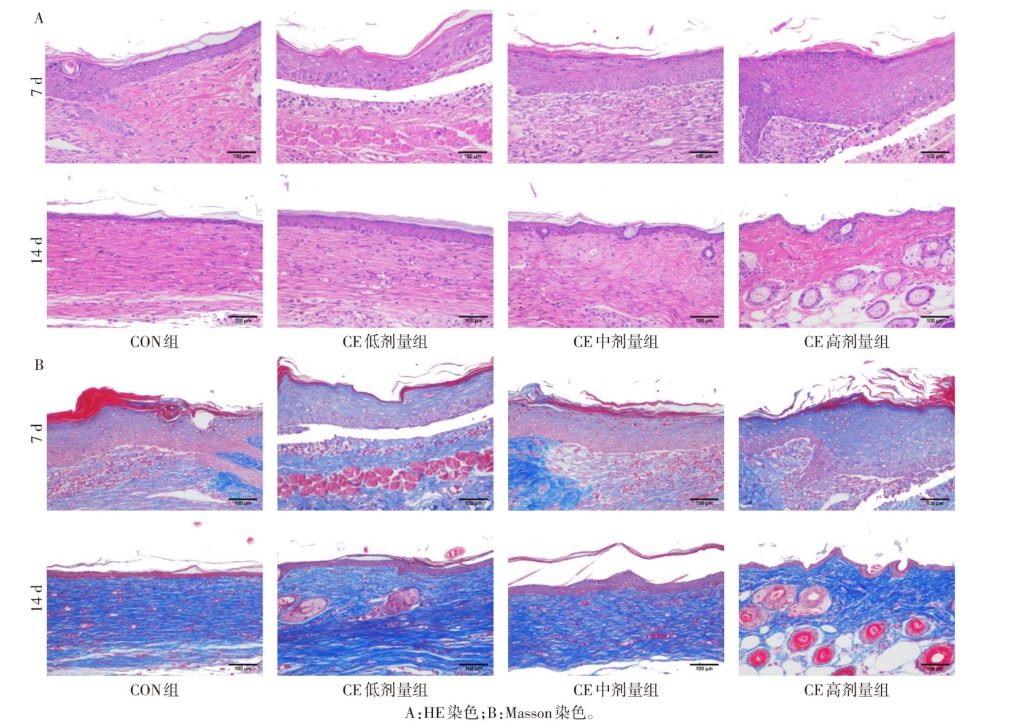
Fig.4 Histomorphology observation of postoperative wound skin in each group of mice (×200)
| 组别 | α-SMA | CD31 | CD86 | CD206 |
|---|---|---|---|---|
| CON组 | 6.63±1.53 | 38.12±2.06 | 43.17±1.50 | 18.11±1.86 |
| CE低剂量组 CE中剂量组 | 15.33±1.53a 26.67±2.08ab | 45.36±1.81a 58.67±2.04ab | 21.37±1.040a 15.26±1.59ab | 24.74±1.77a 35.61±1.28ab |
| CE高剂量组 | 38.67±2.52abc | 68.80±3.80abc | 8.06±1.48abc | 41.40±1.11abc |
| F | 153.700** | 86.090** | 342.000** | 140.000** |
Tab.5 Comparison of expression levels of α-SMA, CD31, CD86 and CD206 in the postoperative traumatic skin tissue between the four groups
| 组别 | α-SMA | CD31 | CD86 | CD206 |
|---|---|---|---|---|
| CON组 | 6.63±1.53 | 38.12±2.06 | 43.17±1.50 | 18.11±1.86 |
| CE低剂量组 CE中剂量组 | 15.33±1.53a 26.67±2.08ab | 45.36±1.81a 58.67±2.04ab | 21.37±1.040a 15.26±1.59ab | 24.74±1.77a 35.61±1.28ab |
| CE高剂量组 | 38.67±2.52abc | 68.80±3.80abc | 8.06±1.48abc | 41.40±1.11abc |
| F | 153.700** | 86.090** | 342.000** | 140.000** |
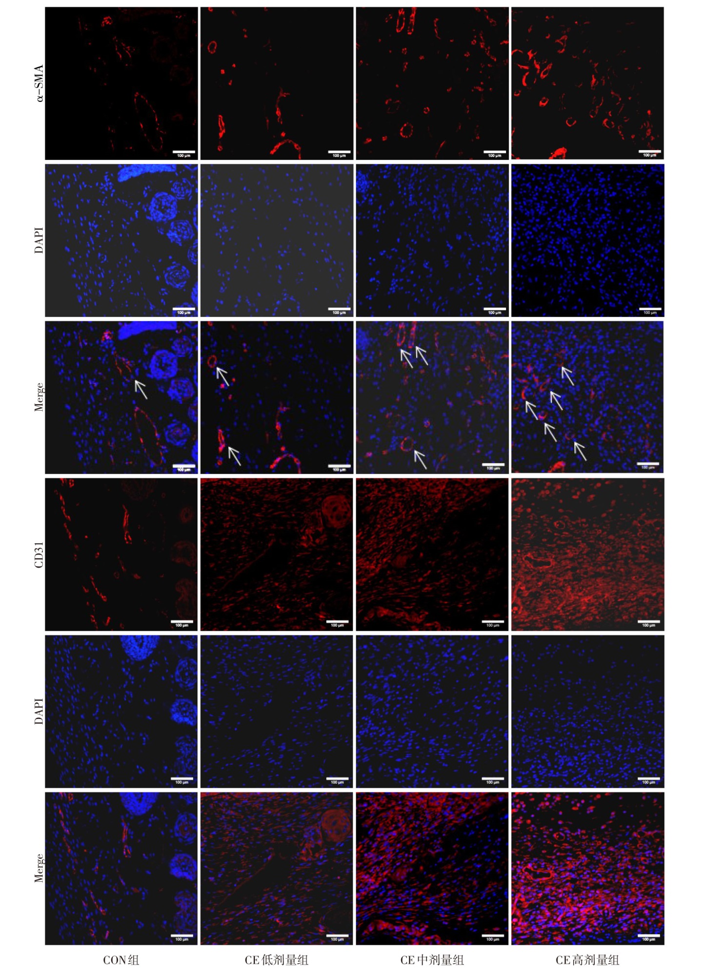
Fig.5 Expression of α-SMA and CD31 in wound skin on day 7 after surgery in each group (immunofluorescence staining, ×200)
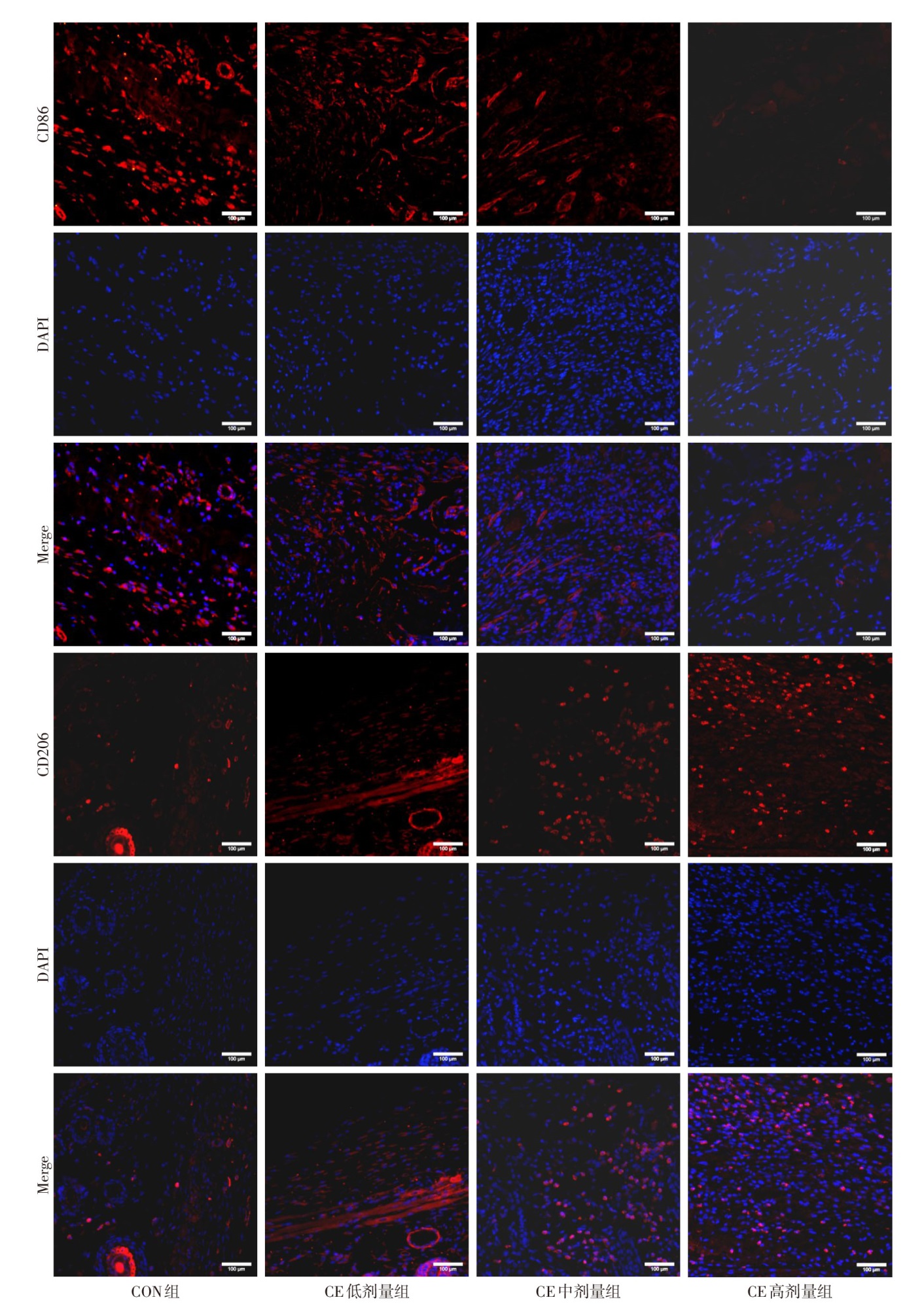
Fig.6 Expression of CD86 and CD206 in wound skin on day 7 after surgery in each group (immunofluorescence staining, ×200)
| [1] | CAO W, PENG S, YAO Y, et al. A nanofibrous membrane loaded with doxycycline and printed with conductive hydrogel strips promotes diabetic wound healing in vivo[J]. Acta Biomater, 2022, 152:60-73. doi:10.1016/j.actbio.2022.08.048. |
| [2] | WANG X, YUAN C X, XU B, et al. Diabetic foot ulcers:Classification,risk factors and management[J]. World J Diabetes, 2022, 13(12):1049-1065. doi:10.4239/wjd.v13.i12.1049. |
| [3] | KIM K, MAHAJAN A, PATEL K, et al. Materials and cytokines in the healing of diabetic foot ulcers[J]. Adv Ther(Weinh), 2021, 4(9):2100075. doi:10.1002/adtp.202100075. |
| [4] | 王一鸣, 朱朝军, 孙旭, 等. 化腐再生法治疗糖尿病足疮面标准操作流程的制定及实践[J]. 中国中西医结合外科杂志, 2024, 30(1):5-8. |
| WANG Y M, ZHU C J, SUN X, et al. Formulation and practice of the standard operating procedures for the treatment of diabetes foot sore with the method of decay and regeneration[J]. Chinese Journal of Integrated Traditional Chinese and Western Medicine Surgery, 2024, 30(1):5-8. doi:10.3969/j.issn.1007-6948.2024.01.001. | |
| [5] | ZHENG S Y, WAN X X, KAMBEY P A, et al. Therapeutic role of growth factors in treating diabetic wound[J]. World J Diabetes, 2023, 14(4):364-395. doi:10.4239/wjd.v14.i4.364. |
| [6] | USLU K, TANSUKER H D, TABARU A, et al. Investigation of the effects of thrombocyte-rich plasma,systemic ozone and hyperbaric oxygen treatment on intraoral wound healing in rats:experimental study[J]. Eur Arch Otorhinolaryngol, 2020, 277(6):1771-1777. doi:10.1007/s00405-020-05872-5. |
| [7] | ZHANG Z, ZHANG W, XU Y, et al. Efficacy of hyperbaric oxygen therapy for diabetic foot ulcers:An updated systematic review and meta-analysis[J]. Asian J Surg, 2022, 45(1):68-78. doi:10.1016/j.asjsur.2021.07.047. |
| [8] | BITO T, OKUMURA E, FUJISHIMA M, et al. Potential of Chlorella as a dietary supplement to promote human health[J]. Nutrients, 2020, 12(9):2524. doi:10.3390/nu12092524. |
| [9] | CHEN S, WANG L, FENG W, et al. Sulfonamides-induced oxidative stress in freshwater microalga Chlorella vulgaris:Evaluation of growth,photosynthesis,antioxidants,ultrastructure,and nucleic acids[J]. Sci Rep, 2020, 10(1):8243. doi:10.1038/s41598-020-65219-2. |
| [10] | ZAHRAN E, ELBAHNASWY S, RISHA E, et al. Antioxidative and immunoprotective potential of Chlorella vulgaris dietary supplementation against chlorpyrifos-induced toxicity in Nile tilapia[J]. Fish Physiol Biochem, 2020, 46(4):1549-1560. doi:10.1007/s10695-020-00814-8. |
| [11] | ZHOU J, WANG M, BÄUERL C, et al. The impact of liquid-pressurized extracts of Spirulina,Chlorella and Phaedactylum tricornutum on in vitro antioxidant,antiinflammatory and bacterial growth effects and gut microbiota modulation[J]. Food Chem, 2023, 401:134083. doi:10.1016/j.foodchem.2022.134083. |
| [12] | ZHOU X, GUO Y, YANG K, et al. The signaling pathways of traditional Chinese medicine in promoting diabetic wound healing[J]. JEthnopharmacol, 2022, 282:114662. |
| [13] | YANG J, CHU Z, JIANG Y, et al. Multifunctional hyaluronic acid microneedle patch embedded by Cerium/Zinc-based composites for accelerating diabetes wound healing[J]. Adv Healthc Mater, 2023, 22:e2300725. doi:10.1002/adhm.202300725. |
| [14] | WU H, YANG P, LI A, et al. Chlorella sp.-ameliorated undesirable microenvironment promotes diabetic wound healing[J]. Acta Pharm Sin B, 2023, 13(1):410-424. doi:10.1016/j.apsb.2022.06.012. |
| [15] | QIAN B, LI J, GUO K, et al. Antioxidant biocompatible composite collagen dressing for diabetic wound healing in rat model[J]. Regen Biomater, 2021, 8(2):rbab003. doi: 10.1093/rb/rbab003. |
| [16] | LI H, CHENG K, WONG C, et al. Evaluation of antioxidant capacity and total phenolic content of different fractions of selected microalgae[J]. Food Chemistry, 2007, 102(3):771-776. |
| [17] | MONTERO-LOBATO Z, VÁZQUEZ M, NAVARRO F, et al. Chemically-induced production of anti-inflammatory molecules in microalgae[J]. Mar Drugs, 2018, 16(12):478. |
| [18] | ZHANG R, CHEN J, MAO X, et al. Anti-inflammatory and anti-aging evaluation of pigment-protein complex extracted from Chlorella pyrenoidosa[J]. Mar Drugs, 2019, 17(10):586. |
| [19] | HWANG H R, LEE E S, KANG S M, et al. Effect of antimicrobial photodynamic therapy with Chlorella and Curcuma extract on Streptococcus mutans biofilms[J]. Photodiagnosis Photodyn Ther, 2021, 35:102411. doi:10.1016/j.pdpdt.2021.102411. |
| Viewed | ||||||
|
Full text |
|
|||||
|
Abstract |
|
|||||