Tianjin Medical Journal ›› 2025, Vol. 53 ›› Issue (4): 439-443.doi: 10.11958/20242130
• Applied Research • Previous Articles Next Articles
HU Ziyue1( ), ZHENG Ruyu1, LIU Dan1, TANG Shan1, KAN Yanmin1,△(), JING Xiang1, LI Qian2
Received:2024-12-05
Revised:2025-02-20
Published:2025-04-15
Online:2025-04-17
Contact:
△ E-mail:HU Ziyue, ZHENG Ruyu, LIU Dan, TANG Shan, KAN Yanmin, JING Xiang, LI Qian. Predictive value of proximal angle of atherosclerosis carotid plaque and distribution of neovascularization in evaluating the recurrence of cerebral infarction[J]. Tianjin Medical Journal, 2025, 53(4): 439-443.
CLC Number:
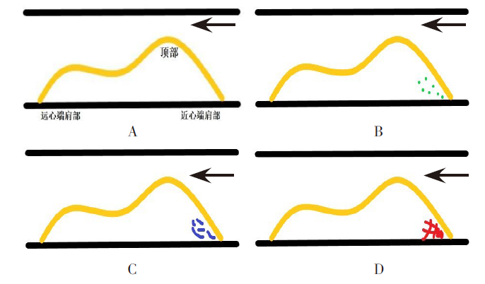
Fig.1 Classification of contrast degree enhancement in the proximal shoulder of the plaque
| 组别 | n | 性别 (男/女) | 年龄/ 岁 | BMI/ (kg/m2) | SBP/ mmHg | |||||
|---|---|---|---|---|---|---|---|---|---|---|
| 非脑梗死组 | 45 | 28/17 | 64.2±7.8 | 25.8±1.5 | 127.4±6.1 | |||||
| 脑梗死组 | 43 | 27/16 | 65.8±7.1 | 26.4±1.3 | 130.0±9.0 | |||||
| χ2或t | 0.956 | 0.997 | 1.786 | 1.563 | ||||||
| 组别 | DBP/ mmHg | LDL-C/ (mmol/L) | FPG/(mmol/L) | 吸烟指数/ 年·支 | ||||||
| 非脑梗死组 | 79.3±6.5 | 3.2±0.6 | 5.6±0.8 | 61.0(0,149.5) | ||||||
| 脑梗死组 | 81.4±7.7 | 3.3±0.5 | 5.8±1.4 | 65.0(0,173.0) | ||||||
| t或Z | 1.366 | 1.128 | 0.703 | 0.084 | ||||||
Tab.1 非脑梗死组与脑梗死组患者临床资料比较
| 组别 | n | 性别 (男/女) | 年龄/ 岁 | BMI/ (kg/m2) | SBP/ mmHg | |||||
|---|---|---|---|---|---|---|---|---|---|---|
| 非脑梗死组 | 45 | 28/17 | 64.2±7.8 | 25.8±1.5 | 127.4±6.1 | |||||
| 脑梗死组 | 43 | 27/16 | 65.8±7.1 | 26.4±1.3 | 130.0±9.0 | |||||
| χ2或t | 0.956 | 0.997 | 1.786 | 1.563 | ||||||
| 组别 | DBP/ mmHg | LDL-C/ (mmol/L) | FPG/(mmol/L) | 吸烟指数/ 年·支 | ||||||
| 非脑梗死组 | 79.3±6.5 | 3.2±0.6 | 5.6±0.8 | 61.0(0,149.5) | ||||||
| 脑梗死组 | 81.4±7.7 | 3.3±0.5 | 5.8±1.4 | 65.0(0,173.0) | ||||||
| t或Z | 1.366 | 1.128 | 0.703 | 0.084 | ||||||
| 组别 | n | 斑块 厚度/mm | 斑块 长度/mm | 斑块近心端 角度/° | 新生血管 评分/分 |
|---|---|---|---|---|---|
| 非脑梗死组 | 45 | 2.5±0.3 | 16.4±2.8 | 17.4±2.3 | 1.0(0.0,1.0) |
| 脑梗死组 | 43 | 3.1±0.2 | 17.3±2.7 | 23.6±2.2 | 2.0(2.0,3.0) |
| t或Z | 11.649** | 1.481 | 13.087** | 4.825** |
Tab.2 非脑梗死组与脑梗死组斑块超声测量比较
| 组别 | n | 斑块 厚度/mm | 斑块 长度/mm | 斑块近心端 角度/° | 新生血管 评分/分 |
|---|---|---|---|---|---|
| 非脑梗死组 | 45 | 2.5±0.3 | 16.4±2.8 | 17.4±2.3 | 1.0(0.0,1.0) |
| 脑梗死组 | 43 | 3.1±0.2 | 17.3±2.7 | 23.6±2.2 | 2.0(2.0,3.0) |
| t或Z | 11.649** | 1.481 | 13.087** | 4.825** |
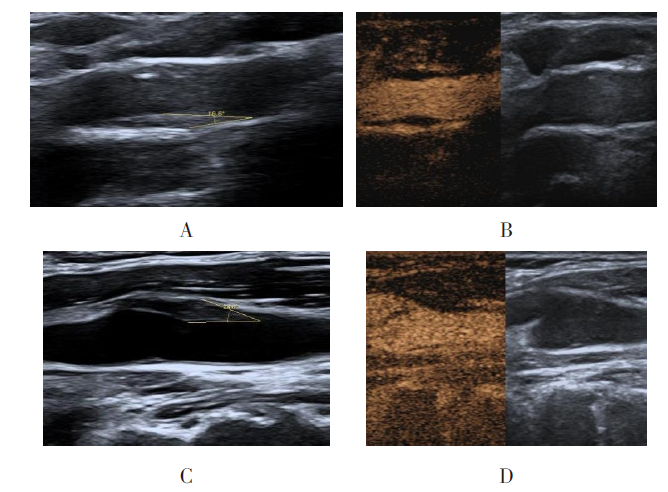
Fig.2 Proximal angle of plaque and distribution of neovascularization in two groups
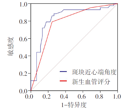
Fig.3 ROC curves of carotid plaque proximal angle and neovascularization score for predicting cerebral infarction
| [1] | 李培远, 王刚, 石军峰. 急性脑梗死患者血浆LncRNA CEBPA-AS1、miR-139-5p表达及与认知功能障碍的相关性分析[J]. 天津医药, 2025, 53(1):75-79. |
| LI P Y, WANG G, SHI J F. The expression of plasma LncRNA CEBPA-AS1 and miR-139-5p in patients with acute cerebral infarction and their correlation with cognitive dysfunction[J]. Tianjin Med J, 2025, 53(1):75-79. doi:10.11958/20241422. | |
| [2] | WANG F, HUA S, ZHANG Y, et al. Association between small vessel disease markers,medial temporal lobe atrophy and cognitive impairment after stroke:a systematic review and meta-analysis[J]. J Stroke Cerebrovasc Dis, 2021, 30(1):105460. doi:10.1016/j.jstrokecerebrovasdis.2020.105460. |
| [3] | GBD 2019 STROKE COLLABORATORS. Global,regional,and national burden of stroke and its risk factors,1990-2019:a systematic analysis for the Global Burden of Disease Study 2019[J]. Lancet Neurol,2021, 20(10):795-820. doi:10.1016/S1474-4422(21)00252-0. |
| [4] | HENEIN M Y, FAGGIANO P, VANCHERI F. Limited accuracy of carotid progression of intima-media thickness inpredicting clinical cardiovascular outcome[J]. Pol Arch Intern Med, 2019, 129(1):1-3. doi:10.20452/pamw.4430. |
| [5] | 聂红军, 李芬穗, 胡珏, 等. 颈动脉斑块超声造影及hs-CRP与急性脑梗死体积关系的研究[J]. 中国超声医学杂志, 2020, 36(8):687-690. |
| NIE H J, LI F S, HU J, et al. The relationship between carotid plaque contrast-enhanced ultrasound and hs-CRP and the volume of acute cerebral infarction[J]. Chinese J Ultrasound Med, 2020, 36(8):687-690. doi:10.3969/j.issn.1002-0101.2020.08.005. | |
| [6] | 李怡, 何文. 颈动脉粥样硬化易损斑块超声标志与脑血管疾病关系的研究进展[J]. 中国卒中杂志, 2021, 16(11):1183-1188. |
| LI Y, HE W. Progress of relationship between ultrasound biomarkers of vulnerable carotid atherosclerotic plaque and cerebrovascular disease[J]. Chinese Journal of Stroke, 2021, 16(11):1183-1188. doi:10.3969/j.issn.1673-5765.2021.11.017. | |
| [7] | THONDAPU V, MAMON C, POON E, et al. High spatial endothelial shear stress gradient independently predicts site of acute coronary plaque rupture and erosion[J]. Cardiovasc Res, 2021, 117(8):1974-1985. doi:10.1093/cvr/cvaa251. |
| [8] | 中国医师协会超声医师分会. 中国超声造影临床应用指南[M]. 北京: 人民卫生出版社,2017:65. |
| Ultrasound Doctor Branch Of Chinese Medical Doctor Association. Chinese guideline for clinical application of contrast-enhanced ultrasound[M]. Beijing: People's Health Publishing House,2017:65. | |
| [9] | WANG Y J, LI Z X, GU H Q. China stroke statistics:an update on the 2019 report from the national center for healthcare quality management in neurological diseases,China national clinical research center for neurological diseases,the Chinese stroke association,national center for chronic and non-communicable disease control and prevention,Chinese center for disease control and prevention and institute for global neuroscience and stroke collaborations[J]. Stroke Vasc Neurol, 2022, 7(5):415-450. doi:10.1136/svn-2021-001374. |
| [10] | YANG J, DUAN J, LI M, et al. Aldehyde dehydrogenase isoform 1 predicts a poor prognosis of acute cerebral infarction[J]. Contrast Media Mol Imaging, 2022,2022:8199917. doi:10.1155/2022/8199917. |
| [11] | HUANG Z, CHENG X Q, LIU Y N, et al. Value of intraplaque neovascularization on contrast-enhanced ultrasonography in predicting ischemic stroke recurrence in patients with carotid atherosclerotic plaque[J]. Korean J Radiol, 2023, 24(4):338-348. doi:10.3348/kjr.2022.0977. |
| [12] | PARITALA P K, YARLAGADDA T, MENDIETA J B, et al. Plaque longitudinal heterogeneity in morphology, property, and mechanobiology[J]. Cerebrovasc Dis, 2021, 50(5):510-519. doi:10.1159/000515690. |
| [13] | WANG Y, WANG T, LUO Y, et al. Identification markers of carotid vulnerable plaques:an update[J]. Biomolecules, 2022, 12(9):1192. doi:10.3390/biom12091192. |
| [14] | RAFAILIDIS V, LI X, SIDHU P S, et al. Contrast imaging ultrasound for the detection and characterization of carotid vulnerable plaque[J]. Cardiovasc Diagn Ther, 2020, 10(4):965-981. doi:10.21037/cdt.2020.01.08. |
| [15] | CUI L, XING Y, WANG L, et al. Carotid intraplaque neovascularization and future vascular events in patients with asymptomatic carotid stenosis[J]. Front Pharmacol, 2022,13:804810. doi:10.3389/fphar.2022.804810. |
| [16] | EVANS P C, FRAGIADAKI M, MORRIS P D, et al. Shear stress:the dark energy of atherosclerotic plaques[J]. Cardiovasc Res, 2021, 117(8):1811-1813. doi:10.1093/cvr/cvaa315. |
| [17] | 中国医师协会超声医师分会. 超声评价颈动脉易损斑块中国专家共识(2023版)[J]. 中华超声影像学杂志, 2023, 32(8):645-655. |
| Ultrasound Doctor Branch Of Chinese Medical Doctor Association. Chinese expert consensus on ultrasound evaluation of vulnerable carotid plaque(2023 edition)[J]. Chin J Ultrasonogr, 2023, 32(8):645-655. doi:10.3760/cma.j.cn131148-20230418-00215. | |
| [18] | 彭佩燕, 张泽鑫, 吴壮雄. 颈动脉斑块最大厚度值和Lp-PLA2水平与颈动脉易损斑块的相关性[J]. 中国超声医学杂志, 2024, 40(1):16-19. |
| PENG P Y, ZHANG Z X, WU Z X. Correlation between the maximum carotid plaque thickness,Lp-PLA2 level and the vulnerable carotid plaque in patients[J]. Chinese J Ultrasound Med, 2024, 40(1):16-19. doi:10.3969/j.issn.1002-0101.2024.01.005. | |
| [19] | MA T, SHI X, YUAN C, et al. Contrast-enhanced ultrasound combined with 2D strain imaging and histopathological multimodal assessment of carotid plaque vulnerability[J]. Ultrasound Med Biol, 2023, 49(7):1595-1601. doi:10.1016/j.ultrasmedbio.2023.03.005. |
| [20] | ZAMANI M, SKAGEN K, SCOTT H, et al. Carotid plaque neovascularization detected with superb microvascular imaging ultrasound without using contrast media[J]. Stroke, 2019, 50(11):3121-3127. doi:10.1161/STROKEAHA.119.025496. |
| Viewed | ||||||
|
Full text |
|
|||||
|
Abstract |
|
|||||