
Tianjin Medical Journal ›› 2026, Vol. 54 ›› Issue (10): 1093-1098.doi: 10.11958/20261200
• Applied Research • Previous Articles Next Articles
DENG Yuanlin1( ), TIAN Hexian1, ZHANG Zhang1, YANG Zhenwen2, LI Fengtan1, LI Dong1, YANG Fan1,△()
Received:2026-05-07
Revised:2026-07-10
Published:2026-10-15
Online:2026-10-10
Contact:
△E-mail:dr_yangfan0201@163.com
DENG Yuanlin, TIAN Hexian, ZHANG Zhang, YANG Zhenwen, LI Fengtan, LI Dong, YANG Fan. The relationship between the left ventricular geometric remodeling and clinical worsening in patients with pulmonary arterial hypertension[J]. Tianjin Medical Journal, 2026, 54(10): 1093-1098.
CLC Number:
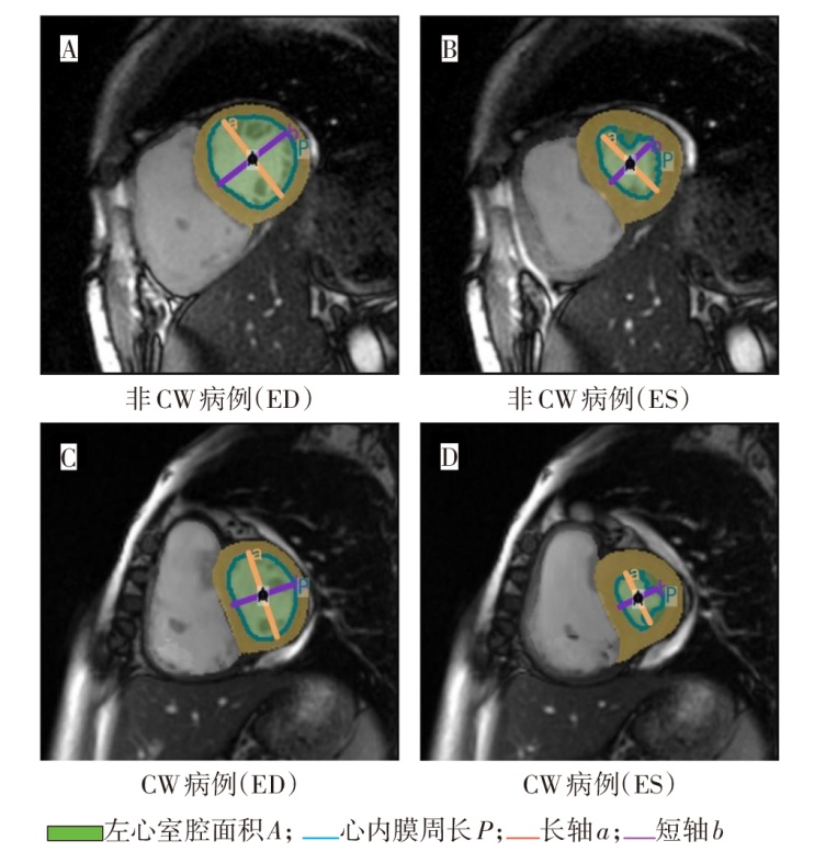
Fig.1 Representative CMR example of left ventricular segmentation and geometric measurements
| 组别 | n | 年龄/岁 | 女性 | 身高/cm | 体质量/kg | |||
|---|---|---|---|---|---|---|---|---|
| 非CW组 | 133 | 40(31,57) | 107(80.5) | 162(160,165) | 60(55,67) | |||
| CW组 | 61 | 51(37,60) | 46(75.4) | 162(160,170) | 62(53,69) | |||
| Z或χ² | 2.121* | 0.372 | 0.354 | 0.635 | ||||
| 组别 | WHO-FC (Ⅰ/Ⅱ/Ⅲ/Ⅳ) | 6MWD/m | BNP/(ng/L) | |||||
| 非CW组 | 5/85/39/4 | 382(298,457) | 102.50(4.32,332.00) | |||||
| CW组 | 1/23/30/7 | 238(43,378) | 508.50(8.83,931.00) | |||||
| χ²或Z | 15.691** | 5.282** | 4.431** | |||||
Tab.1 Comparison of clinical characteristics between the two groups
| 组别 | n | 年龄/岁 | 女性 | 身高/cm | 体质量/kg | |||
|---|---|---|---|---|---|---|---|---|
| 非CW组 | 133 | 40(31,57) | 107(80.5) | 162(160,165) | 60(55,67) | |||
| CW组 | 61 | 51(37,60) | 46(75.4) | 162(160,170) | 62(53,69) | |||
| Z或χ² | 2.121* | 0.372 | 0.354 | 0.635 | ||||
| 组别 | WHO-FC (Ⅰ/Ⅱ/Ⅲ/Ⅳ) | 6MWD/m | BNP/(ng/L) | |||||
| 非CW组 | 5/85/39/4 | 382(298,457) | 102.50(4.32,332.00) | |||||
| CW组 | 1/23/30/7 | 238(43,378) | 508.50(8.83,931.00) | |||||
| χ²或Z | 15.691** | 5.282** | 4.431** | |||||
| 组别 | n | mRAP/mmHg | sPAP/mmHg |
|---|---|---|---|
| 非CW组 | 133 | 5.00(3.00,7.00) | 70.00(55.25,84.00) |
| CW组 | 61 | 7.00(5.00,11.00) | 88.50(69.25,99.50) |
| Z | 4.592** | 3.933** | |
| 组别 | dPAP/mmHg | mPAP/mmHg | PVR/WU |
| 非CW组 | 24.00(19.00,30.00) | 41.00(35.00,52.00) | 8.83(6.34,12.50) |
| CW组 | 31.00(25.00,39.75) | 53.00(43.00,63.00) | 13.69(10.25,18.60) |
| Z | 4.159** | 4.767** | 4.725** |
Tab.2 Comparison of RHC parameters between the two groups
| 组别 | n | mRAP/mmHg | sPAP/mmHg |
|---|---|---|---|
| 非CW组 | 133 | 5.00(3.00,7.00) | 70.00(55.25,84.00) |
| CW组 | 61 | 7.00(5.00,11.00) | 88.50(69.25,99.50) |
| Z | 4.592** | 3.933** | |
| 组别 | dPAP/mmHg | mPAP/mmHg | PVR/WU |
| 非CW组 | 24.00(19.00,30.00) | 41.00(35.00,52.00) | 8.83(6.34,12.50) |
| CW组 | 31.00(25.00,39.75) | 53.00(43.00,63.00) | 13.69(10.25,18.60) |
| Z | 4.159** | 4.767** | 4.725** |
| 组别 | n | RVEDVI/(mL/m²) | RVESVI/(mL/m²) | ||||||||||
|---|---|---|---|---|---|---|---|---|---|---|---|---|---|
| 非CW组 | 133 | 81.64(62.36,102.22) | 48.62(37.14,62.86) | ||||||||||
| CW组 | 61 | 120.06(96.70,147.40) | 82.38(65.49,107.26) | ||||||||||
| Z | 6.003** | 7.109** | |||||||||||
| 组别 | RVSVI/(mL/m²) | RVEF/% | RVMMI/(g/m²) | ||||||||||
| 非CW组 | 31.54(22.88,38.68) | 38.44±8.93 | 16.56(12.15,17.33) | ||||||||||
| CW组 | 33.50(27.78,42.37) | 28.51±8.01 | 32.66(25.69,43.87) | ||||||||||
| Z或t | 1.463 | 7.733** | 4.282** | ||||||||||
| 组别 | RVCI/[L/(min·m²)] | LVEDVI/(mL/m²) | |||||||||||
| 非CW组 | 2.60(2.13,3.10) | 67.63(55.62,77.99) | |||||||||||
| CW组 | 2.13(1.59,2.89) | 62.79(50.53,72.41) | |||||||||||
| Z | 2.892** | 1.731 | |||||||||||
| 组别 | LVESVI/(mL/m²) | LVSVI/(mL/m²) | LVMMI/(g/m²) | ||||||||||
| 非CW组 | 25.75(20.93,33.38) | 38.49±12.04 | 45.86(40.13,52.85) | ||||||||||
| CW组 | 27.66(20.57,34.17) | 35.79±10.37 | 39.81(35.14,47.46) | ||||||||||
| Z或t | 0.171 | 1.726 | 2.861** | ||||||||||
| 组别 | LVCI/[L/(min·m²)] | LVEF/% | |||||||||||
| 非CW组 | 2.77(2.53,3.48) | 59.80(52.80,64.00) | |||||||||||
| CW组 | 2.47(2.31,2.90) | 56.00(44.10,63.00) | |||||||||||
| Z | 2.474* | 1.833 | |||||||||||
Tab.3 Comparison of conventional CMR functional parameters between the two groups
| 组别 | n | RVEDVI/(mL/m²) | RVESVI/(mL/m²) | ||||||||||
|---|---|---|---|---|---|---|---|---|---|---|---|---|---|
| 非CW组 | 133 | 81.64(62.36,102.22) | 48.62(37.14,62.86) | ||||||||||
| CW组 | 61 | 120.06(96.70,147.40) | 82.38(65.49,107.26) | ||||||||||
| Z | 6.003** | 7.109** | |||||||||||
| 组别 | RVSVI/(mL/m²) | RVEF/% | RVMMI/(g/m²) | ||||||||||
| 非CW组 | 31.54(22.88,38.68) | 38.44±8.93 | 16.56(12.15,17.33) | ||||||||||
| CW组 | 33.50(27.78,42.37) | 28.51±8.01 | 32.66(25.69,43.87) | ||||||||||
| Z或t | 1.463 | 7.733** | 4.282** | ||||||||||
| 组别 | RVCI/[L/(min·m²)] | LVEDVI/(mL/m²) | |||||||||||
| 非CW组 | 2.60(2.13,3.10) | 67.63(55.62,77.99) | |||||||||||
| CW组 | 2.13(1.59,2.89) | 62.79(50.53,72.41) | |||||||||||
| Z | 2.892** | 1.731 | |||||||||||
| 组别 | LVESVI/(mL/m²) | LVSVI/(mL/m²) | LVMMI/(g/m²) | ||||||||||
| 非CW组 | 25.75(20.93,33.38) | 38.49±12.04 | 45.86(40.13,52.85) | ||||||||||
| CW组 | 27.66(20.57,34.17) | 35.79±10.37 | 39.81(35.14,47.46) | ||||||||||
| Z或t | 0.171 | 1.726 | 2.861** | ||||||||||
| 组别 | LVCI/[L/(min·m²)] | LVEF/% | |||||||||||
| 非CW组 | 2.77(2.53,3.48) | 59.80(52.80,64.00) | |||||||||||
| CW组 | 2.47(2.31,2.90) | 56.00(44.10,63.00) | |||||||||||
| Z | 2.474* | 1.833 | |||||||||||
| 组别 | n | ED左心室偏心率 | ED左心室轴比 | ED左心室圆形度 | |||||
|---|---|---|---|---|---|---|---|---|---|
| 非CW组 | 133 | 0.59(0.47,0.66) | 1.24(1.14,1.34) | 0.62(0.60,0.63) | |||||
| CW组 | 61 | 0.71(0.62,0.77) | 1.41(1.28,1.56) | 0.59(0.55,0.62) | |||||
| Z | 5.221** | 5.223** | 5.110** | ||||||
| 组别 | ED LVGI | ES左心室偏心率 | |||||||
| 非CW组 | -0.63(-2.15,0.60) | 0.61(0.49,0.74) | |||||||
| CW组 | 1.32(-0.09,2.79) | 0.71(0.59,0.78) | |||||||
| Z | 5.523** | 2.395* | |||||||
| 组别 | ES 左心室轴比 | ES左心室圆形度 | ES LVGI | ||||||
| 非CW组 | 1.26(1.15,1.47) | 0.61(0.56,0.64) | -0.85(-2.39,1.69) | ||||||
| CW组 | 1.42(1.23,1.61) | 0.57(0.54,0.60) | 0.75(-0.32,2.05) | ||||||
| Z | 2.394* | 3.611** | 2.998** | ||||||
Tab.4 Comparison of left ventricular geometric indices between the two groups
| 组别 | n | ED左心室偏心率 | ED左心室轴比 | ED左心室圆形度 | |||||
|---|---|---|---|---|---|---|---|---|---|
| 非CW组 | 133 | 0.59(0.47,0.66) | 1.24(1.14,1.34) | 0.62(0.60,0.63) | |||||
| CW组 | 61 | 0.71(0.62,0.77) | 1.41(1.28,1.56) | 0.59(0.55,0.62) | |||||
| Z | 5.221** | 5.223** | 5.110** | ||||||
| 组别 | ED LVGI | ES左心室偏心率 | |||||||
| 非CW组 | -0.63(-2.15,0.60) | 0.61(0.49,0.74) | |||||||
| CW组 | 1.32(-0.09,2.79) | 0.71(0.59,0.78) | |||||||
| Z | 5.523** | 2.395* | |||||||
| 组别 | ES 左心室轴比 | ES左心室圆形度 | ES LVGI | ||||||
| 非CW组 | 1.26(1.15,1.47) | 0.61(0.56,0.64) | -0.85(-2.39,1.69) | ||||||
| CW组 | 1.42(1.23,1.61) | 0.57(0.54,0.60) | 0.75(-0.32,2.05) | ||||||
| Z | 2.394* | 3.611** | 2.998** | ||||||
| 参数 | β | SE | Wald χ2 | P | HR(95%CI) |
|---|---|---|---|---|---|
| ED LVGI | 0.315 | 0.147 | 4.613 | 0.032 | 1.370(1.028~1.827) |
| ES LVGI | 0.130 | 0.134 | 0.941 | 0.332 | 1.139(0.876~1.480) |
| mRAP | 0.304 | 0.125 | 5.911 | 0.015 | 1.356(1.061~1.732) |
| PVR | 0.062 | 0.188 | 0.108 | 0.743 | 1.064(0.736~1.536) |
| RVEF | -0.720 | 0.166 | 18.831 | <0.001 | 0.487(0.352~0.674) |
| RVEDVI | 0.158 | 0.120 | 1.741 | 0.187 | 1.171(0.926~1.481) |
Tab.5 Multivariate Cox regression analysis of the association between LVGI and clinical worsening
| 参数 | β | SE | Wald χ2 | P | HR(95%CI) |
|---|---|---|---|---|---|
| ED LVGI | 0.315 | 0.147 | 4.613 | 0.032 | 1.370(1.028~1.827) |
| ES LVGI | 0.130 | 0.134 | 0.941 | 0.332 | 1.139(0.876~1.480) |
| mRAP | 0.304 | 0.125 | 5.911 | 0.015 | 1.356(1.061~1.732) |
| PVR | 0.062 | 0.188 | 0.108 | 0.743 | 1.064(0.736~1.536) |
| RVEF | -0.720 | 0.166 | 18.831 | <0.001 | 0.487(0.352~0.674) |
| RVEDVI | 0.158 | 0.120 | 1.741 | 0.187 | 1.171(0.926~1.481) |
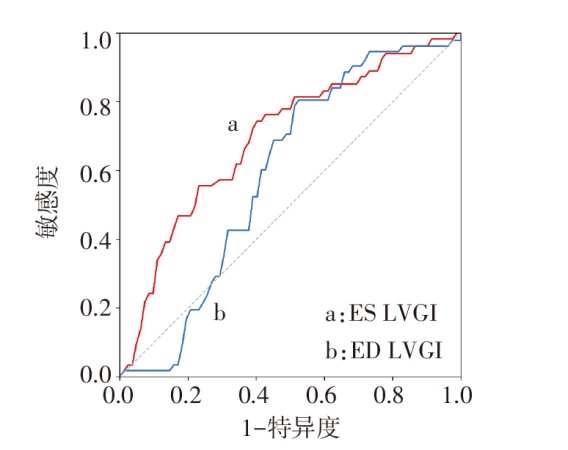
Fig.2 ROC curve of LVGI for predicting clinical worsening
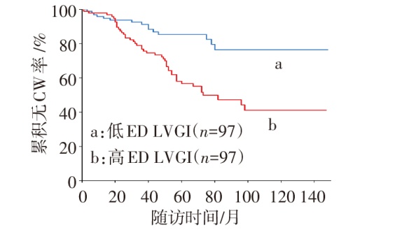
Fig.3 Kaplan-Meier curves for clinical worsening events in patients with different ED LVGI levels
| 参数 | mRAP | mPAP | PVR | RVEF | RVEDVI | LVCI |
|---|---|---|---|---|---|---|
| ED LVGI | 0.122 | 0.436** | 0.421** | -0.247** | 0.399** | -0.227** |
| ES LVGI | 0.007 | 0.363** | 0.418** | -0.219** | 0.293** | -0.055 |
Tab.6 Correlation analysis between LVGI and selected hemodynamic and cardiac functional parameters
| 参数 | mRAP | mPAP | PVR | RVEF | RVEDVI | LVCI |
|---|---|---|---|---|---|---|
| ED LVGI | 0.122 | 0.436** | 0.421** | -0.247** | 0.399** | -0.227** |
| ES LVGI | 0.007 | 0.363** | 0.418** | -0.219** | 0.293** | -0.055 |
| [1] | Kovacs G, Bartolome S, Denton C P, et al. Definition,classification and diagnosis of pulmonary hypertension[J]. Eur Respir J, 2024, 64(4):2401324. doi:10.1183/13993003.01324-2024. |
| [2] | 阿里旦·艾尔肯, 李鸣远, 米娜瓦尔·胡加艾合买提. 右心室重构特征与肺动脉高压患者治疗反应性的相关性[J]. 中华全科医学, 2026, 24(3):403-406. |
| Aierken A D, Li M Y, Hujiaaimaiti M. Correlation between right ventricular remodeling characteristics and treatment responsiveness in patients with pulmonary hypertension[J]. Chin J Gen Pract, 2026, 24(3):403-406. doi:10.16766/j.cnki.issn.1674-4152.004406. | |
| [3] | McCoy D, Movahed M R. Review of D-shape left ventricle seen on magnetic resonance imaging(MRI)or computed tomography(CT),similar to the Movahed sign seen on cardiac gated single-photon emission computed tomography(SPECT)as an indicator for right ventricular overload[J]. J Clin Med, 2025, 14(17):6041. doi:10.3390/jcm14176041. |
| [4] | Celant L R, Wessels J N, Marcus J T, et al. Toward the implementation of optimal cardiac magnetic resonance risk stratification in pulmonary arterial hypertension[J]. Chest, 2024, 165(1):181-191. doi:10.1016/j.chest.2023.07.028. |
| [5] | Humbert M, Kovacs G, Hoeper M M, et al. 2022 ESC/ERS guidelines for the diagnosis and treatment of pulmonary hypertension[J]. Eur Heart J, 2022, 43(38):3618-3731. doi:10.1093/eurheartj/ehac237. |
| [6] | Weatherald J, Fleming T R, Wilkins M R, et al. Clinical trial design,end-points,and emerging therapies in pulmonary arterial hypertension[J]. Eur Respir J, 2024, 64(4):2401205. doi:10.1183/13993003.01205-2024. |
| [7] | Namazi M, Eftekhar S P, Mosaed R, et al. Pulmonary hypertension and right ventricle:a pathophysiological insight[J]. Clin Med Insights Cardiol, 2024, 18:11795468241274744. doi:10.1177/11795468241274744. |
| [8] | 武才鑫, 闫彦, 邓瑗琳, 等. 心脏磁共振评估结缔组织病伴重度肺动脉高压的价值[J]. 天津医药, 2024, 52(7):691-695. |
| Wu C X, Yan Y, Deng Y L, et al. Value of cardiac magnetic resonance in evaluating severe pulmonary hypertension associated with connective tissue disease[J]. Tianjin Med J, 2024, 52(7):691-695. doi:10.11958/20231770. | |
| [9] | Fang H, Wang J, Shi R, et al. Biventricular dysfunction and ventricular interdependence in patients with pulmonary hypertension:a 3.0-T cardiac MRI feature tracking study[J]. J Magn Reson Imaging, 2024, 60(1):350-362. doi:10.1002/jmri.29044. |
| [10] | Ahmad A, Zhang S, Ren Q, et al. Transthoracic echocardiography of left ventricular underfilling improves risk stratification in pulmonary arterial hypertension[J]. Sci Rep, 2025, 15(1):45718. doi:10.1038/s41598-025-28206-z. |
| [11] | Lindholm A, Kjellström B, Rådegran G, et al. Right ventricular dyssynchrony predicts outcome in pulmonary arterial hypertension when assessed in multiple cardiac magnetic resonance views[J]. J Cardiovasc Magn Reson, 2024, 26(2):101103. doi:10.1016/j.jocmr.2024.101103. |
| [12] | Chiou F R, Sung S H, Chiu J H, et al. Cardiac magnetic resonance parameters for prognostic stratification in pulmonary hypertension:a longitudinal study[J]. J Chin Med Assoc, 2025, 88(11):858-867. doi:10.1097/JCMA.0000000000001292. |
| [13] | Li W, Zhang X C, Qian Y, et al. Prognostic value of cardiac MRI-derived left ventricular layer-specific strain in pulmonary arterial hypertension[J]. Radiol Cardiothorac Imaging, 2025, 7(4):e240334. doi: 10.1148/ryct.240334. |
| [14] | Burkett D A, Patel S S, Mertens L, et al. Relationship between left ventricular geometry and invasive hemodynamics in pediatric pulmonary hypertension[J]. Circ Cardiovasc Imaging, 2020, 13(5):e009825. doi:10.1161/CIRCIMAGING.119.009825. |
| [15] | Haarman M G, Coenraad I, Hagdorn Q A J, et al. Cardiac magnetic resonance derived left ventricular eccentricity index and right ventricular mass measurements predict outcome in children with pulmonary arterial hypertension[J]. Children(Basel), 2023, 10(4):756. doi:10.3390/children10040756. |
| [16] | Kallianos K, Brooks G C, Mukai K, et al. Cardiac magnetic resonance evaluation of left ventricular myocardial strain in pulmonary hypertension[J]. Acad Radiol, 2018, 25(1):129-135. doi:10.1016/j.acra.2017.07.009. |
| [17] | Guo J, Wang J, Wang L, et al. Left ventricular underfilling in PAH: a potential indicator for adaptive-to-maladaptive transition[J]. Pulm Circ, 2023, 13(4):e12309. doi:10.1002/pul2.12309. |
| [18] | Ahmad A, Zou Y, Zhang P, et al. Non-invasive imaging techniques for early diagnosis of bilateral cardiac dysfunction in pulmonary hypertension: current crests,future peaks[J]. Front Cardiovasc Med, 2024, 11:1393580. doi:10.3389/fcvm.2024.1393580. |
| Viewed | ||||||
|
Full text |
|
|||||
|
Abstract |
|
|||||
