Tianjin Medical Journal ›› 2023, Vol. 51 ›› Issue (5): 487-490.doi: 10.11958/20221258
• Experimental Research • Previous Articles Next Articles
XUE Li( ), HAN Hong, ZHANG Li
Received:2022-08-11
Revised:2022-11-23
Published:2023-05-15
Online:2023-05-05
XUE Li, HAN Hong, ZHANG Li. Effects of Morin on neuronal apoptosis in cerebral ischemia-reperfusion rats by inhibiting TXNIP/NLRP3/Caspase-1 signaling pathway[J]. Tianjin Medical Journal, 2023, 51(5): 487-490.
CLC Number:
| 组别 | IL-1β | IL-18 |
|---|---|---|
| 假手术组 | 10.27±1.03 | 75.15±7.52 |
| 模型组 | 29.12±2.92a | 138.52±13.86a |
| Morin-L组 | 22.04±2.21b | 106.37±10.64b |
| Morin-H组 | 15.34±1.54bc | 84.15±8.42bc |
| Morin-H+pLVX-NC组 | 15.27±1.53 | 84.28±8.43 |
| Morin-H+pLVX-TXNIP组 | 24.35±2.44de | 115.68±11.57de |
| F | 115.929** | 54.256** |
Tab.1 Comparison of serum levels of IL-1β and IL-18 between the six groups
| 组别 | IL-1β | IL-18 |
|---|---|---|
| 假手术组 | 10.27±1.03 | 75.15±7.52 |
| 模型组 | 29.12±2.92a | 138.52±13.86a |
| Morin-L组 | 22.04±2.21b | 106.37±10.64b |
| Morin-H组 | 15.34±1.54bc | 84.15±8.42bc |
| Morin-H+pLVX-NC组 | 15.27±1.53 | 84.28±8.43 |
| Morin-H+pLVX-TXNIP组 | 24.35±2.44de | 115.68±11.57de |
| F | 115.929** | 54.256** |
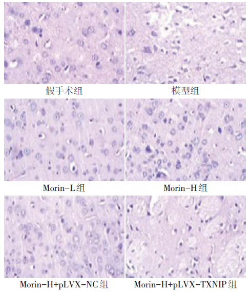
Fig.1 Histopathological changes of cerebral penumbra in each group of rats (HE, ×400)
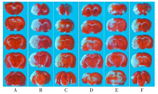
Fig.2 Changes in infarct size of brain tissue (TTC staining, ×100)
| 组别 | 梗死面积(%) | 脑含水量(%) |
|---|---|---|
| 假手术组 | 0.00±0.00 | 65.12±3.01 |
| 模型组 | 21.05±2.11a | 85.34±3.55a |
| Morin-L组 | 13.21±1.33b | 75.66±2.76b |
| Morin-H组 | 8.51±0.86bc | 67.43±2.34bc |
| Morin-H+pLVX-NC组 | 8.48±0.85 | 68.32±2.28 |
| Morin-H+pLVX-TXNIP组 | 15.64±1.57de | 82.54±2.88de |
| F | 92.531** | 26.751** |
Tab.2 Comparison of infarct size of brain tissue and brain water content between six groups of rats
| 组别 | 梗死面积(%) | 脑含水量(%) |
|---|---|---|
| 假手术组 | 0.00±0.00 | 65.12±3.01 |
| 模型组 | 21.05±2.11a | 85.34±3.55a |
| Morin-L组 | 13.21±1.33b | 75.66±2.76b |
| Morin-H组 | 8.51±0.86bc | 67.43±2.34bc |
| Morin-H+pLVX-NC组 | 8.48±0.85 | 68.32±2.28 |
| Morin-H+pLVX-TXNIP组 | 15.64±1.57de | 82.54±2.88de |
| F | 92.531** | 26.751** |
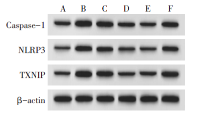
Fig.3 Expression of TXNIP, NLRP3 and Caspase-1 proteins in each group (Western blot assay)
| 组别 | TXNIP | NLRP3 | Caspase-1 |
|---|---|---|---|
| 假手术组 | 0.25±0.03 | 0.17±0.02 | 0.34±0.04 |
| 模型组 | 1.19±0.12a | 0.85±0.09a | 1.12±0.12a |
| Morin-L组 | 0.63±0.07b | 0.54±0.06b | 0.77±0.08b |
| Morin-H组 | 0.34±0.04bc | 0.24±0.03bc | 0.39±0.04bc |
| Morin-H+pLVX-NC组 | 0.37±0.04 | 0.25±0.03 | 0.37±0.04 |
| Morin-H+pLVX-TXNIP组 | 0.71±0.08de | 0.66±0.07de | 0.74±0.08de |
| F | 96.979** | 95.663** | 72.103** |
Tab.3 Comparison of TXNIP, NLRP3 and Caspase-1 protein expression between six groups of rats
| 组别 | TXNIP | NLRP3 | Caspase-1 |
|---|---|---|---|
| 假手术组 | 0.25±0.03 | 0.17±0.02 | 0.34±0.04 |
| 模型组 | 1.19±0.12a | 0.85±0.09a | 1.12±0.12a |
| Morin-L组 | 0.63±0.07b | 0.54±0.06b | 0.77±0.08b |
| Morin-H组 | 0.34±0.04bc | 0.24±0.03bc | 0.39±0.04bc |
| Morin-H+pLVX-NC组 | 0.37±0.04 | 0.25±0.03 | 0.37±0.04 |
| Morin-H+pLVX-TXNIP组 | 0.71±0.08de | 0.66±0.07de | 0.74±0.08de |
| F | 96.979** | 95.663** | 72.103** |
| [1] | SHAO Z Q, DOU S S, ZHU J G, et al. Apelin-13 inhibits apoptosis and excessive autophagy in cerebral ischemia/reperfusion injury[J]. Neural Regen Res, 2021, 16(6):1044-1051. doi:10.4103/1673-5374.300725. |
| [2] | 刘海颖, 冯子人, 孙辉, 等. 大鼠脑缺血再灌注早期过氧化物酶体增殖物激活受体γ活化与细胞焦亡的关系[J]. 天津医药, 2020, 48(1):34-37. |
| LIU H Y, FENG Z R, SUN H, et al. Relationship between activation of peroxisome proliferator-activated receptor γ and cell apoptosis in the early stage of cerebral ischemia-reperfusion in rats[J]. Tianjin Med J, 2020, 48(1):34-37. doi:10.11958/20192153. | |
| [3] | LIANG T Y, PENG S Y, MA M, et al. Protective effects of sevoflurane in cerebral ischemia reperfusion injury:a narrative review[J]. Med Gas Res, 2021, 11(4):152-154. doi:10.4103/2045-9912.318860. |
| [4] | HAN L, XI G, GUO N, et al. Expression and mechanism of TXNIP/NLRP3 inflammasome in sciatic nerve of type 2 diabetic rats[J]. Dis Markers, 2022, 2022:9696303. doi:10.1155/2022/9696303. |
| [5] | HEEBA G H, RABIE E M, ABUZEID M M, et al. Morin alleviates fructose-induced metabolic syndrome in rats viaameliorating oxidative stress,inflammatory and fibrotic markers[J]. Korean J Physiol Pharmacol, 2021, 25(3):177-187. doi:10.1002/jnr.24385. |
| [6] | XING F, LIU Y, DONG R, et al. miR-374 improves cerebral ischemia reperfusion injury by targeting Wnt5a[J]. Exp Anim, 2021, 70(1):126-136. doi:10.1538/expanim.20-0034. |
| [7] | 张泽莲, 刘培俊, 陈娟, 等. 桑黄素通过抑制HMGB1/TLR4/NF-κB通路改善重症肺炎大鼠肺损伤[J]. 免疫学杂志, 2022, 38(6):478-486. |
| ZHANG Z L, LIU P J, CHEN J, et al. Morgenin improves lung injury in severe pneumonia rats by inhibiting HMGB1/TLR4/NF-κB pathway[J]. Immunol J, 2022, 38(6):478-486. | |
| [8] | 潘琼, 郭科, 李亚倩, 等. TXNIP介导的氧化应激在雌激素延缓阿尔兹海默病中的作用[J]. 中南大学学报(医学版), 2019, 44(12):1360-1366. |
| PAN Q, GUO K, LI Y Q, et al. The role of TXNIP-mediated oxidative stress in estrogen delaying Alzheimer's disease[J]. J Cent South Univ(Med Sci), 2019, 44(12):1360-1366. | |
| [9] | GUO H, ZHU L, TANG P, et al. Carthamin yellow improves cerebral ischemia-reperfusion injury by attenuating inflammation and ferroptosis in rats[J]. Int J Mol Med, 2021, 47(4):52. doi:10.3892/ijmm.2021.4885. |
| [10] | LI J, PENG L, BAI W, et al. Biliverdin protects against cerebral ischemia/reperfusion injury by regulating the miR-27a-3p/rgs1 axis[J]. Neuropsychiatr Dis Treat, 2021, 17:1165-1181. doi:10.2147/NDT.S300773. |
| [11] | ZHANG C, ZHEN L, FANG Z, et al. Adiponectin treatment attenuates cerebral ischemia-reperfusion injury through HIF-1α-mediated antioxidation in mice[J]. Oxid Med Cell Longev, 2021, 2021:5531048. doi:10.1155/2021/5531048. |
| [12] | 植天道, 黄齐慧. 桑色素的研究进展[J]. 中国中医药现代远程教育, 2009, 7(3):112-115. |
| ZHI T D, HUANG Q H. Research progress of morin[J]. Chin Med Mod Distance Educat Chin, 2009, 7(3):112-115. | |
| [13] | PARK H J, PARK J N, YOON S Y, et al. Morin disrupts cytoskeletonreorganization in osteoclasts through an ROS/SHP1/c-Src axis and grants protection from LPS-induced bone loss[J]. Antioxidants (Basel), 2022, 11(5):963. doi:10.3390/antiox11050963. |
| [14] | CHEN Y, LI Y, XU H, et al. Morin mitigates oxidative stress,apoptosis and inflammation in cerebral ischemic rats[J]. Afr J Tradit Complement Altern Med, 2017, 14(2):348-355. doi:10.21010/ajtcam.v14i2.36. |
| [15] | KHAMCHAI S, CHUMBOATONG W, HATA J, et al. Morin protects the blood-brain barrier integrity against cerebral ischemia reperfusion through anti-inflammatory actions in rats[J]. Sci Rep, 2020, 10(1):13379. doi:10.1038/s41598-020-70214-8. |
| [16] | ZHANG C R, ZHU W N, TAO W, et al. Moxibustion against cyclophosphamide-induced premature ovarian failure in rats through inhibiting NLRP3-/Caspase-1-/GSDMD-Dependent pyroptosis[J]. Evid Based Complement Alternat Med, 2021, 2021:8874757. doi:10.1155/2021/8874757. |
| [17] | CHEN D, DIXON B J, DOYCHEVA D M, et al. IRE1α inhibition decreased TXNIP/NLRP3 inflammasome activation through miR-17-5p after neonatal hypoxic-ischemic brain injury in rats[J]. J Neuroinflammation, 2018, 15(1):32. doi:10.1186/s12974-018-1077-9. |
| [18] | ZHOU Y, CHEN Z, YANG X, et al. Morin attenuates pyroptosis of nucleus pulposus cells and ameliorates intervertebral disc degeneration via inhibition of the TXNIP/NLRP3/Caspase-1/IL-1β signaling pathway[J]. Biochem Biophys Res Commu, 2021, 559:106-112. doi:10.1016/j.bbrc.2021.04.090. |
| Viewed | ||||||
|
Full text |
|
|||||
|
Abstract |
|
|||||